

 PDF
PDF ePub
ePub Citation
 Print
Print


Abstract
The normal healing of a cutaneous wound is achieved via well-orchestrated integration of complex biological and molecular events of cell migration, proliferation, extracellular matrix deposition and tissue remodeling. Chronic wounds fail to progress through the normal stages of healing, and enter a state of pathologic inflammation. Complicated diabetic patients show delayed wound healing caused by multiple factors including vascular insufficiency, abnormalities of the biochemical environment and hyperglycemia per se. Novel technologies including growth factor therapy, gene therapy, stem cell technologies, synthetic skins and hyperbaric oxygen treatment are under development. In the near future, these therapeutic strategies will be clinically available.
Figures and Tables
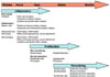
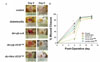
Fig. 2
Minicircle-VEGF165 gene delivery via sonoporation enhanced wound closure and increased blood perfusion in the wound tissue of treated diabetic mice. A. Macroscopic pictures of the wounds. B. Average area (in pixels) of the wounds of the minicircle-VEGF165 treated diabetic mice was significantly small as compared with those of diabetic control and pβ-VEGF165 treated diabetic mice. Minicircle-VEGF165 treatment promoted diabetic wound closure (*P < 0.05). (adapted form reference 58).
References
1. Falanga V. Wound healing and its impairment in the diabetic foot. Lancet. 2005. 366:1736–1743.

2. Nwomeh BC, Yager DR, Cohen IK. Physiology of the chronic wound. Clin Plast Surg. 1998. 25:341–356.
3. Li J, Chen J, Kirsner R. Pathophysiology of acute wound healing. Clin Dermatol. 2007. 25:9–18.
4. Kirsner RS, Eaglstein WH. The wound healing process. Dermatol Clin. 1993. 11:629–640.
5. Singer AJ, Clark RAF. Cutaneous wound healing. N Engl J Med. 1999. 341:738–746.
6. Postlethwaite AE, Kang AH. Collagen- and collagen peptide induced chemotaxis of human blood monocytes. J Exp Med. 1976. 143:1299–1307.
7. Lewis JS, Lee JA, Underwood JC, Harris AL, Lewis CE. Macrophage responses to hypoxia: relevance to disease mechanisms. J leukoc biol. 1999. 66:889–900.
8. Falanga V. The chronic wound: impaired healing and solutions in the context of wound bed preparation. Blood Cells Mol Dis. 2004. 32:88–94.
9. Hell E, Lawrence JC. The initiation of epidermal wound healing in cuts and burns. Br J Exp Pathol. 1979. 60:171–179.
10. Santoro MM, Gaudino G. Cellular and molecular facets of keratinocyte reepithelization during wound healing. Exp Cell Res. 2005. 304:274–286.
11. Giancotti FG, Ruoslahti E. Integrin signaling. Science. 1999. 285:1028–1032.
12. Sepp NT, Li LJ, Lee KH, Brown EJ, Caughman SW, Lawley TJ, Swerlick RA. Basic fibroblast growth factor increases expression of the alpha v beta 3 integrin complex on human microvascular endothelial cells. J Invest Dermatol. 1994. 103:295–299.
13. Parks WC. Matrix metalloproteinases in repair. Wound Repair Regen. 1999. 7:423–432.
14. Kurkinen M, Vaheri A, Roberts PJ, Stenman S. Sequential appearance of fibronectin and collagen in experimental granulation tissue. Lab Invest. 1980. 43:47–51.
15. Woodley DT, O'Keefe EJ, Prunieras M. Cutaneous wound healing: a model for cell-matrix interactions. J Am Acad Dermatol. 1985. 112:420–433.
16. Clark RA. Basics of cutaneous wound repair. J Dermatol Surg Oncol. 1993. 19:693–706.
17. Welch MP, Odland GF, Clark RA. Temporal relationships of F-actin bundle formation, collagen and fibronectin matrix assembly, and fibronectin receptor expression to wound contraction. J Cell Biol. 1990. 110:133–145.
18. Booth BA, Polak KL, Uitto J. Collagen biosynthesis by human skin fibroblasts: I. Optimization of the culture conditions for synthesis of type I and type III procollagens. Biochim Biophys Acta. 1980. 607:145–160.
19. Abercrombie M, Flint MH, James DW. Wound contraction in relation to collagen formation in scorbutic guinea pigs. J Embryol Exp Morph. 1956. 4:167–175.
20. Menke NB, Ward KR, Witten TM, Bonchev DG, Diegelmann RF. Impaired wound healing. Clin Dermatol. 2007. 25:19–25.
21. Nwomeh BC, Liang HX, Cohen IK, Yager DR. MMP-8 is the predominant collagenase in healing wounds and nonhealing ulcers. J Surg Res. 1999. 81:189–195.
22. Nwomeh BC, Liang HX, Diegelmann RF, Cohen IK, Yager DR. Dynamics of the matrix metalloproteinases MMP-1 and MMP-8 in acute open human dermal wounds. Wound Repair Regen. 1998. 6:127–134.
23. Wysocki AB, Staiano-Coico L, Grinnell F. Wound fluid from chronic leg ulcers contains elevated levels of metalloproteinases MMP-2 and MMP-9. J Invest Dermatol. 1993. 101:64–68.
24. Yager DR, Zhang LY, Liang HX, Diegelmann RF, Cohen IK. Wound fluids from human pressure ulcers contain elevated matrix metalloproteinase levels and activity compared to surgical wound fluids. J Invest Dermatol. 1996. 107:743–748.
25. Lobmann R, Ambrosch A, Schultz G, Waldmann K, Schiweck S, Lehnert H. Expression of matrix-metalloproteinases and their inhibitors in the wounds of diabetic and non-diabetic patients. Diabetologia. 2002. 45:1011–1016.
26. Bullen EC, Longaker MT, Updike DL, Benton R, Ladin D, Hou Z, Howard EW. Tissue inhibitor of metalloproteinases-1 is decreased and activated gelatinases are increased in chronic wounds. J Invest Dermatol. 1995. 104:236–240.
27. Mast B, Schultz G. Interactions of cytokines, growth factors, and proteases in acute and chronic wounds. Wound Repair Regen. 1996. 4:420–441.
28. Cooper DM, Yu EZ, Hennessey P, Ko F, Robson MC. Determination of endogenous cytokines in chronic wounds. Ann Surg. 1994. 219:688–691.
29. Brem H, Tomic-Canic M. Cellular and molecular basis of wound healing in diabetes. J Clin Invest. 2007. 117:1219–1222.
30. Galkowska H, Wojewodzka U, Olszewski WL. Chemokines, cytokines, and growth factors in keratinocytes and dermal endothelial cells in the margin of chronic diabetic foot ulcers. Wound Repair Regen. 2006. 14:558–565.
31. Goren I, Muller E, Pfeilschifter J, Frank S. Severely impaired insulin signaling in chronic wounds of diabetic ob/ob mice: a potential role of tumor necrosis factor-alpha. Am J Pathol. 2006. 168:765–777.
32. Galiano RD, Tepper OM, Pelo CR, Bhatt KA, Callaghan M, Bastidas N, Bunting S, Steinmetz HG, Gurtner GC. Topical vascular endothelial growth factor accelerates diabetic wound healing through increased angiogenesis and by mobilizing and recruiting bone marrow-derived cells. Am J Pathol. 2004. 164:1935–1947.
33. Maruyama K, Asai J, Ii M, Thorne T, Losordo DW, D'Amore PA. Decreased macrophage number and activation lead to reduced lymphatic vessel formation and contribute to impaired diabetic would healing. Am J Pathol. 2007. 170:1178–1191.
34. Gibran NS, Jang YC, Isik FF, Greenhalgh DG, Muffley LA, Underwood RA, Usui ML, Larsen J, Smith DG, Bunnett N, Ansel JC, Olerud JE. Diminished neuropeptide levels contribute to the impaired cutaneous healing response associated with diabetes mellitus. J Surg Res. 2002. 108:122–128.
35. Blakytny R, Jude E. The molecular biology of chronic wounds and delayed healing in diabetes. Diabet Med. 2006. 23:594–608.
36. Tsuboi R, Shi CM, Sato C, Cox GN, Ogawa H. Co-administration of insulin-like growth factor (IGF)-I and IGF-binding protein-1 stimulates wound healing in animal models. J Invest Dermatol. 1995. 104:199–203.
37. Roberts AB. Transforming growth factor-β: activity and efficacy in animal models of wound healing. Wound Rep Regen. 1995. 3:408–418.
38. Galeano M, Deodato B, Altavilla D, Cucinotta D, Arsic N, Marini H, Torre V, Giacca M, Squadrito F. Adeno-associated viral vector-mediated human vascular endothelial growth factor gene transfer stimulates angiogenesis and wound healing in the genetically diabetic mouse. Diabetologia. 2003. 46:546–555.
39. Greenhalgh DG, Sprugel KH, Murray MJ, Ross R. PDGF and FGF stimulate wound healing in the genetically diabetic mouse. Am J Pathol. 1990. 136:1235–1246.
40. Brown DL, Kao WW-Y, Greenhalgh DG. Apoptosis downregulates inflammation under the advancing epithelial wound edge: delayed patterns in diabetes and improvement with topical growth factors. Surgery. 1997. 121:372–380.
41. Matsuda H, Koyama H, Sato H, Sawada J, Itakura A, Tanaka A, Matsumoto M, Konno K, Ushio H, Matsuda K. Role of nerve growth factor in cutaneous wound healing: accelerating effects in normal and healing-impaired diabetic mice. J Exp Med. 1998. 187:297–306.
42. Cantürk NZ, Vural B, Esen N, Cantürk Z, Oktay G, Kirkali G, Solakoglu S. Effects of granulocyte-macrophage colony-stimulating factor on incisional wound healing in an experimental diabetic rat model. Endocr Res. 1999. 25:105–116.
43. Ishikawa T, Terai H, Yamamoto T, Harada K, Kitajima T. Delivery of a growth factor fusion protein having collagen-binding activity to wound tissues. Artif Organs. 2003. 27:147–154.
44. Yoshida S, Matsumoto K, Tomioka D, Bessho K, Itami S, Yoshikawa K, Nakamura T. Recombinant hepatocyte growth factor accelerates cutaneous wound healing in a diabetic mouse model. Growth Factors. 2004. 22:111–119.
45. Bennett SP, Griffiths GD, Schor AM, Leese GP, Schor SL. Growth factors in the treatment of diabetic foot ulcers. Br J Surg. 2003. 90:133–146.
46. Loot MA, Kenter SB, Au FL, van Galen WJ, Middelkoop E, Bos JD, Mekkes JR. Fibroblasts derived from chronic diabetic ulcers differ in their response to stimulation with EGF, IGF-I, bFGF and PDGF-AB compared to controls. Eur J Cell Biol. 2002. 81:153–160.
47. Hasan A, Murata H, Falabella A, Ochoa S, Zhou L, Badiavas E, Falanga V. Dermal fibroblasts from venous ulcers are unresponsive to the action of transforming growth factor-beta 1. J Dermatol Sci. 1997. 16:59–66.
48. Kim BC, Kim HT, Park SH, Cha JS, Yufit T, Kim SJ, Falanga V. Fibroblasts from chronic wounds show altered TGF-beta-ignaling and decreased TGF-beta type II receptor expression. J Cell Physiol. 2003. 195:331–336.
49. Altavilla D, Saitta A, Cucinotta D, Galeano M, Deodato B, Colonna M, Torre V, Russo G, Sardella A, Urna G, Campo GM, Cavallari V, Squadrito G, Squadrito F. Inhibition of lipid peroxidation restores impaired vascular endothelial growth factor expression and stimulates wound healing and angiogenesis in the genetically diabetic mouse. Diabetes. 2001. 50:667–674.
50. Frank S, Hubner G, Breier G, Longaker MT, Greenhalgh DG, Werner S. Regulation of vascular endothelial growth factor expression in cultured keratinocytes. J Biol Chem. 1995. 270:12607–12613.
51. Boulton AJ, Kirsner RS, Vileikyte L. Clinical practice. Neuropathic diabetic foot ulcers. N Engl J Med. 2004. 351:48–55.
52. Hinchliffe RJ, Valk GD, Apelqvist J, Armstrong DG, Bakker K, Game FL, Hartemann-Heurtier A, Löndahl M, Price PE, van Houtum WH, Jeffcoate WJ. A systematic review of the effectiveness of interventions to enhance the healing of chronic ulcers of the foot in diabetes. Diabetes Metab Res Rev. 2008. 24:suppl 1. S119–S144.
53. Duzgun AP, Satir HZ, Ozozan O, Saylam B, Kulah B, Coskun F. Effect of hyperbaric oxygen therapy on healing of diabetic foot ulcers. J Foot Ankle Surg. 2008. 47:515–519.
54. Papanas N, Maltezos E. Becaplermin gel in the treatment of diabetic neuropathic foot ulcers. Clin Interv Aging. 2008. 3:233–240.
55. Hong JP, Jung HD, Kim YW. Recombinant human epidermal growth factor (EGF) to enhance healing for diabetic foot ulcers. Ann Plast Surg. 2006. 56:394–398.
56. Hanft JR, Pollak RA, Barbul A, van Gils C, Kwon PS, Gray SM, Lynch CJ, Semba CP, Breen TJ. Phase I trial on the safety of topical rhVEGF on chronic neuropathic diabetic foot ulcers. J Wound Care. 2008. 17:30–32.
57. Yoon CS, Jung HS, Kim TK, Kwon MJ, Kim MK, Lee M, Koh KS, Rhee BD, Park JH. Comparison of the efficiency and toxicity of sonoporation with branched polyethylenimine-mediated gene transfection in various cultured cell lines. J Drug Target. 2008. 16:773–779.
58. Yoon CS, Jung HS, Kwon MJ, Lee SH, Kim CW, Kim MK, Lee M, Park JH. Sonoporation of the Minicircle-VEGF(165) for Wound Healing of Diabetic Mice. Pharm Res. 2009. 26:794–801.
 XML Download