



 PDF
 ePub
 Citation
 Print


Introduction
In our previous study1, patients with post-tuberculosis emphysema (PTE) were found to have a higher PaCO2 and a lower right ventricular ejection fraction (RVEF) than patients with pure pulmonary emphysema (PE) at rest, whereas their FEV1 and FVC results were similar. No significant difference was found in resting PaO2 and A-a O2 gradient between the PTE and PE patient groups. However, after exercise, A-a O2 gradient was significantly widened in PTE, which strongly suggests that V/Q mismatching increased with exercise due to a combination of changes in pulmonary arterial pressure and total pulmonary perfusion (cardiac output) with clear redistribution of the right ventricular output to poorly ventilated areas. In addition, RVEF diminished with exercise in the PTE group, which showed clear hypercapnia and more severe hypoxia after exercise than the PE group.
Angiotensin II may contribute to hypoxic pulmonary hypertension due to its vasoconstrictor and growth-stimulatory effects on vascular smooth muscle cells. Therefore, the use of angiotensin-converting enzyme (ACE) inhibitors might reduce hypoxic pulmonary hypertension by reducing pulmonary vasomotor tone and/or vascular remodeling2.
Recently, several studies have been reported that ACE inhibitors can reduce exacerbations and mortality, and treat cardiovascular co-morbid conditions in patients with COPD3-5. However, its long-term use in patients with PTE has not been studied.
We hypothesized that right ventricular function and V/Q matching are likely to be altered by long-term ACE inhibition in PTE, since ACE inhibition alters pulmonary arterial pressure and right ventricular afterload, which can be crudely estimated from mean pulmonary arterial pressure. The aim of this prospective study was to assess the effects of long-term ACE inhibition on the heart, and gas exchange both at rest and after exercise in patients with PTE.
Materials and Methods
1. Patient population
Based on medical history, clinical symptoms, and spirometric lung function and radiological findings, PTE patients were enrolled until 20 patients completed the study according to the protocol. All of the patients suffered from functional class II or III dyspnea (as classified by the New York Heart Association), had been in a stable state for 3 months before enrollment, and had not experienced respiratory failure or heart disease. The presence of significant emphysema was confirmed by HRCT; maximal transpulmonary pressure at TLC and coefficient of retraction values were greater than 19 cm H2O and 2.5 cm H2O/L, respectively, thus proving the presence of concomitant pneumosclerosis with emphysema in PTE6.
This study was approved by the Institutional Review Board of the hospital.
2. Study protocol
This study was designed to be minimally invasive, and neither right heart catheterization nor arterial cannulation was performed.
Pulmonary function testing, arterial blood gas analysis, symptom-limited exercise, and echocardiography were performed according to the following methods at baseline (before ACE inhibition) and then after 6 months of treatment with an ACE inhibitor, Ramipril (10 mg once/day).
No change in subject activity occurred during the study period, specifically, they did not participate in any rehabilitation or exercise program.
1) Pulmonary function testing and arterial blood gas analysis
Using a computerized flow-volume device and an automated body plethysmograph (CareFusion, San Diego, CA, USA), FVC, FEV1, FEV1/FVC, VC, RV, FRC, TLC and airway resistance were measured. Esophageal pressure was measured by inserting an intra-esophageal balloon, and lung compliance is defined as the slope of the pressure- volume curve between FRC and 0.5 L. Maximal transpulmonary pressure at TLC was measured using a body plethysmograph, and the coefficient of retraction (defined as the maximal pressure at TLC in cm H2O divided by the volume at TLC in liters) was calculated6. DLCO was measured using the single-breath method, as described by the American Thoracic Society standard7.
Arterial blood was withdrawn by radial puncture at rest and immediately after symptom-limited exercise (nearly simultaneously with the termination of exercise). Blood gases were analyzed using an automatic analyzer (GMI Inc., Ramsey, MN, USA), and A-a O2 gradient was calculated using the formula: [A-a O2 gradient=150-PaCO2/R-PaO2 mm Hg] where R (respiratory quotient)=0.8 for room air.
2) Symptom-limited exercise and echocardiography
Exercise was performed by treadmill using a CASE-8000 ergometer (GE Marquette Medical Systems, Munich, Germany) according to the Bruce (stepwise) protocol, and was terminated when a patient complained of intolerable dyspnea. Maximal heart rate, blood pressure, exercise duration, and metabolic equivalents (METs) were recorded.
Echocardiography was done at rest and immediately after exercise using an Acuson Sequoier C-256 sonograph (Acuson Co., Malvern, PA, USA).
To evaluate right ventricular function, two-dimensional echocardiograms were recorded from apical and subcostal four-chamber views. The echocardiograms, recorded on videotapes, were analyzed in stop-frame mode. The area of the right ventricle was measured at end-diastole (the peak of the R wave by EKG) and at end-systole (the end of T wave by EKG) during the expiratory cycle (since it was difficult for the dyspneic patients to hold breath after exercise). The final end-diastolic and end-systolic values were obtained by averaging 5 or 6 measurements. Right ventricular end-diastolic and end-systolic volumes were obtained from these values by using the modified biplane Simpson's method using the formula V=π/4Σ (I=1-20) (two-chamber area) (four-chamber area) L/20, where L is the right ventricular long axis. RVEF, a measure of the systolic pump performance of the right heart, was calculated from these ventricular volumes.
LVEF and CO were measured from parasternal views, and pulsed, continuous and color-flow Doppler studies were also performed.
Results
1. Demography
Twenty-four patients were enrolled in this study: 23 men and one woman, however, four participants dropped out, because of dizziness in one (only female) case, cough in another one case, and patient's request in the other two cases.
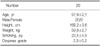
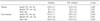
The demographic characteristics of the patients who completed the study are displayed in Table 1.
2. Pulmonary function and arterial blood gas profiles
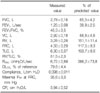
Pulmonary function profiles in PTE are summarized in Table 2. FVC was 65.0±4.2% (of predicted value), FEV1 39.9±2.5%, and the FEV1/FVC ratio 45.3±3.5%. RV increased more than TLC did, and airway resistance was very high. Lung compliance did not increase, but DLCO reduced. After long-term ACE inhibition, no significant change was observed in the above variables.
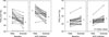
At baseline; pH was 7.38±0.01 at rest and 7.29±0.01 after exercise. After exercise, PaO2 reduced from 74.2±1.9 to 66.6±1.6 mm Hg (p<0.01) and PaCO2 increased from 42.4±0.9 to 44.9±1.1 mm Hg (p<0.01; Figure 1).
At 6 months after ACE inhibition, pH (7.39±0.01 at rest and 7.27±0.18 after exercise) and resting PaCO2 (42.8±0.8 mm Hg; p=0.524) were not altered. However, resting PaO2 (83.8±1.6 mm Hg) was higher than at baseline, and PaO2 reduced to 74.0±1.2 mm Hg after exercise, but remained higher than that at baseline (each p<0.01). Post-exercise PaCO2 (46.3±1.1 mm Hg) was higher than resting PaCO2 and than at baseline (each p<0.01; Figure 1).
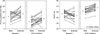
At baseline, A-a O2 gradient was 22.5±1.5 mm Hg at rest and increased to 26.9±1.6 mm Hg after exercise (p<0.05). After ACE inhibition, resting (12.4±1.4 mm Hg) and post-exercise (17.8±1.5 mm Hg) A-a O2 gradient were lower than at baseline (each p<0.01), but A-a O2 gradient increased after exercise (p<0.01; Figure 2).
3. Exercise profiles and hemodynamic changes
Exercise time was 344.9±31.7 seconds at baseline and 357.5±39.2 seconds after ACE inhibition (p=0.387), whereas exercise performance was 7.41±0.59 at baseline and 7.74±0.68 METs after ACE inhibition (p=0.453). Changes in hemodynamic profiles are summarized in Table 3. No clinically significant change was observed in systemic blood pressure or heart rate after ACE inhibition, although there were some statistically significant differences. Pulmonary arterial pressure could not be assessed because of echocardiographic limitations.
At baseline, RVEF was 56.9±1.2% at rest and reduced to 53.5±1.7% after exercise, without significance (p=0.055). After ACE inhibition, resting RVEF (61.5±1.0%) was higher than at baseline, and RVEF increased further to 67.6±1.2% after exercise (each p<0.01), which was also higher than at baseline (p<0.01; Figure 2).
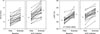
At baseline, CO was 5.42±0.22 L/min at rest and increased to 6.72±0.24 L/min after exercise (p<0.01). After ACE inhibition, resting CO (6.37±0.24 L/min) was higher than at baseline, and CO increased to 8.27±0.34 L/min after exercise (each p<0.01), which was also higher than at baseline (p<0.01; Figure 3).
At baseline, LVEF was 62.7±1.6% at rest and increased to 71.5±1.5% after exercise (p<0.01). After ACE inhibition, resting LVEF (66.7±2.7%) was higher than at baseline (p<0.05), and LVEF increased to 74.5±2.6% after exercise (p<0.01), which was also higher than at baseline (p<0.05; Figure 3).
Discussion
In patients with severe fibrotic pulmonary disease following tuberculosis, the clinical pattern of disease is almost indistinguishable from that of emphysema with severe airway obstruction8. Obstructive airway changes in PTE increase with age, the number of cigarettes smoked, and the extent of the initial tuberculous disease9,10. The impairment of pulmonary function found in this study was similar to that of stage III (FEV1/ FVC <70%; 30% ≤ FEV1 <50% predicted); Severe COPD according to the Global Initiative for Chronic Obstructive Lung Disease (GOLD) guidelines11.
In this study, considering decreased cardiopulmonary reservoir capacity in patients with PTE, symptom-limited exercise was adopted. However, post-exercise heart rates showed that exercise levels reached submaximal levels. Ideally, it is best to sample arterial blood during exercise to ensure steady conditions for gas analysis. However, instead, arterial blood gas analysis was done by direct arterial puncture immediately after exercise, because arterial cannulation was not performed. There was no measurable interval between sampling and exercise cessation.
Rubin suggested following guidelines for vasodilator therapy in cases of right ventricular failure or pulmonary hypertension12: 1) pulmonary vascular resistance is lowered by 20%, 2) cardiac output is increased or unchanged, 3) pulmonary artery pressure is decreased or unchanged, and 4) systemic blood pressure is not significantly reduced (eg, no side effects). After these benefits are established and the patient has begun on a therapeutic regimen of a particular medication, right ventricular catheterization should be repeated after 4 to 6 months of continued therapy to ascertain whether the hemodynamic benefits persist. Adapting Rubin's guidelines, pulmonary function testing, arterial blood gas analysis, and echocardiography were repeated 6 months after ACE inhibition.
The normal response of the heart to exercise is at least a 5% increase in the ejection fraction of each ventricle13. In this study, 17 of 20 patients (85.0%) showed abnormal right ventricular response to exercise, and six patients (30.0%) had abnormal left ventricular performance after exercise at baseline. These findings are consistent with the results of other studies, namely, that the prevalence of right ventricular dysfunction is high in patients with advanced pulmonary disease, but that the prevalence of left ventricular dysfunction is relatively low13,14. After long-term ACE inhibition, abnormal right- or left-ventricular response to exercise was observed in only 2 cases (10.0%), respectively.
The most common mechanism of reduced RVEF in advanced pulmonary disease is augmentation of right ventricular afterload13. RVEF may be depressed in the setting of pulmonary arterial hypertension, even if contractility is normal15. Although the cause of augmented afterload has not been established clearly, due to the potent pulmonary vasoconstrictor effect of hypoxemia, it appears to be a major factor leading to pulmonary hypertension. Pulmonary vascular destruction is an additional factor in these patients. Ultimately, chronic remodeling of the pulmonary vascular bed leads to reduced recruitability of pulmonary blood vessels15,16, and this restricted, relatively nonrecruitable pulmonary vascular bed with inordinate high pulmonary arterial pressure is considered to be the most likely mechanism of abnormal right ventricular response to exercise13.
Therapies with agents that reduce right ventricular afterload have the potential to improve the systolic performance of this chamber by reducing pulmonary vascular resistance and/or pulmonary arterial pressure12. Angiotensin II is a potent vasoconstrictor that induces the growth of vascular smooth muscle cells and increases collagen deposition, which leads to vascular remodeling17, and it also plays a pivotal role in the remodeling of pulmonary arteries in hypoxia18. Therefore, ACE inhibitors have the potential to improve the pump performance of the right ventricle by reducing pulmonary vasomotor tone and/or vascular remodeling2.
ACE inhibitors may have other beneficial effects in COPD, as angiotensin II may have proinflammatory effects19, and polymorphisms of the ACE gene have been linked to increased susceptibility to COPD20. Because ACE inhibitors are routinely used in the management of hypertension, cardiac failure, and diabetes, all of which are common comorbidities of COPD, prospective trials of ACE inhibitors in patients with COPD are now warranted21.
It was suggested that peripheral oxygen delivery and exercise oxygen consumption in patients with COPD are dependent on CO, which, in turn, is related to the right ventricular function22. The most notable findings of this study were an increase in PaO2 and a decrease in A-a O2 gradient associated with an increase in ventricular ejection fraction, especially in RVEF after long-term ACE inhibition in PTE. These findings can be explained as follows: RVEF was depressed at baseline in PTE. After ACE inhibition, the systolic performance of right ventricle increased, as it was augmented by a reduced right ventricular afterload, and total pulmonary perfusion (CO) increased. For a given oxygen consumption, an increase in CO would increase mixed venous oxygen tension and increase PaO2, even if the distributions of V/Q were unchanged. Furthermore, the increment of pulmonary perfusion might be redistributed to relatively well ventilated areas, possibly by changes in vasomotor tone and alterations in vascular remodeling. Therefore, A-a O2 gradient decreased and PaO2 increased at rest after ACE inhibition. However, these parameters were re-aggravated after exercise due to respiratory pump insufficiency and the redistribution of right ventricular output to poorly ventilated, sclerosed portions of the lung in PTE1.
Right ventricular function has been shown to be important in such diseases as COPD, pulmonary thromboembolism, and right ventricular ischemia secondary to coronary artery disease13. Therefore, an ACE inhibitor can be used in these diseases if it favorably alters cardiovascular- pulmonary hemodynamics and right ventricular function. However, its routine use in stable COPD is not recommended11, because it may lower PaO2 by worsening V/Q matching, although it may acutely improve pulmonary hemodynamics22. Nevertheless, an ACE inhibitor might be also tried in selected cases of COPD, which show abnormal right ventricular response to exercise.
In PTE, the pump performance of the right heart and cardiac output were augmented after long-term ACE inhibition. V/Q mismatching was also alleviated (as evidenced by a reduction in A-a O2 gradient) with a resultant increase in peripheral oxygen delivery (as indicated by an increase in PaO2). These findings suggest that ACE inhibitor have the potential to alleviate co-morbid cardiac conditions and benefit the patients with
PTE.
 XML Download