

 PDF
PDF ePub
ePub Citation
 Print
Print


Abstract
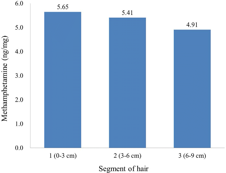
The authors report a case of an otherwise healthy 33-year-old man who presented with intracerebral hemorrhage in the right frontal lobe following chronic methamphetamine use. An autopsy was performed within 2 days after death. The postmortem examination revealed cerebral edema and intracerebral and intraventricular hemorrhage. Microscopic examination revealed endovasculitis in the systemic vessels including the aorta and carotid and coronary arteries, but no aneurysm or arteriovenous malformation. Acute toxicity and chronic methamphetamine use was verified using blood and segmental hair analysis, respectively. Cerebrovascular accidents including stroke and intracerebral and subarachnoid hemorrhage are rare in young persons, but methamphetamine use is a risk factor for cerebrovascular accidents in young adults. Therefore, forensic pathologists should be aware of the acute and chronic harmful effects of methamphetamine. Detailed history taking and toxic screening tests for illicit drug use, especially methamphetamine, as well as a meticulous postmortem examination should be conducted in young patients who died due to cerebrovascular accident.
REFERENCES
1. Chiu ZK, Bennett IE, Chan P, et al. Methamphetamine-related brainstem haemorrhage. J Clin Neurosci. 2016; 32:137–9.

2. McGee SM, McGee DN, McGee MB. Spontaneous intracerebral hemorrhage related to methamphetamine abuse: autopsy findings and clinical correlation. Am J Forensic Med Pathol. 2004; 25:334–7.
3. Yen DJ, Wang SJ, Ju TH, et al. Stroke associated with methamphetamine inhalation. Eur Neurol. 1994; 34:16–22.
4. Lineberry TW, Bostwick JM. Methamphetamine abuse: a perfect storm of complications. Mayo Clin Proc. 2006; 81:77–84.
5. Shibata S, Mori K, Sekine I, et al. Subarachnoid and intracerebral hemorrhage associated with necrotizing angiitis due to methamphetamine abuse: an autopsy case. Neurol Med Chir (Tokyo). 1991; 31:49–52.
6. Matick H, Anderson D, Brumlik J. Cerebral vasculitis associated with oral amphetamine overdose. Arch Neurol. 1983; 40:253–4.
7. Ho EL, Josephson SA, Lee HS, et al. Cerebrovascular complications of methamphetamine abuse. Neurocrit Care. 2009; 10:295–305.
8. Islam MN, Kuroki H, Hongcheng B, et al. Cardiac lesions and their reversibility after long term administration of methamphetamine. Forensic Sci Int. 1995; 75:29–43.
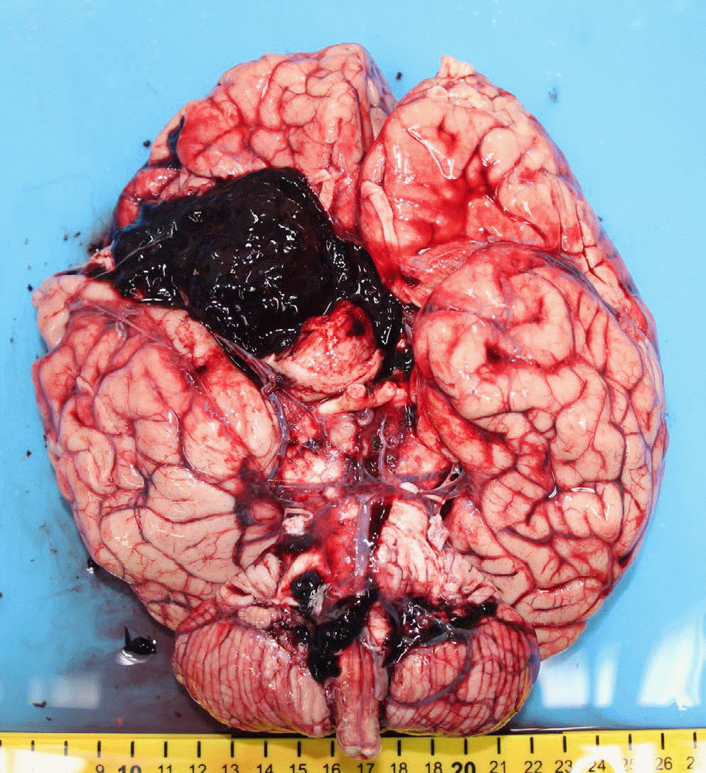
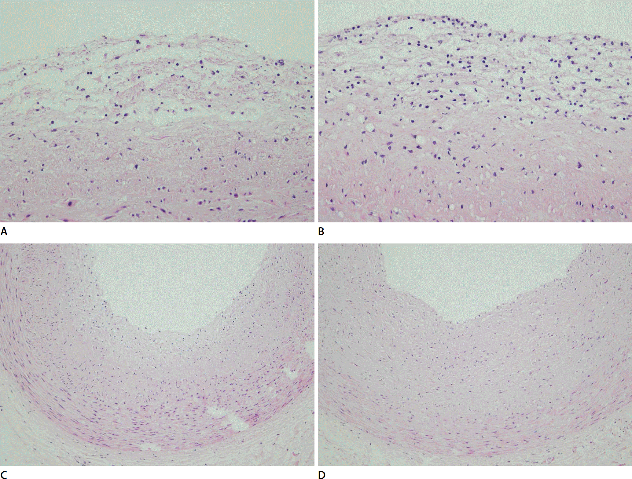
Fig. 2.
Microscopic examination verified the edema and chronic lymphocytic infiltration in the intima of the descending aorta (A) and carotid artery (B) (A and B, ×200) and thickening and infiltration of the lymphocyte in the intima of the left descending coronary artery (C) and right coronary artery (D) (C and D, × 100).
 XML Download