

 PDF
PDF ePub
ePub Citation
 Print
Print


Abstract
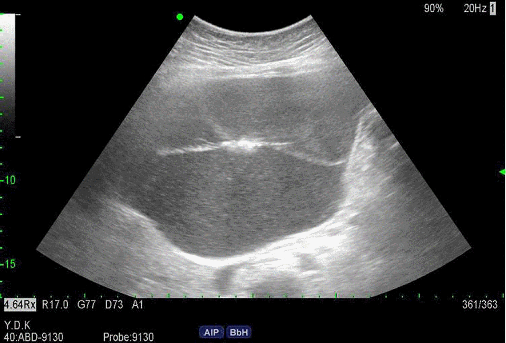
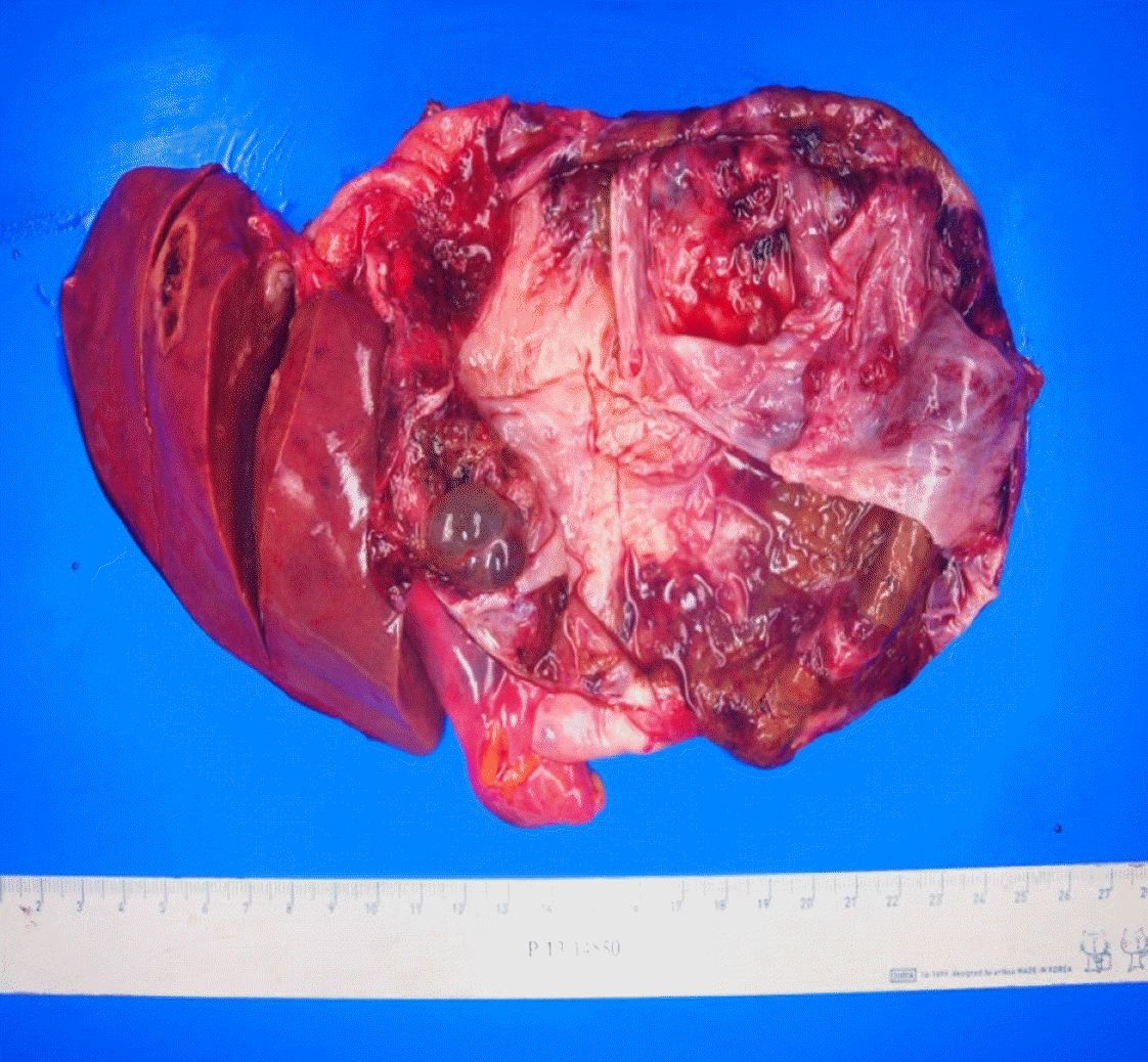
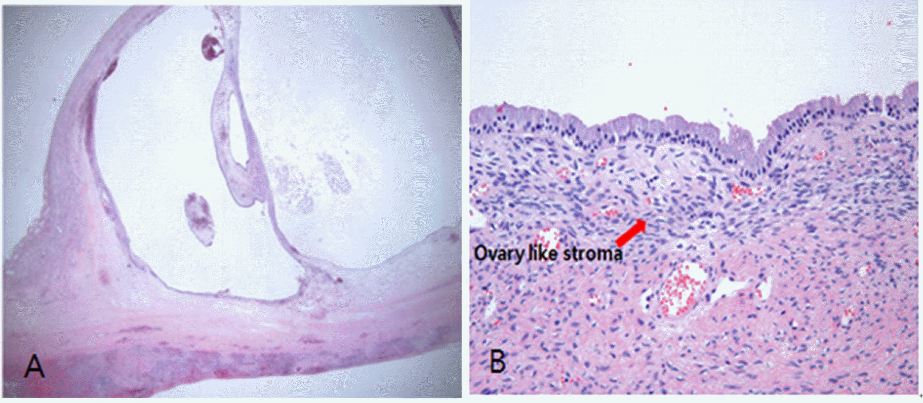
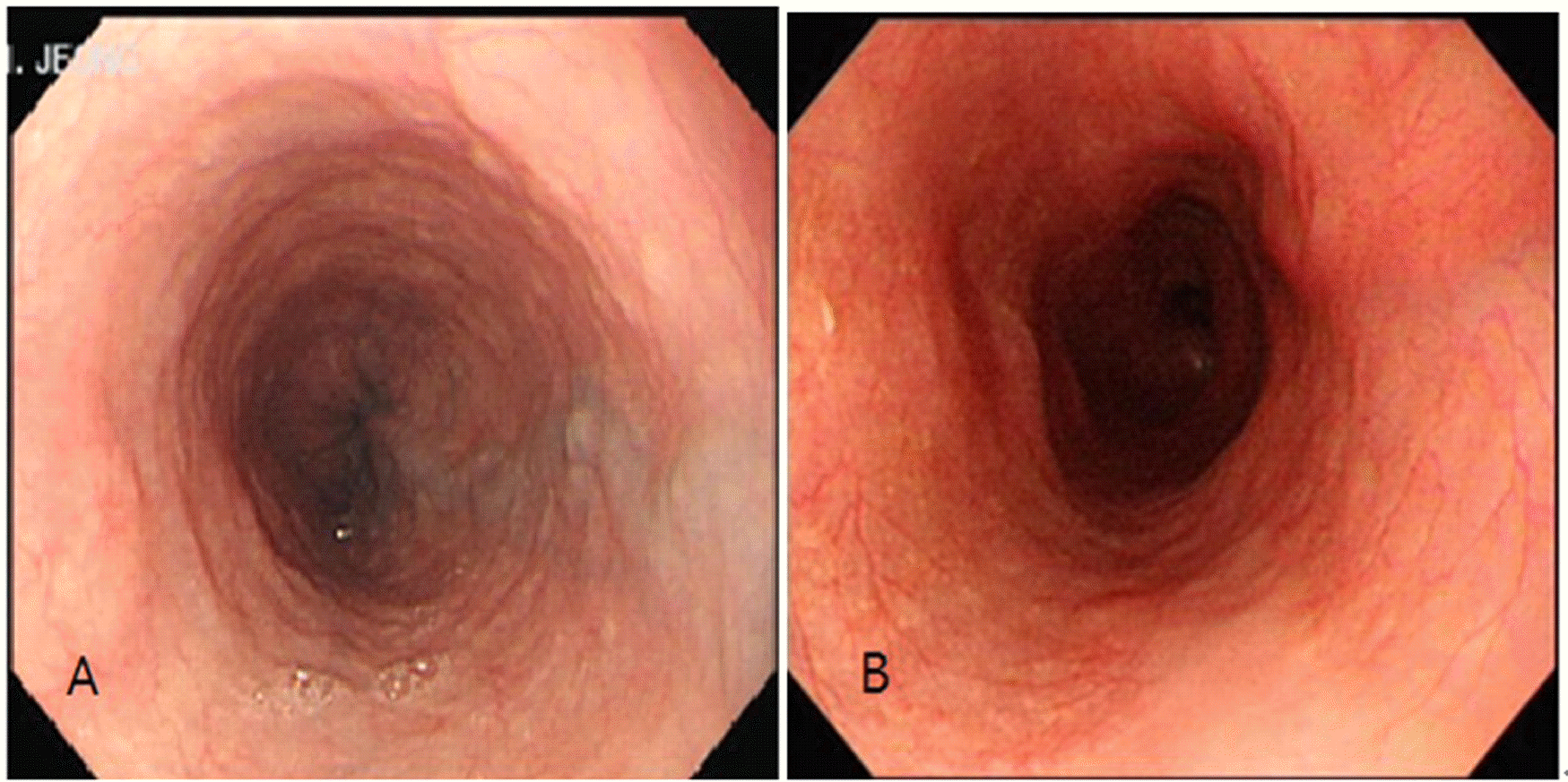
Biliary cystadenomas are benign but potentially malignant cystic neoplasm. The preferred treatment is radical resection because it is difficult to differentiate a benign from a malignant biliary cystadenoma. A 40 year-old woman presented with moderate abdominal discomfort. Esophageal varix was found up to mid-esophagus on endoscopy. She has no prior history of liver disease or chronic alcohol ingestion. About 15cm sized biliary cystadenoma was diagnosed by ultrasonography, computed tomography and magnetic resonance imaging. Serum level of bilirubin, alanine aminotransferase, alkaline phosphatase, gamma-glutamyl transpeptidase and tumor marker were elevated. The patient underwent US-guided aspiration. Tumor markers from the aspirated fluid are increased. Left hepatectomy was performed to completely remove the cyst. Histology of the resected specimen confirmed a biliary cystadenoma of the liver with ovary-like stroma. Without prior history of liver disease or chronic alcoholic ingestion, incidental finding of esophageal varix could show an important clue for diagnosis of biliary cystadenoma.
References
1. Del Poggio P, Buonocore M. Cystic tumors of the liver: a practical approach. World J Gastroenterol. 2008; 14:3616–20.
2. Ratti F, Ferla F, Paganelli M, Cipriani F, Aldrighetti L, Ferla G. Biliary cystadenoma: short- and longterm outcome after radical hepatic resection. Updates Surg. 2012; 64:13–8.

3. Grubor NM, Colovic RB, Atkinson HD, Micev MT. Giant biliary mucinous cystadenoma of the liver. Ann Hepatol. 2013; 12:979–83.
4. Erdogan D, Kloek J, Lamers WH, Offerhaus GJ, Busch OR, Gouma DJ, et al. Mucinous cystadenomas in liver: management and origin. Dig Surg. 2010; 27:19–23.
5. Gamblin TC, Holloway SE, Heckman JT, Geller DA. Laparoscopic resection of benign hepatic cysts: a new standard. J Am Coll Surg. 2008; 207:731–6.
6. Choi HK, Lee JK, Lee KH, Lee KT, Rhee JC, Kim KH, et al. Differential diagnosis for intrahepatic biliary cystadenoma and hepatic simple cyst: significance of cystic fluid analysis and radiologic findings. J Clin Gastroenterol. 2010; 44:289–93.
7. Ryu KH, Lee TH, Kwon TG. Vascular lesion in an adult mimicking esophageal varix. Gastroenterololy. 2013; 144:35.

 XML Download