

 PDF
PDF ePub
ePub Citation
 Print
Print


Introduction
Synchronous tumors/lesions represent simultaneous occurrence of different tumors/lesions in more than one organ at the same time. Synchronous tumors of the endometrium and ovary account for 50% to 70% of all synchronous female genital tract malignancies.1 Usual combination is that of endometrioid adenocarcinoma of endometrium and ovarian endometrioid adenocarcinoma. Synchronous tumors of completely different histological types and of different histogenesis are very rare. We report a case of endometrioid adenocarcinoma of the endometrium which is a malignant epithelial tumor, benign cystic teratoma in one ovary belonging to the germ cell tumors of ovary, fibrothecoma in the other ovary which is under the category of sex cord stromal tumor, and adenomyosis of uterus. This case constitutes a potpourri of tumors with unrelated histogenesis in a single organ system. Extensive search of the English medical literature did not reveal case with a combination of such tumors in the female genital organ.
Case Report
A 59-year-old female patient presented with postmenopausal bleeding of 1 year duration. The bleeding was occasional and mild. Patient is gravida 2, para 2, live 2; both childbirths were full term normal, last 35 years back. Not sterilised. Menopause was attained 6 years back, Menarche was at the age of 13 years. Patient was on treatment for type 2 diabetes since last 7 years. There was no history of any other illness. No family history of malignancy. All investigations were within normal limits. On clinical examination, a soft palpable mass was felt in the left iliac fossa 6.0 × 6.0 cm. Uterus was atrophic. Ultrasonogram showed a cystic lesion with internal hyper echoic area similar to fat in the left adnexa possibly a dermoid cyst. Endometrial thickness assessed was 7 mm. An endometrial biopsy was done preoperatively which was not diagnostic. The report was descriptive as mainly blood clot with a few closely packed atypical endometrial glands suspicious of carcinoma. So with a provisional clinical diagnosis of left ovarian teratoma and suspicious endometrial carcinoma, total abdominal hysterectomy with bilateral salpingo-oophorectomy was decided by the gynaecologist.
Per operatively, cystic mass lesion was observed in the left ovary. Uterus was atrophic. Right ovary was normal in size. Patient underwent total abdominal hysterectomy with bilateral salpingo-oophorectomy.
We received the specimen in our pathology department. Uterus measured 8.0 × 4.0 × 3.0 cm. Left ovary was cystic and measured 8.5 × 7.0 × 4.0 cm. Left fallopian tube measured 3.0 cm ine nlgth. Right ovary measured 3.5 × 3.0 × 1 .0 cm. Right tube measured 4.5 cm in elngth.
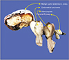
On cutting open, the uterus showed an irregular friable grey white neoplasm measuring 4.0 × 1.3 × 4.0 cm in the endometrial cavity with attachment to one side of the wall invading into the myometrium and lower uterine segment. No lymph nodes were identified in the parametrium. Opening the left ovarian cyst showed a unilocular cyst containing pultaceous material and tufts of hair. Inner surface was smooth. No solid areas or papillary projections noted. Cut section of the right fallopian tube was unremarkable. Cut section of the right ovary showed a circumscribed pale white fibrous neoplasm measuring 1.0 × 1.0 cm with focal pale yellow spots. Portion of ovarian tissue was seen surrounding the neoplasm. Cut section of right fallopian tube was unremarkable (Fig. 1).
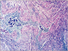
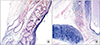
Microscopy showed a neoplasm in the endometrial cavity composed of tumor cells arranged in closely packed glandular pattern. The cells were columnar with moderate amount of cytoplasm and round/oval hyperchromatic moderately pleomorphic nuclei. Mitosis 3 to 4 per high power field. Glandular lumen was filled with neutrophils and necrotic debri (Fig. 2A, 2B). Areas of necrosis were noted. The tumor is seen infiltrating to less than half thickness of the myometrium and the upper part of lower uterine segment. Uterus also showed multiple foci of adenomyosis (Fig. 3). Both parametrium were free of tumor and no lymph nodes were identified.
Sections from left ovarian cyst showed a neoplasm with varying tissues of the 3 germ cell layers -stratified squamous/ mucinous/respiratory epithelial lined cyst wall, sebaceous glands, hair follicles, mature cartilage, smooth muscle tissue, and thyroid follicles etc. No neuro-epithelial or other immature tissue noted (Fig. 4A, 4B). Sections from the right ovary showed a circumscribed neoplasm composed of intersecting short fascicles of spindle cells in a collagenous stroma and interspersed with luteinized theca cells (Fig. 5A–5C). Both fallopian tubes were histologically unremarkable.
Discussion
In the present case, histology showed multiple synchronous lesions in the genital tract- grade 1 endometrioid adenocarcinoma of the endometrium-an epithelial malignancy, benign cystic teratoma of the left ovary - a benign germ cell tumor, fibrothecoma of right ovary - a sex cord stromal tumor of benign pure stromal tumor category and adenomyosis of the uterus which is ectopic placement of endometrial tissue in the myometrium.
About 1% to 2% of women with gynaecological cancers are found to have 2 or more simultaneous independent primary malignancies.2 The typical histology of Synchronous cancer is endometrioid adenocarcinoma in both the endometrium and the ovary, which is found in > 70% of cases.3 But in our case, the endometrial carcinoma was not associated with endometrioid carcinoma of the ovary but a histogenetically different benign tumor in both ovaries.
Many other combinations of endometrial carcinoma with tumors of the ovary like granulosa cell tumor, Brenner tumor, benign cystic teratoma, fibroma etc. have been reported. Sharma et al.4 reported a case of a coexisting Brenner tumor and well-differentiated endometrial carcinoma in a 55-year-old nulliparous postmenopausal woman. Comunoglu et al.5 has reported a case of an ovarian hemangioma occurring synchronously with contralateral mature cystic teratoma in an 81-year-old woman. Pekin et al.6 reported in a 62-year-old patient a synchronous combination of cervical squamous cell carcinoma, right ovarian Brenner tumor, left ovarian granulosa cell tumor and endometrial polyps. But no report has been found as in our case with benign cystic teratoma in one ovary and fibrothecoma in the contralateral ovary in association with endometrial carcinoma and adenomyosis.
Combination tumors are more common in hereditary mutation syndromes like nonpolyposis colon cancer (Lynch) syndrome, breast ovarian cancer syndrome, Li-Fraumeni syndrome etc. Our case had no clinical evidence of any syndromic association.
Kim7 reported an unusual clinical manifestation of the torsion of a dermoid cyst and fibrothecoma in the right ovary with postmenopausal bleeding. This was in the same ovary as a collision tumor and not a synchronous tumor. Collision tumor is coexistence of 2 distinct tumors in the same organ without any histological intermixing.8 Singh and Singh9 reported 4 cases of collision tumor of ovary-combination of mucinous cystadenoma with teratoma in 2 cases, teratoma with serous cystadenoma/carcinoma in the other 2 cases. One of the authors of this article has reported a rare collision tumor of fibroma with serous cystadenoma in the ovary.10 In the present case, the tumors in the ovary was not collision but synchronous, with benign cystic teratoma in one ovary and a fibrothecoma in the other.
Fibromas of the ovary are usually hormonally inactive. Thecomas usually present with estrogenic manifestation. In this case, the fibro-thecoma in the ovary was very small in size (1.0 cm) with a minor theca cell component, which was clinically and sonologically undetected. Hence an estrogenic stimulation of the endometrium to cause endometrial carcinoma is very unlikely. Moreover, the endometrial carcinoma was larger in size. Benign cystic teratoma in the other ovary is also unrelated to the pathogenesis of endometrial carcinoma. The incidence of this synchronous tumors from sites having different embryological origin and histological appearance is unexplainable. This probably may be a coincidental combination. The patient is under close follow up and is free of disease till this date.
Conclusion
Hereby, we are reporting an unusual case of a combination of tumors in the female genital organs in a 59-yearold patient detected synchronously. This patient had endometrioid adenocarcinoma of the endometrium-a malignant epithelial tumor, benign cystic teratoma of one ovary - a benign germ cell tumor, fibrothecoma of the other ovary belonging to the pure ovarian benign stromal tumor group. Apart from these tumors, patient also had adenomyosis of the uterus. To the best of our knowledge, such a combination is not reported in the English medical literature and this is the first case to be erported.


 XML Download