

 PDF
PDF ePub
ePub Citation
 Print
Print


Introduction
The pathophysiologic link between adipose tissue and bone metabolism is not entirely understood, but adipokines seem to play an important role.12 Levels of several adipokines have been found to correlate with bone mass.34
Irisin is a recently identified myokine secreted by skeletal muscle and adipose tissue.5 Serum irisin levels were found to be lower in women with previous osteoporotic fractures compared to controls.67 Chemerin, a more recently discovered adipose tissue specific adipokine, has a crucial role in adipocyte differentiation and development, as well as in glucose and lipid metabolism.8 Apolipoprotein M (apoM) has atheroprotective effects attributed to high-density lipoprotein (HDL) and has an important role in insulin and glucose metabolism.9 In the present study, we evaluated the links among serum chemerin, irisin and apoM levels in women with postmenopausal osteoporosis. Such knowledge may lead to new insights into the understanding of different cross-talks and treatment of people suffering osteoporosis.
The objective of this study is to describe the levels of chemerin, irisin and apoM in women with postmenopausal osteoporosis.
Materials and Methods
A total of 176 women who had been postmenopausal for at least 12 months and visited Bozok University Menopause Clinic for the evaluation and treatment of osteoporosis were enrolled in this cross-sectional study.
The study included 88 women with postmenopausal osteoporosis. The current analysis is a case-control study in postmenopausal women with T-score > -1 were selected as controls (n = 88) and matched on age (within 2 years), body mass index (BMI) (within 1.0 kg/m2) in a 1:1 ratio to cases. Bone mineral density (BMD) was measured using dual energy X-ray absorptiometry (DXA). Measurements included BMD of the hip (femoral neck) and lumbar spine (L1-L4) and were performed in the anteroposterior (AP) view for the lumbar spine. According to World Health Organization criteria, a T-score of ≥ -1 denotes normal bone, a T-score between -1 and -2.5 denotes osteopenia, and a T-score of ≤ -2.5 denotes osteoporosis. The BMI was calculated as weight (kg)/square height (m2).Women with systemic disorders (diabetes mellitus, rheumatoid arthritis...), who had taken drugs that could affect bone metabolism, with premature menopause (< 40 years of age) were excluded. The protocol used was approved by the Committee on Ethics of the university and funded by the university (BAP Project).
After an overnight fasting, venous blood samples were collected. All samples were obtained during the spring (March to May 2014). Within an hour of venipuncture, whole blood samples were centrifuged and separated, and serum portions were frozen at -80℃ for future analysis. ApoM, irisin and chemerin levels were determined using a commercially available enzyme-linked immunosorbent assay (ELISA) kit (BioVendor Laboratory Medicine, Brno, Czech Republic). All other parameters were measured using standard laboratory methods in the core laboratory. Limit of detection of chemerin kit is 0.1 ng/mL. The intra-assay and inter-assay coefficient of variation (CV) were less than 7%. For irisin, the lowest level of irisin that can be detected by the assay is 1 ng/mL. Intra-assay CV (%) was 4.86 to 6.75 and inter-assay CV (%) was 6.2 to 9.66. The lowest level of apoM that can be detected by this assay is 0.07 ng/mL. Intra-assay CV (%) was 4.90 to 5.22 and inter-assay CV (%) was 5.70 to 5.80.
All analyses were conducted using SPSS Windows version 15.0 software (SPSS Inc., Chicago, IL, USA). We conducted a power analysis according to the results of our pilot study, and we determined suitable sample size as 60 patients (α = 0.05, β = 0.80 and effect size d = 0.66). Continuous variables were expressed as the mean ± standard deviation (SD). The normality of the distribution was evaluated with the Kolmogorov-Smirnov test. Parameter comparisons were performed with Student's t-test and the Mann-Whitney U test. The correlation between variables was assessed with Pearson's and Spearman's correlation tests. P value of < 0.05 was considered to be statistically significant.
Results
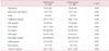
The results from the 88 patients diagnosed with osteoporosis and the 88 patients who consisted of the control group were depicted in Table 1. There were no significant differences in age, BMI, parity, cholesterol and apoM levels between the two groups.
C-reactive protein levels were significantly increased in women with osteoporosis. Serum chemerin levels (240.1 ± 46.1 vs. 261.5 ± 50.8 ng/mL) were significantly lower in the women with osteoporosis compared to the controls (Table 1). In contrast serum irisin levels were also decreased in women with osteoporosis (0.7 ± 0.2 vs. 0.8 ± 0.2 ng/mL; P = 0.007).
We found a significant negative correlation between chemerin and irisin levels (r = -0.962, P = 0.0001).
Discussion
In the present study, serum chemerin and irisin concentrations have been shown to be decreased in patients with osteoporosis. Irisin is a myokine with a possible protective effect on metabolic disorders. Similar to our results, serum irisin concentrations have also been shown to be decreased in patients with osteoporosis.67 Zhang et al.8 revealed that irisin increased bone trabecular density and cortical thickness in mice by activating osteoblasts. Irisin also induces osteoblast differentiation.9
Chemerin is synthesized by the enlarged adipose tissue and released into the blood. Since the discovery of chemerin, there are numerous reports on the effects of chemerin which plays a role in the development of obesity and metabolic syndrome. Chemerin also induces insulin resistance.10 The mechanisms that regulate the serum levels of chemerin in postmenopausal women with osteoporosis are not known. There aren't many published reports about its role in osteogenesis and osteoclastogenesis. In contrast to our results, in a recent study, He et al.11 studied -for the first time- chemerin levels in patients with osteoporosis and found a higher level of serum chemerin. The study population is completely different from ours including men. The decrease of chemerin levels in osteoporosis found in our study is interesting and difficult to explain.
On the other hand, plasma apoM levels are found to be reduced in type II diabetes mellitus patients compared with controls.12 Luegmayr et al.13 revealed that osteoclasts depend on lipoproteins to modulate cellular cholesterol levels. In the present study, there was no significant difference in apoM levels between the two groups.
The strengths of this study are that participants were recruited from consecutive patients; standardized institutional methods were used for collecting self-reported data, and blood samples.
In the present study, osteoporosis is found to be associated with decreased circulating chemerin and irisin. The study of He et al.11 and the present one revealed that chemerin might play a role in the pathogenesis of osteoporosis. Further studies are required to find out the cross-talk between adipokines and bone.
 XML Download