

 PDF
PDF ePub
ePub Citation
 Print
Print


Abstract
The aim of this study is to evaluate the contractile properties of thigh muscles in bodybuilders through tensiomyography (TMG). Our hypothesis is that maximal displacement (Dm) in bodybuilders would be lower than in controls because Dm is increased when the muscle is stiffed or hypertrophied. Nine bodybuilder athletes and 15 university students were assessed by TMG. The biceps femoris (BF), semitendinosus (ST), vastus lateralis, vastus medialis (VM), and rectus femoris (RF) were evaluated. The TMG parameters obtained for each muscle were Dm, contraction time (Tc). And we calculated contraction velocity (Vc) as the rate of the radial displacement occurring during the time period of Tc with respect to Tc. Dm values of all muscles in bodybuilders were significantly higher compared to the control group. There were no significant differences in Tc values of most muscles except right BF and ST muscles. Vc values of VM, RF, and ST muscles were lower in bodybuilders than in the control group. This is the first report about TMG assessment of muscle hypertrophy. We found that Dm was most effective in detecting muscle hypertrophy and muscle stiffness secondary to muscle hypertrophy could induce decrease in Dm and Vc.
References
1. McCall GE, Byrnes WC, Dickinson A, Pattany PM, Fleck SJ. Muscle fiber hypertrophy, hyperplasia, and capillary density in college men after resistance training. J Appl Physiol (1985). 1996; 81:2004–12.
2. MacDougall JD, Ward GR, Sale DG, Sutton JR. Biochemical adaptation of human skeletal muscle to heavy resistance training and immobilization. J Appl Physiol Respir Environ Exerc Physiol. 1977; 43:700–3.

3. Gonyea W, Ericson GC, Bonde-Petersen F. Skeletal muscle fiber splitting induced by weight-lifting exercise in cats. Acta Physiol Scand. 1977; 99:105–9.
4. MacDougall JD, Sale DG, Elder GC, Sutton JR. Muscle ultrastructural characteristics of elite powerlifters and bodybuilders. Eur J Appl Physiol Occup Physiol. 1982; 48:117–26.
5. Kim C, Chai JH, Kim BK, Kim CH, Bae SW. A novel method for the assessment of muscle injuries. Korean J Sports Med. 2015; 33:59–66.
6. Ekstrand J, Healy JC, Walden M, Lee JC, English B, Hagglund M. Hamstring muscle injuries in professional football: the correlation of MRI findings with return to play. Br J Sports Med. 2012; 46:112–7.
7. Ma MY. MMG sensor for muscle activity detection: low cost design, implementation and experimentation [dissertation]. Auckland: Massey University;2009.
8. Garcia-Manso JM, Rodriguez-Ruiz D, Rodriguez-Matoso D, de Saa Y, Sarmiento S, Quiroga M. Assessment of muscle fatigue after an ultra-endurance triathlon using tensiomyography (TMG). J Sports Sci. 2011; 29:619–25.
9. Rey E, Lago-Penas C, Lago-Ballesteros J, Casais L. The effect of recovery strategies on contractile properties using tensiomyography and perceived muscle soreness in professional soccer players. J Strength Cond Res. 2012; 26:3081–8.
10. Garcia-Garcia O. Preseason neuromuscular profile of knee extensor and flexor muscles in elite amateur road cyclist's assessment through tensiomyography. Ann Sports Med Res. 2015; 2:1024.
11. Gil S, Loturco I, Tricoli V, et al. Tensiomyography parameters and jumping and sprinting performance in Brazilian elite soccer players. Sports Biomech. 2015; 14:340–50.
12. Dahmane R, Valen i V, Knez N, Er en I. Evaluation of the ability to make non-invasive estimation of muscle contractile properties on the basis of the muscle belly response. Med Biol Eng Comput. 2001; 39:51–5.
13. Pisot R, Narici MV, Simunic B, et al. Whole muscle contractile parameters and thickness loss during 35-day bed rest. Eur J Appl Physiol. 2008; 104:409–14.
14. Garcia-Manso JM, Rodriguez-Matoso D, Sarmiento S, et al. Effect of high-load and high-volume resistance exercise on the tensiomyographic twitch response of biceps brachii. J Electromyogr Kinesiol. 2012; 22:612–9.
15. Rodriguez-Ruiz D, Garcia-Manso JM, Rodriguez-Matoso D, Sarmiento S, Da Silva-Grigoletto M, Pisot R. Effects of age and physical activity on response speed in knee flexor and extensor muscles. Eur Rev Aging Phys Act. 2013; 10:127–32.
16. Evetovich TK, Housh TJ, Stout JR, Johnson GO, Smith DB, Ebersole KT. Mechanomyographic responses to concentric isokinetic muscle contractions. Eur J Appl Physiol Occup Physiol. 1997; 75:166–9.
17. Valencic V, Knez N, Simunic B. Tenziomiography: detection of skeletal muscle response by means of radial muscle belly displacement. Biomed Eng. 2001; 1:1–10.
18. Dahmane R, Djordjevic S, Simunic B, Valencic V. Spatial fiber type distribution in normal human muscle Histochemical and tensiomyographical evaluation. J Biomech. 2005; 38:2451–9.
19. Tesch PA, Larsson L. Muscle hypertrophy in bodybuilders. Eur J Appl Physiol Occup Physiol. 1982; 49:301–6.
20. Jurimae J, Abernethy PJ, Quigley BM, Blake K, McEniery MT. Differences in muscle contractile characteristics among bodybuilders, endurance trainers and control subjects. Eur J Appl Physiol Occup Physiol. 1997; 75:357–62.
21. Kesidis N, Metaxas TI, Vrabas IS, et al. Myosin heavy chain isoform distribution in single fibres of bodybuilders. Eur J Appl Physiol. 2008; 103:579–83.
22. Shoepe TC, Stelzer JE, Garner DP, Widrick JJ. Functional adaptability of muscle fibers to longterm resistance exercise. Med Sci Sports Exerc. 2003; 35:944–51.
23. Widrick JJ, Stelzer JE, Shoepe TC, Garner DP. Functional properties of human muscle fibers after short-term resistance exercise training. Am J Physiol Regul Integr Comp Physiol. 2002; 283:R408–16.
24. D'Antona G, Lanfranconi F, Pellegrino MA, et al. Skeletal muscle hypertrophy and structure and function of skeletal muscle fibres in male body builders. J Physiol. 2006; 570:611–27.
25. Wickiewicz TL, Roy RR, Powell PL, Perrine JJ, Edgerton VR. Muscle architecture and force-velocity relationships in humans. J Appl Physiol Respir Environ Exerc Physiol. 1984; 57:435–43.
26. Gray V, Rice CL, Garland SJ. Factors that influence muscle weakness following stroke and their clinical implications: a critical review. Physiother Can. 2012; 64:415–26.
Fig. 1.
System components of tensiomyography (TMG) include electrical stimulator, electrodes, TMG sensor, and user interface.
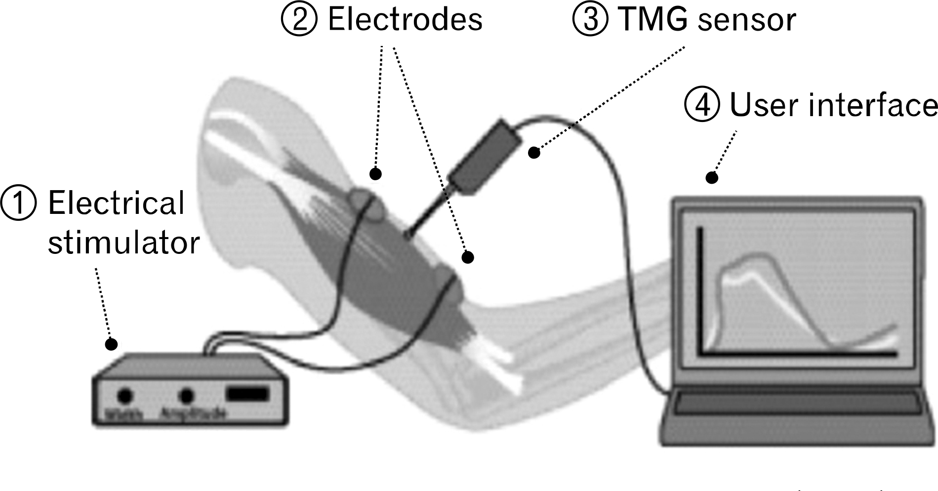
Fig. 2.
Tensiomyography record with parameters' definitions. (A) Basic principle of tensiomyography. Arrow: direction of muscle contraction. (B) Basic result of tensiomyograph. Tc: contraction time, Tr: relaxation time, Ts: sustain time, Dm: maximal displacement, Td: delay time.
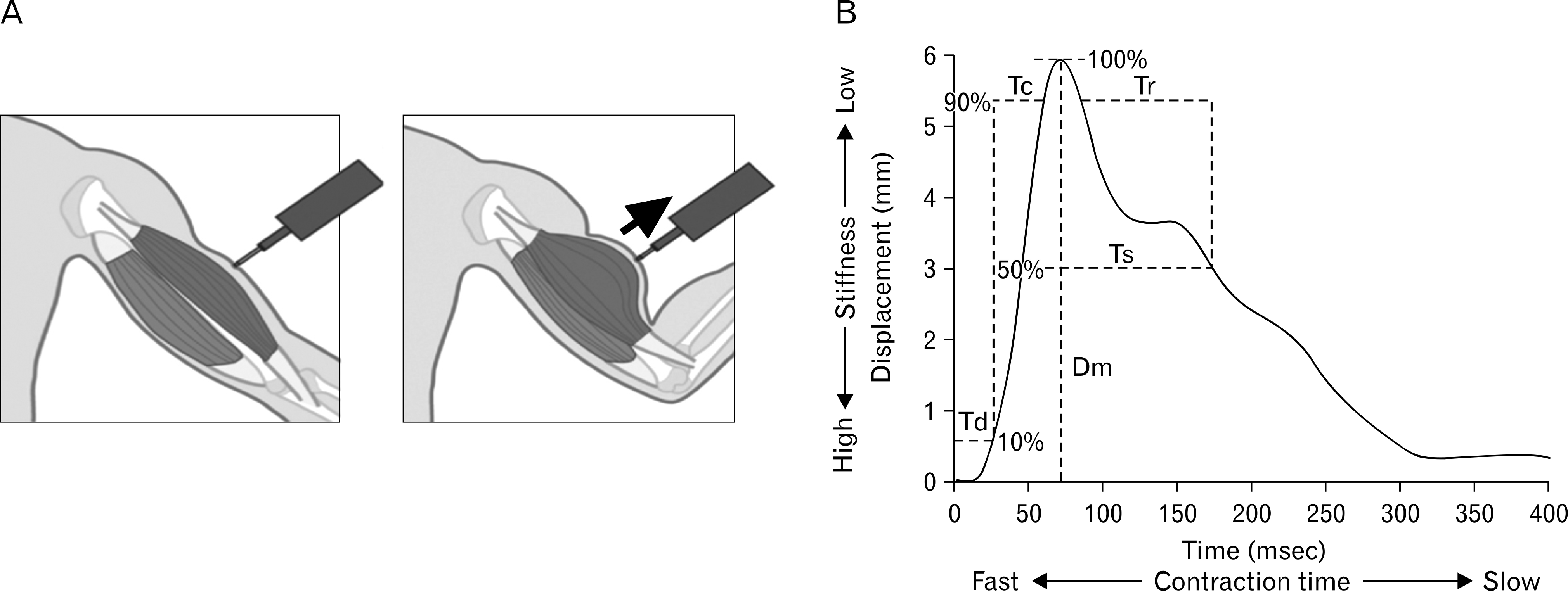
Fig. 3.
Representative tensiomyography records of bodybuilder and control groups. (A, B) Biceps femoris. (C, D) Rectus femoris.
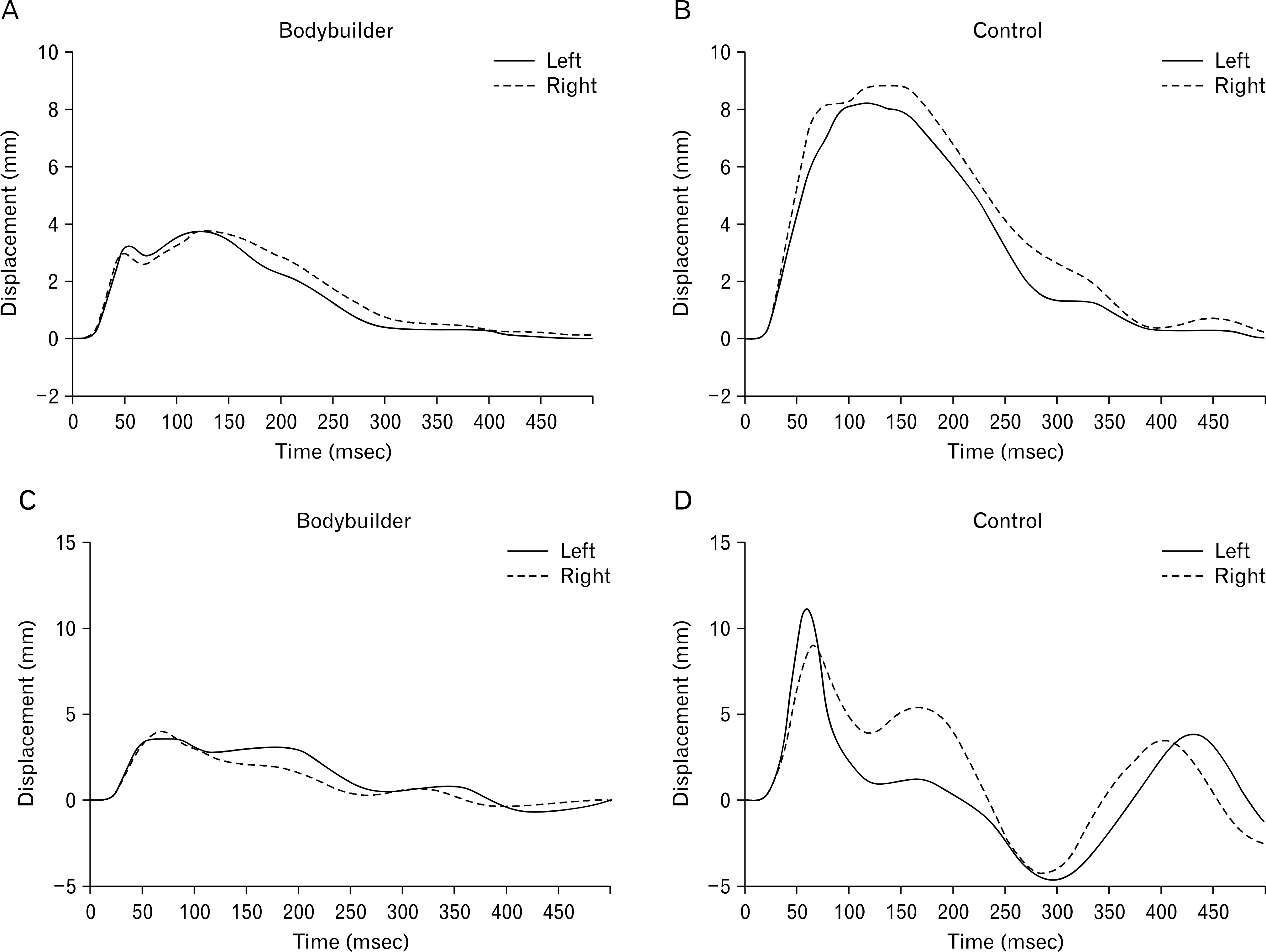
Table 1.
Dm values for the analyzed muscles in each group
| Variable | BB (n=9) | Con (n=15) | p-value |
|---|---|---|---|
| Sum | |||
| BF | 9.47±3.44∗∗ | 15.14±4.93 | 0.006 |
| ST | 9.51±3.17∗∗ | 18.71±5.10 | 0.001 |
| VL | 9.73±3.12∗ | 13.42±3.89 | 0.025 |
| VM | 11.91±2.51∗∗ | 16.18±3.00 | 0.002 |
| RF | 9.72±2.35∗∗ | 16.08±4.81 | 0.001 |
| Right leg | |||
| BF | 4.86±2.17∗ | 7.45±3.06 | 0.037 |
| ST | 3.95±1.70∗∗ | 8.63±2.59 | 0.001 |
| VL | 4.52±1.69∗ | 6.41±2.12 | 0.033 |
| VM | 5.35±2.04∗∗ | 8.40±1.68 | 0.001 |
| RF | 4.79±1.28∗∗ | 7.21±1.85 | 0.002 |
| Left leg | |||
| BF | 4.61±1.56∗∗ | 7.69±2.85 | 0.007 |
| ST | 5.56±2.20∗∗ | 10.08±3.21 | 0.001 |
| VL | 5.21±1.64∗ | 7.00±2.07 | 0.038 |
| VM | 6.56±1.05 | 7.78±1.72 | 0.068 |
| RF | 4.93±1.94∗∗ | 8.87±3.32 | 0.001 |
Table 2.
Tc values for the analyzed muscles in each group
| Variable | BB (n=9) | Con (n=15) | p-value |
|---|---|---|---|
| Sum | |||
| BF | 57.27±25.68∗∗ | 97.77±26.90 | 0.001 |
| ST | 55.70±23.66∗∗ | 80.91±16.06 | 0.005 |
| VL | 50.06±7.36 | 51.16±5.07 | 0.666 |
| VM | 49.79±6.02∗ | 45.20±3.17 | 0.022 |
| RF | 57.66±8.56 | 57.28±4.49 | 0.886 |
| Right leg | |||
| BF | 25.86±11.58∗∗ | 52.79±18.75 | 0.001 |
| ST | 22.49±11.45∗∗ | 39.80±8.83 | 0.005 |
| VL | 26.14±6.68 | 25.45±4.52 | 0.766 |
| VM | 25.32±2.66∗ | 22.92±1.75 | 0.014 |
| RF | 30.29±9.16 | 29.93±3.35 | 0.91 |
| Left leg | |||
| BF | 31.42±14.75 | 44.98±16.05 | 0.051 |
| ST | 33.22±15.43 | 41.11±9.85 | 0.194 |
| VL | 23.92±4.15 | 25.71±2.83 | 0.221 |
| VM | 24.48±3.65 | 22.28±1.80 | 0.06 |
| RF | 27.37±5.20 | 27.35±3.58 | 0.992 |
Table 3.
Vc values (mm/sec) for the analyzed muscles in each group
| Variable | e BB (n=9) | Con (n=15) | p-value |
|---|---|---|---|
| Sum | |||
| BF | 137.88±33.30 | 130.26±47.36 | 0.677 |
| ST | 143.64±32.73∗ | 186.62±41.84 | 0.015 |
| VL | 161.32±70.44 | 212.29±63.97 | 0.082 |
| VM | 191.25±37.25∗∗ | 287.66±56.79 | 0.001 |
| RF | 137.29±39.47∗∗ | 225.55±65.17 | 0.001 |
| Right leg | |||
| BF | 153.46±49.96 | 123.11±56.99 | 0.2 |
| ST | 145.12±45.13 | 176.00±41.25 | 0.1 |
| VL | 148.88±80.36 | 205.86±70.10 | 0.081 |
| VM | 165.92±61.83∗∗ | 295.71±68.11 | 0.001 |
| RF | 133.18±50.49∗∗ | 195.94±53.18 | 0.009 |
| Left leg | |||
| BF | 126.46±34.51 | 150.44±67.49 | 0.263 |
| ST | 141.35±28.09∗∗ | 98.07±50.19 | 0.005 |
| VL | 180.30±70.58 | 220.42±67.17 | 0.178 |
| VM | 218.71±45.26∗ | 279.93±61.13 | 0.03 |
| RF | 146.38±53.80∗∗ | 260.34±91.63 | 0.001 |
 XML Download