

 PDF
PDF ePub
ePub Citation
 Print
Print


Introduction
Supernumerary teeth are more often found in the maxillary anterior region than in any other area of either dental arch.1 Unless supernumerary teeth are diagnosed early and managed properly, they may exert a variety of pathological effects on the developing permanent dentition.2 It has been reported that if a significant delay or ectopic or asymmetric eruption of the maxillary permanent incisors is observed clinically, the presence of mesiodens should be suspected.3
Supernumerary teeth, particularly those in the anterior maxilla, can cause failure of eruption, displacement, rotation of the permanent maxillary incisors, and median diastema.234 Other rare findings that are thought to be due to the presence of supernumerary teeth are root resorption of permanent teeth,5 cyst formation,6 and eruption of the supernumerary tooth into the nasal cavity.7 Early diagnosis of a supernumerary tooth is vital to reduce possible complications, the extent of surgery, and orthodontic treatment.8
Supernumerary teeth are detected via a thorough clinical examination and routine radiological examinations.9 Radiographic examinations are used to assess the number, location, path, and sagittal position of the impacted supernumerary tooth.10 Radiographic imaging plays an important role in deciding on an appropriate treatment plan and when to intervene, especially when removal of the supernumerary tooth is required.11
The optimal timing for the surgical removal of unerupted maxillary anterior supernumeraries has often been the subject of debate.210 Some researchers have argued for immediate surgical removal after diagnosis to permit spontaneous eruption of the permanent incisors and to avoid possible orthodontic problems,12 while others have advocated a delayed approach in order to avoid iatrogenic damage to the developing adjacent teeth.1314 The immediate surgical removal of impacted supernumeraries is indicated when the chances of developing complications if the supernumerary tooth was left untreated are significant.3 The surgical removal of supernumerary teeth based on accurate radiographic localization minimizes the risk of devitalization of the permanent teeth when performed before the maturation of their apices.15
The aim of this study was to examine the radiographic features associated with impacted premaxillary supernumerary teeth, to determine the relationship between their characteristics and the effects on permanent incisors, and to investigate the types of orthodontic treatment that patients received after the extraction of impacted supernumerary teeth.
Materials and Methods
The Institutional Review Board of Pusan National University Dental Hospital approved this study. The subjects of this study were selected from patients who visited Pusan National University Dental Hospital and underwent conebeam computed tomography (CBCT) and panoramic radiography to remove impacted premaxillary supernumerary teeth between 2012 and 2013. The clinical records and radiographs of 193 patients who had supernumerary teeth removed were retrospectively reviewed. The patients comprised 144 boys and 49 girls, with a mean age of 7.41 years at the time of supernumerary tooth extraction (age range, 4-12 years).
CBCT scans were acquired with a PaX-Zenith 3D scanner (Vatech Co., Hwasung, Korea). The scanning parameters were set at 105 kVp, 5.6 mA, 24 seconds, a voxel size of 0.2 mm, and field of view of 12×9 cm. The CBCT volumetric data were reconstructed using Ez3D Plus Professional CBCT software (Vatech Co., Hwasung, Korea). All panoramic radiographs were taken with a Proline XC machine (Planmeca Co., Helsinki, Finland).
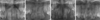
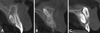
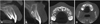
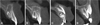
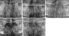
The CBCT and panoramic images were examined to determine the number, location, sagittal position, orientation, and morphology of impacted supernumerary teeth. The location was classified as midline, central incisor, lateral incisor, or between the central and lateral incisors (Fig. 1). The sagittal position of the supernumerary teeth relative to the permanent incisors was classified as palatal, within the arch, or labial (Fig. 2). The orientation was categorized as normal, inverted, transverse, or horizontal (Fig. 3). The shape of the supernumerary tooth was described as conical, tuberculate, supplemental, or odontoma-type (Fig. 4).
Based on patients' clinical records, panoramic radiographs, and CBCT images, the following effects of supernumerary teeth on the permanent upper incisors were examined: median diastema, delayed eruption, displacement, rotation, and resorption (Fig. 5). The types of subsequent orthodontic treatment after the removal of supernumerary teeth were also investigated. The data were analyzed using the chi-square test and the Fisher exact test. P-values <.05 were considered to indicate statistically significant differences. All statistical analyses were performed using SPSS version 23.0 (IBM Corp., Armonk, NY, USA).
Results
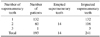
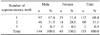
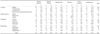
The 193 patients had 14 erupted and 241 impacted supernumerary teeth. The 14 erupted supernumeraries were excluded, and the 241 impacted premaxillary supernumerary teeth were examined (Table 1). Of the 193 children, 132 (68.4%) had 1 supernumerary tooth, 60 (31.1%) had 2 supernumeraries, and 1 (0.5%) had 3 supernumeraries. There were 144 male and 49 female patients, and the sex distribution was in favor of males in the ratio of 2.9 : 1. No statistically significant difference was found in the number of supernumeraries according to sex (Table 2).
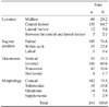
Premaxillary supernumerary teeth were most frequently observed in the central incisor region (64.7%) and palatal position (76.8%). An inverted orientation (60.6%) and conical shape (75.5%) were most common. The labial position was very rarely observed, appearing in only 1 case (0.4%) (Table 3).
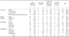
The main complications caused by supernumerary teeth were median diastema (17.8%), delayed eruption of incisors (12.9%), displacement (14.5%), and rotation (6.6%) of the adjacent teeth. Root resorption was noted in 1 case (0.4%) (Table 4).
The relationships among the characteristics of supernumeraries and complications were investigated. Median diastema was more frequently observed when the supernumerary was positioned in the midline, within the arch, or transversely (P<.05). Delayed eruption of adjacent incisors was more frequently observed in the vertical orientation than the inverted orientation (P<.05). Ten (71.4%) of the 14 odontomas showed delayed eruption. Displacement was more common for the tuberculate and supplement types than for the conical type (P<.05). Rotated incisors were more frequently noted in supernumeraries within the arch than in palatally positioned supernumeraries (P<.05) (Table 4).
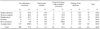
The distribution of cases according to the type of orthodontic treatment after the extraction of supernumerary teeth was examined. Orthodontic traction was performed in 7 cases (50%) of the odontoma type. Orthodontic closure of median diastema was most frequently performed after extraction of supernumerary teeth located in the midline (Table 5).
Fifteen (34.9%) of 43 cases with median diastema were closed orthodontically after removal of the supernumerary tooth. Orthodontic traction was applied to 8 cases (25.8%) of incisors affected by delayed eruption. Twelve (34.3%) displaced incisors and 10 cases (62.5%) of rotated incisors were corrected by the orthodontic creation of sufficient space. Using orthodontic creation of space, 32 (13.3%) maxillary incisors were brought into a good position in the dental arch after the surgical removal of the unerupted supernumerary tooth. In 186 cases (77.2%), permanent incisors erupted without orthodontic treatment after the removal of the supernumerary tooth (Table 6).
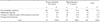
Permanent incisors were more likely to erupt spontaneously when the supernumerary teeth were extracted from the primary dentition, although this trend was not statistically significant (P=.066). When supernumerary teeth were removed from the primary dentition, orthodontic traction was not required for unerupted maxillary incisors (Table 7).
Discussion
The present study included 193 patients in whom the surgical removal of impacted supernumerary teeth in the premaxillary area was performed. It was observed that premaxillary supernumerary teeth occurred more frequently in boys than in girls.2101617 In this study, the male-to-female ratio was 2.9 : 1, indicating a male predominance. Multiple occurrences of premaxillary supernumerary teeth have been reported in the literature, ranging from 8% to 34% of mesiodens cases.235 In our study, a single supernumerary tooth was found in 132 cases (68.4%), and 2 were found in 60 cases (31.1%). Triple or quadruple occurrence was extremely rare,10 with only a single such case (0.5%) observed in the patients within the present study.
A greater predilection was observed for supernumeraries to be positioned palatally in this study, similarly to previous studies.18192021 Several studies101719202223 have reported that the vertical orientation was most frequent, although others218 have shown inverted supernumeraries to be the most common. In our study, most of the supernumerary teeth (60.6%) were found in the inverted position, followed by the vertical position (21.2%) and transverse position (16.6%). Conical supernumeraries have been found to be the most common,182021232425 which corresponded well with the results of our study.
Maxillary midline diastema has been reported in 10%-38% of patients with supernumerary teeth.24102526 These widely differing results could be due to the manner in which median diastema was diagnosed.4 In the present study, the most common complication was median diastema (17.8%), and it was most frequent in midline supernumerary teeth. Vertically oriented supernumeraries were more frequently associated with delayed eruption of the permanent incisors than inverted types.2226 In this study, the delayed eruption of adjacent incisors was more frequently observed in the vertical position than in the inverted position. Previous studies have reported that morphology was associated with the delayed eruption of teeth.91626 Tuberculate supernumerary teeth were more frequently associated with delayed eruption than the conical type,9162728 which corresponds to the findings of our study. It has been reported that the most common cause of the impaction of the upper central incisors was odontomashaped supernumerary teeth.29 In our study, 71.4% of odontoma-shaped teeth were associated with the delayed eruption of permanent incisors.
Orthodontic traction may be required to bring unerupted maxillary incisors into a good position after the surgical removal of impacted supernumerary teeth.162024 In this study, 8 cases of unerupted incisors erupted after orthodontic traction, and 7 of the 8 cases were incisors associated with the odontoma type. The majority of delayed permanent teeth erupted spontaneously if sufficient space was available or was created at the time of impacted supernumerary removal.2327 Insufficient space for the unerupted incisor may obstruct the eruption of permanent upper incisors after the removal of supernumerary teeth.2427 Rotation or displacement of the permanent incisors has been found to be caused by a lack of space due to the presence of supernumerary teeth.4 In our study, the percentage of displaced or rotated incisors was high when space for the incisors was insufficient, and 34.3% displaced incisors and 62.5% rotated incisors were corrected via the orthodontic creation of space after the removal of supernumerary teeth.
Irrespective of whether or not an immediate or delayed surgical approach is adopted, early diagnosis is critical.3103031 The early diagnosis of mesiodens minimizes the treatment required and prevents the development of associated complications.832 If malalignment of the adjacent permanent teeth is anticipated, this is an indication for the early surgical removal of a supernumerary tooth.1033 Surgical removal of impacted supernumerary teeth could be delayed when no complications due to supernumerary teeth are found to prevent damage to the adjacent unerupted developing teeth.2325 Supernumerary teeth should be followed up radiographically25 because a delay in treatment can create the need for more complex surgical and orthodontic management.32 In this study, permanent incisors were more likely to erupt without orthodontic treatment when the supernumerary teeth were extracted from the primary dentition than from mixed dentition.
In conclusion, premaxillary supernumerary teeth were most frequently observed in the central incisor region, palatal position and inverted orientation, and the conical shape was the most common. Median diastema was more frequently observed in the midline supernumerary teeth. The percentage of delayed eruption was high in the supernumerary teeth with a vertical orientation and odontoma shape. Displacement of the incisors was more frequently observed in tuberculate or supplemental shapes. Orthodontic traction was most frequently performed after removal of odontoma-type supernumerary teeth. The early removal of impacted premaxillary supernumerary teeth might be advantageous when complications caused by the supernumerary teeth in the maxillary anterior regions are expected.
 XML Download