

 PDF
PDF ePub
ePub Citation
 Print
Print


Introduction
Small-cell carcinoma is one of the most aggressive, poorly differentiated, and highly malignant neuroendocrine tumors and includes small-cell lung cancer and extrapulmonary small cell carcinoma (EPSCC). It is estimated that 10% of patients with small cell lung cancer have syndrome of inappropriate secretion of antidiuretic hormone (SIADH) [1]. EPSCC is a rare disease, accounting for 2.5% to 5.0% of all small cell carcinoma [2,3].
Small cell carcinoma of the uterine cervix (SmCC) is a very rare disease representing only 2% of all uterine cervical cancers and is extremely aggressive subtype of uterine cervical cancer [4]. In the currently used World Health Organization histological classification of tumors of the uterine cervix (2003), Small cell carcinoma is classifi ed as a neuroendocrine tumor.
Compared to more common sqaumous cell carcinoma of the cervix, small cell carcinoma is more likely to show lymphovascular invasion, metastasize to lymph nodes and recur [5-7].
The clinical characteristics of cervical small cell carcinoma are not fully understood, and reports of paraneoplastic syndromes, especially SIADH, are rare. We report a case of ADH producing cervical small cell carcinoma with SIADH as a paraneoplastic syndrome.
Case report
A 41-year-old Korean woman with parity of 3-0-4-3 who had no significant findings in a Pap smear conducted 9 months earlier than she came to the hospital. She had suffered vaginal spotting and blood tinged vaginal discharge for 5 months and experienced vaginal bleeding the day before she went to see a doctor in February 2012.
Physical examination revealed no abnormal findings, and gynecological examination showed exocervical protruded cervical mass measured about 4.5 cm in diameter and parametrium was not involved. Colposcopy confirmed the polypoid mass arising from the exocervical side of the cervix. Biopsy of this tumor was reported histologically as small cell carcinoma of the cervix. On T2-weighted imaging of pelvic magnetic resonance imaging (MRI), an enhancing tumor of the uterine cervix, sized 3.5×3.6×4.7 cm, was found and normal cervical stromal hyposignal intensity was almost missing (Fig. 1A). Irregular spiculated margin of the tumor mass was noted. Vaginal invasion was not observed but two prominent lymph nodes were noted along the both iliac chains (Fig. 1B). Chest X-ray did not show any significant findings. Since the tumor was over 4 cm in size and limited to the cervix without findings of vaginal or parametrial invasion, we clinically diagnosed her illness as cervical cancer IB2.
Her blood test showed the following results: hemoglobin 12.1 g/dL, white blood cell 6,000/µL, and platelet 359,000/µL. Liver function tests, renal function tests and coagulation profile were all normal while urinalysis was normal except hematuria, and urine specific gravity was 1.015. She was positive for high-risk human papillomavirus (HPV) 18, and carcinoembryonic antigen (CEA) was 3.81 ng/mL higher than normal levels (under 3 ng/mL), and the level of squamous cell carcinoma antigen was 1.14 ng/mL (normal range, 0-2 ng/mL). There were no abnormal findings from an esophagogastroscopy, colonoscopy and cystoscopy. Before surgery, we found the hyponatremia ,124 mEq/L (normal range, 135-145 mEq/L) and low chloride 97 mEq/L (normal range, 98-110 mEq/L), although the serum potassium concentration was normal, 4.5 mEq/L (normal range, 3.5-5.5 mEq/L). After the administration of intravenous hydration, the levels of sodium and chloride dropped even further to 122 mEq/L and 91 mEq/L, respectively. The diagnosis of SIADH was confirmed by diagnostic criteria, i.e., decreased serum osmolality (252 mOsm/kg) and elevated urine osmolality (384 mOsm/kg). There were no significant findings from thyroid function tests and adrenal functions were normal. Under the diagnosis of SIADH, we treated her with water restriction and 3% hypertonic saline. Her blood sodium and chloride levels return to normal (136 mEq/L and 105 mEq/L, respectively), and proceeded with surgery. The types of surgical procedures were a radical hysterectomy, bilateral salpingectomy, pelvic lymph node dissection and para-aortic lymph node dissection.
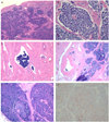
During operation, the size of uterus was normal and exocervical mass measured about 4.5 cm in size was found. Grossly, invasion into parametrium, paracolpium or vagina was not found. Both adnexa were normal, but we found both external iliac lymph nodes slightly swollen. Microscopic findings showed that tumor cells were small and round- or spindle-shaped with hyperchromatic nuclei and scant cytoplasm (Fig. 2A, B). Tumor size was 4 × 3.5 × 3.5 cm and subtle parametrial invasion was present, but the depth was less than 0.1 cm (Fig. 2C). We observed myometrial invasion (Fig. 2D) and metastases to both external iliac lymph nodes (Fig. 2E) as well. The immunocytochemical profile demonstrated marker positivity for neurone-specific enolase and positive for chromogranin (Fig. 2F). They were partially positive for cytokeratin and negative for leukocyte common antigen, so we diagnosed her with small cell carcinoma.
On 48 hours after surgery, the serum sodium were increased and showed persistent normal range (over 135 mEq/L). Potassium and chloride were also normal (3.8 mEq/L and 106 mEq/L).
She was discharged 11days after surgery without any significant complications. Considering the high risk factors of the pathology, the patient received concurrent chemoradiotherapy (CCRT) on 30 days after operation. There is no definite abnormal finding on follow-up pelvic computed tomography (CT) taken 3 months after the operation. However, 4 months after surgery, the patient was admitted to our hospital because of anorexia and fatigue. She had hyponatremia (133 mEq/L) and elevated CRP (c-reactive protein) (7.88 mg/dL; normal, 0-0.3 mg/dL). On abdominal and pelvic CT scanning showed multiple enlarged para-aortic lymph nodes. These data strongly suggested the recurrence of small cell cervical carcinoma. with SIADH. After the completion of carboplatin-paclitaxel chemotherapy (×3 cycles), positron emission tomography (PET)-CT scanning conducted.
But, metastases to Lt (left) common iliac chain, retrocaval and Lt para-aortic areas were existed. Those enlarged lymph nodes were removed at 8 months after the first surgery. CEA, a tumor marker of this case, had been within a normal range after the first surgery but rose to 4.92 ng/mL before removal of recurrent lymph nodes. But after the lymph node excision, CEA level returned to normal again (1.62 ng/mL). However, after 1year, the first surgery which means 4 months after the lymph node excision, the CEA level has been elevated up to 4.41 ng/mL and PET-CT results showed that there is metastasis of para-aortic lymph nodes, supraclavicular lymph nodes and right femur lymph nodes. Therefore the patient underwent radiation therapy and now is having etoposide-cisplatin chemotherapy.
Discussion
Small cell carcinoma of the uterine cervix is a rare disorder taking up only 0.5% to 5% of the types of cervical cancer. The incidence rate is 0.06 per 100,000 people per year and 45-year-old adults are most commonly affected by the cancer [7]. According to the Annual Report of Gynecologic Cancer Registry Program in Korea for 2004, squamous cell carcinoma takes up 81.1% while adenocarcinoma and small cell carcionoma amount to 10.6% and 0.4%, respectively.
One of the causes of small cell carcinoma of the uterine cervix is HPV infection, and in particular, HPV type 18 is closely associated [8]. Clinically, small cell carcinoma of the uterine cervix has similar characteristics with small cell lung carcinoma [6]. About 60% of SmCC is diagnosed in International Federation of Gynecology and Obstetrics (FIGO) stages I and II. However, because of early lymph node metastases or hematogenous metastases, many patients are diagnosed with advanced cancer [4]. Lymph node metastases and lymphovascular space invasion occur frequently and the recurrence rate is also higher than squamous cell carcinoma of cervix. The rates of lymph node involvement by FIGO stage for each histology are 27.5% in stage I of small cell carcinoma that was higher than 10.9% of squamous cell carcinoma [9] report, the rate of lymph node metastases in stage IB1 was 20%, and in stage≥IB2, at least half had lymph node metastases. It progresses quickly and shows frequent metastases to distant organs such as the bones, brain, liver, and bone marrow. Hematogeneous metastasis was also very high. In addition, the tumors are remarkably resistant to conventional treatment modalities. For these reasons, small cell carcinoma of the uterine cervix is notorious for its poor prognosis [8]. The light microscopy and immunohistochemical staining are used for diagnosis of the disease and the tumor cells behave similarly to the lung [6]. When immunohistochemically stained, they are positive for general neuroendocrine markers including synaptophysin, chromogranin A, neuron-specific enolase, and CD 56 [10].
Small cell carcinoma has often neuroendocrine characteristics. Neuroendocrine tumors may have the ability to synthesise and secrete biologically active substances characteristic of the cell of origin that can cause distinct clinical syndromes. The term 'paraneoplastic syndromes' is used to denote syndromes secondary to substances secreted from tumors not related to their specific organ or tissue of origin ; such syndromes are mainly associated with hormonal and neurological symptoms. Small cell carcinoma of lung is the most frequent cause of paraneoplastic syndromes. These syndromes should be actively excluded whenever a patient presents with any of their associated features. The most frequent endocrine syndromes are the SIADH and Cushing's syndrome. However, the SIADH is rare in small cell carcinoma of the uterine cervix.
We described a patient with cervical small cell carcinoma accompanied by severe hyponatremia who had rapid recurrence after surgery. SIADH was diagnosed on the basis of diagnostic criteria.
The serum sodium level had shown the normal range after radical hysterectomy operation. Kuji et al. [9] report of 52 small cell carcinoma of the uterine cervix in Japan showed that hyponatremia was 4% in stage I and 11% in stage II-IV. The excessive ADH production interferes with renal excretion of water, resulting in hyponatremia and concentrated urine. Symptoms of hyponatremia appear when the serum sodium level goes under 125 to 130 mmol/L and these symptoms are usually neurological and include nausea and malaise. If the serum sodium level drops under 115 mmol/L, a patient begins to suffer from headache, altered mental status, seizures, coma and then respiratory arrest. SIADH is diagnosed by hyponatremia, low serum osmolality, increased urinary sodium, osmolality, and/or specific gravity. If underlying diseases are treated, SIADH may be corrected. Serum sodium was useful for post-treatment surveillance in small cell lung cancer patients who presented with SIADH, with 71% (10/14) developing SIADH again at the time of recurrence. SIADH is a poor prognostic factor for small cell lung cancer [11].
The treatment of small cell carcinoma of the uterine cervix is a radical hysterectomy, pelvic lymph node dissection, and para-aortic lymph node dissection, and adjuvant chemotherapy and radiation therapy are added after surgery [6]. Small cell cervical cancer (SCCC) carries poor prognosis due to its propensity for early hematogeneous and lymphatic spread. Treatment for SCCC have been largely extrapolated from the experience with small cell lung cancer (SCLC). SCLC is highly responsive to multiple chemotherapeutic drugs, and chemotherapy dramatically prolong survival compared to best supportive care. In multiple randomized trials, the platinum/etoposide regimen appears to be at least as effective as older regimens such as vincristine, doxorubicine, and cyclophosphamide and has less toxicity for SCLC [12]. The Society of Gynecologic Oncology also proposed that combination chemotherapy with radiation (CCRT-EP) should be considered for non-surgical candidates (IIB-IVB) with neuroendocrine carcinoma of the uterine cervix [13].
Although the Korean Society of Obstetrics and Gynecology and the National Cancer Center recommend the Pap smear to women over age 30, the effectiveness of the Pap smear for diagnosis of small cell carcinoma is relatively low as in adenocarcinoma [14]. Therefore, it could be helpful for elevating the sensitivity of Pap smear to HPV testing.
For women over 40, the Pap smear and HPV testing at the same time would be helpful to improve the accuracy of diagnosis. Since cases in which small cell carcinoma of the uterine cervix is involved with SIADH, a paraneoplastic syndrome, are relatively rare and no case has been reported so far in Korea, we think it is meaningful to report our case herein with brief review of literature.

 XML Download