

 PDF
PDF ePub
ePub Citation
 Print
Print


Abstract
We report a case that mixed germ cell tumor developed in an 18-year-old girl with 45,X/46,X,+mar mosaic Turner's syndrome. Molecular biological studies showed that the patient's DNA contained a fragment of Y chromosome. The patient underwent an operation and chemotherapy, and until now there is no evidence of recurrence. A presence of Y chromosome component should be evaluated in case a marker chromosome is found in a woman with Turner's syndrome.
REFERENCES
1. Kim SC, Park SS, Kim KH, Park DY, Yoon MS. A case of coincidental gonadoblastoma and dysgerminoma in 45X/47XYY mosaic Turner variant. Korean J Obstet Gynecol. 2005; 48:1805–10.
2. Cho YY, Cho CH, Yu SD, Kim HM, Park JB. A case of variants of Turner syndrome showing 46,X,inv(Y)/45,X karyotype with Y chromosome microdeletion. Korean J Obstet Gynecol. 2006; 49:892–8.
3. Korean Society of Obstetrics and Gynecology. Gynecology. 4th ed.Seoul: Korean Medical Book Publisher;2007.
4. Ito K, Kawamata Y, Osada H, Ijichi M, Takano H, Sekiya S. Pure yolk sac tumor of the ovary with mosaic 45X/46X + mar Turner's syndrome with a Y-chromosomal fragment. Arch Gynecol Obstet. 1998; 262:87–90.
5. Sinisi AA, Perrone L, Quarto C, Barone M, Bellastella A, Fag-giano M. Dysgerminoma in 45,X Turner syndrome: report of a case. Clin Endocrinol (Oxf). 1988; 28:187–93.

6. Pierga JY, Giacchetti S, Vilain E, Extra JM, Brice P, Espie M, et al. Dysgerminoma in a pure 45,X Turner syndrome: report of a case and review of the literature. Gynecol Oncol. 1994; 55:459–64.
7. Oliveira RM, Verreschi IT, Lipay MV, Eca LP, Guedes AD, Bianco B. Y chromosome in Turner syndrome: review of the literature. Sao Paulo Med J. 2009; 127:373–8.
Fig. 1.
Patient's axillary and pubic hair was absent, but her breast development was normal. (A) Tanner stage III.
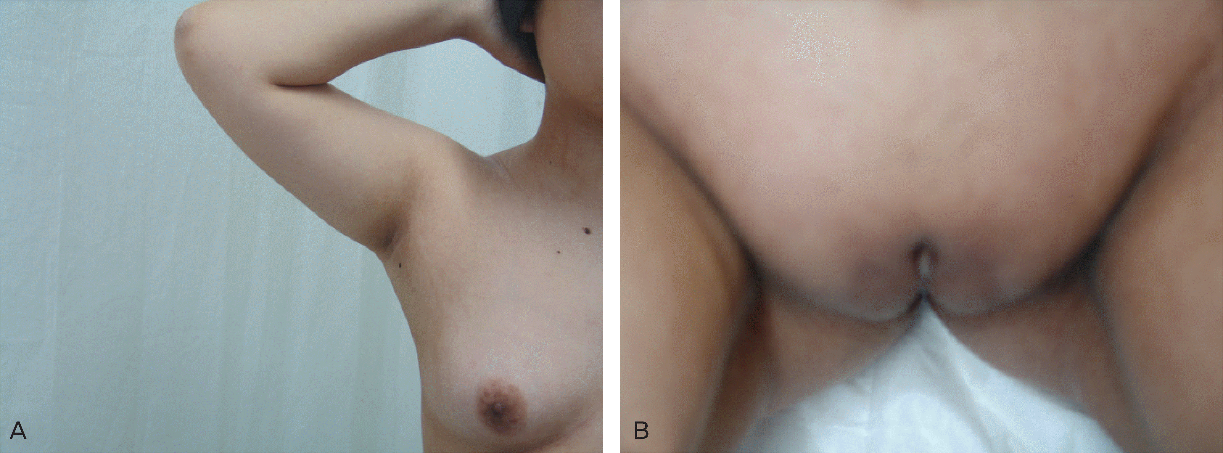
Fig. 2.
Chromosomal analysis showed mosaic Turner's syndrome [46,X,+mar] (A) and SRY gene of marker chromosome was identified (B).
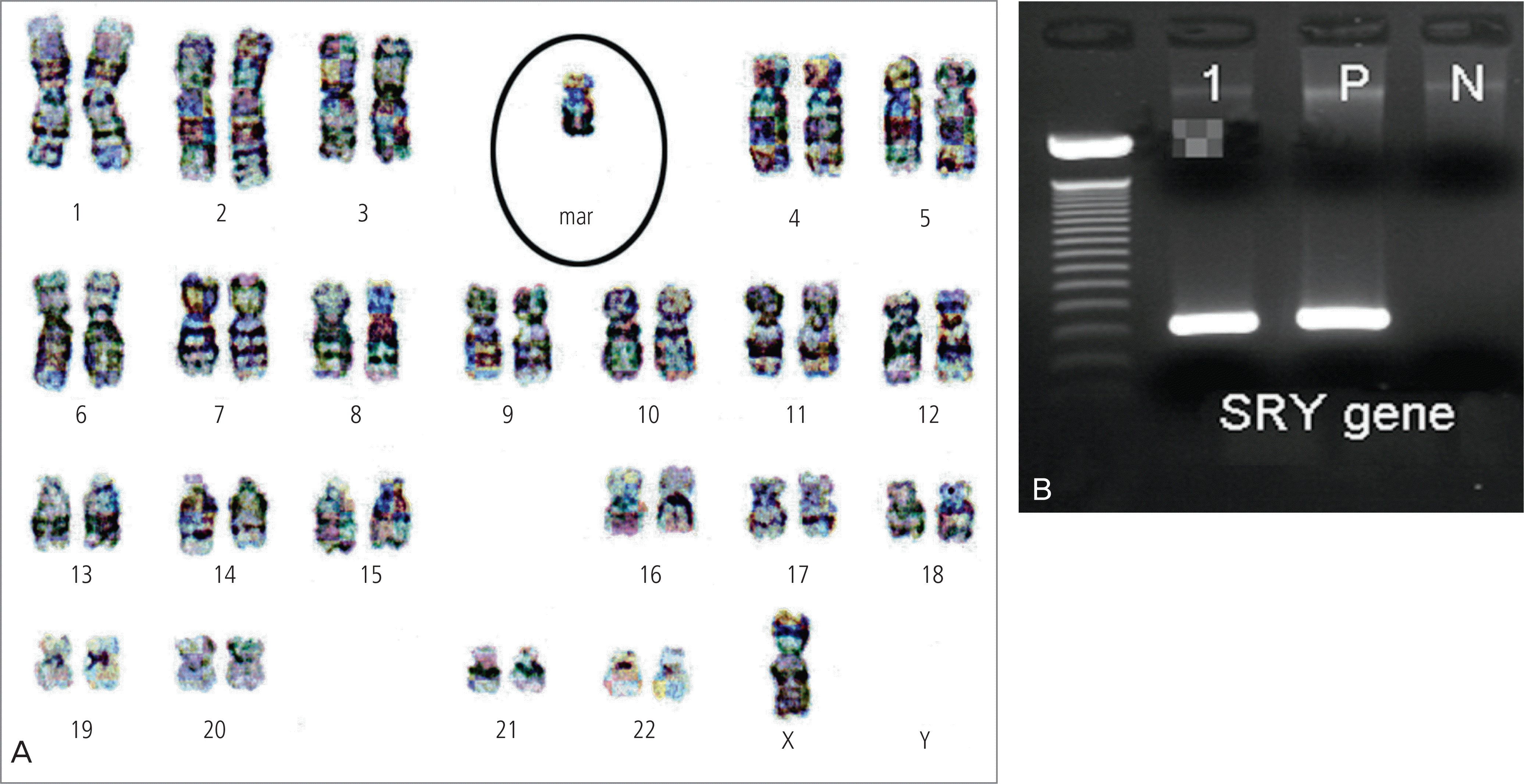
Fig. 3.
The representatives of MRI imaging showed large multiseptated cystic mass with solid component and thick septal wall, small and immature uterus, and no visible normal ovaries (A) horizontal view, (B) sagittal view.

Fig. 4.
The resected right ovary shows solid and partially cystic tumor with areas of necrosis and hemorrhage (gross view).
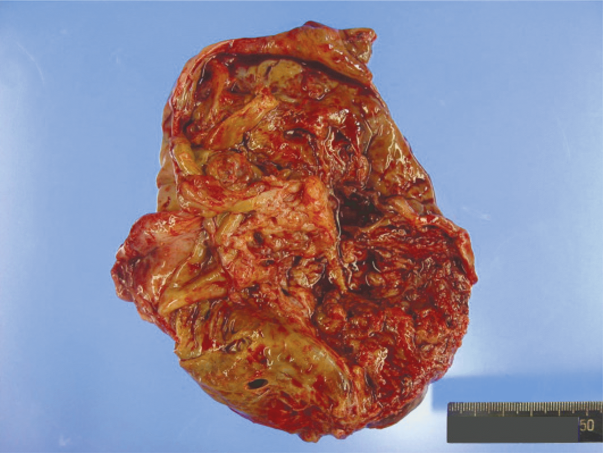
Fig. 5.
Representative microphotographs of right (A to E) and left (F) ovary. (A) Varying sized and shaped immature cartilage island embedded in immature mesenchymal stroma seen in immature teratoma portion (H&E, ×40). (B) Scattered primitive germ cells and lymphoplasma cells in dysgerminoma portion (H&E, ×100). (C) Abortive glands and papillae of primitive germ cells seen in embryonal carcinoma portion (H&E, ×40). (D) Reticular and microcystic structures in loose connective tissue of the yolk sac tumor portion (H&E, ×100), (E) Scattered multinucleated syncytial giant cells seen in the choriocarcinoma portion (H&E, ×100), (F) Multiple mineralized cartilage islands and nests of primitive germ cells in left ovary (H&E, ×40). (G) Positive immunohistochemical stain of alpha-fetoprotein in yolk-sac tumor component (×100). (H) Positive immunohistochemical stain of beta human chorionic gonadotropin in syncytial giant cells in choriocarcinoma component (×100). (I) Positive immunohistochemical stain of CD30 in primitive epithelial cells of embryonal carcinoma component (×100).

 XML Download