

 PDF
PDF ePub
ePub Citation
 Print
Print


INTRODUCTION
Exercise-induced bronchospasm (EIB) describes acute airway narrowing that occurs as a result of exercise [1]. Symptoms that are often associated with vigorous exercise, such as shortness of breath, cough, wheeze, and mucus production, are neither sensitive nor specific for identifying those with EIB. These symptoms may only be provoked by exercise or may only occur in specific environments, such as ice rinks or indoor swimming pools [2, 3]. The prevalence of EIB in asthmatics has been reported to vary from 40-90% [4, 5, 6, 7]. Eosinophils are important in the pathophysiology of EIB. Also, in vitro studies have demonstrated that eosinophils generate and release cysteinyl leukotrienes when subjected to a hyperosmolar stimulus, which is an important condition that provokes EIB. The severity of EIB is significantly correlated to eosinophil levels measured in the blood and sputum of asthmatic patients [8]. The diagnosis of EIB is established by changes in lung function after exercise, not on the basis of symptoms. The airway response is expressed as the percent fall in forced expiratory volume in one second (FEV1) from the baseline value. The difference between the pre-exercise FEV1 value and the lowest FEV1 value recorded within 30 minutes after exercise is expressed as a percentage of the pre-exercise value. The criterion for the percent fall in FEV1 used to diagnose EIB is >10% [1]. Other spirometric parameters, such as forced expiratory flow between 75% and 25% of vital capacity (FEF25-75%), forced expiratory flow at 50% of the vital capacity (FEF50%), peak expiratory flow (PEF), have been less studied in children with asthma in the diagnosis of EIB [9, 10]. In the asthmatic patients, FEF25-75% and PEF are the second most used spirometric parameters after FEV1 value in the evaluation of small and large airways function respectively. In addition, FEF50% can be used instead of FEF25-75%. The purpose of this study is to evaluate the association of FEV1 and FEF50%, PEF parameters, and blood eosinophil counts in EIB in children in mild asthma.
MATERIALS AND METHODS
Subjects
Sixty-seven children (aged 5.5-16.5 years) with mild asthma were included in this study (Table 1). Asthma is a chronic inflammatory disorder of airways. The chronic inflammation is associated with airway hyperresponsiveness that leads to episodic wheezing, breathlessness, chest tightness, and coughing. Airway obstruction is of ten reversible either spontaneously or with the treatment of broncho-dilatator drugs. Airway hyperresponsiveness is defined as the methacholine concentration that caused a 20% decrease in FEV1 from baseline (PC20). PC20 is usually less than 16 mg/dL in children. Also, bronchodilator response to the broncho-dilatator drugs is commonly accepted as a 12% or greater and 200 mL or greater change in FEV1 from baseline for asthma definition in children. Mild asthma was defined as "low-dose inhaled corticosteroid or other low-intensity treatment (e.g., leukotriene receptor antagonist)" required achieving patient's best level of asthma [11, 12, 13]. Inhaled corticosteroids were stopped 1 month before the exercise challenge test. None of the patients had received oral corticosteroids in the previous 1 month. Also, none of the subjects had suffered from clinically apparent upper respiratory tract infections or asthma exacerbations in the previous 1 month. Using β2-agonists were stopped 12 a least hours before the study.
Study design
The study took place in an air-conditioned room (temperature under 25℃ and relative humidity around 45%) and was performed between 10 AM-4 PM. After measurement of baseline lung function, the patients were asked to exercise for 6 minutes on a treadmill at 85% of their maximum heart rate (220-age), measured by a heart rate monitor. Following the exercise challenge, we measured lung function at 1, 5, 10, and 20 minutes. Exercise response for FEV1, FEF50%, and PEF were recorded as the greatest fall in FEV1, FEF50%, and PEF following exercise, expressed as a percentage of the baseline FEV1, FEF50%, and PEF. A positive response to exercise was defined as a fall in FEV1 of 10% or greater, or a fall in FEF50% (instead of FEF25-75%) of 26% or greater, or a fall in PEF of 17.5% or greater since these values represent twice the standard deviation of FEV1, FEF25-75%, and PEF respectively, as shown by Custovic et al. [10]. FEF50% is not identical to FEF25-75%. However, FEF25-75% and FEF50% are highly correlated, and the ratio of the two is fairly constant. There is a linear relationship between the two parameters. Either one of these parameters can be chosen to evaluate EIB [14]. FEF50% was chosen instead of FEF25-75% in this study due to software program. Jaeger 2004 spirometer was used for spirometric evaluations. Our protocol was approved by the Institutional Ethics Committee of school of medicine of Erciyes University in Kayseri, Turkey. Informed consent was obtained from the children and their parents.
Statistical analysis
Statistical analyses were carried out using the IBM SPSS Statistics ver. 22.0 (IBM Co., Armonk, NY, USA). Comparisons between values of FEV1 and FEF50%, PEF, and blood eosinophil counts were done by linear regression. A p value less than 0.05 was evaluated as statistically significant.
RESULTS
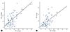
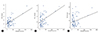
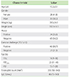
Table 1 shows the age, gender distribution, weight, height, atopy, eosinophil count, and IgE of the children studied. The children were between 5.5 and 16.5 years old (mean ± SD: 10.3 ± 3.1 years). The median falls in FEV1, FEF50%, and PEF were between 14% (9-18%), 23% (11-41%), and 15% (7-29%) respectively. The median eosinophil counts and IgE levels were 200/mm3 (90-340/mm=) and 46 IU/mL (15-143 IU/mL) respectively (Table 1). Forty six patients (68.7%) had a FEV1 fall of ≥10%. Twenty-six (38.8%) and thirty-one (46.3%) patients had a fall FEF50% of ≥26% and a PEF fall of ≥17.5% respectively. There was a positive correlation between maximal FEV1 with FEF50%, and PEF changes after the exercise test (p<0.05; FEF50%, r=0.68; PEF, r=0.65) (Fig. 1). Also, there was found a positive correlation between blood eosinophil counts and maximal FEV1, FEF50%, and PEF changes after the exercise test (p<0.05; FEV1, r=0.54; FEF50%, r=0.42; PEF, r=0.26) (Fig. 2). Moreover, in the exercise negative group for FEV1 (≤10), the FEF50% and PEF values decreased more than the cutoff values (FEF50% ≥ 26%, PEF ≥ 17.5%) in 3, and 2 patients respectively.
DISCUSSION
Although nonspecific bronchial responsiveness is usually evaluated with bronchoactive agonists such as histamine or methacholine, the bronchial response to exercise has also been used. Exercise challenge has been considered more specific for asthma than histamine or methacholine challenges. The exercise challenge was easy to perform and did not cause side effects, even in children with severe persistent asthma [15, 16]. EIB is more frequent in children and young adults, probably because of their high level of physical activity. Using a decrease greater than two standard deviations from the mean as a positive response, Custovic et al. [10] observed in children with mild to moderate asthma that EIB was detected by FEV1 in 98%, by FEF25-75% in 95%, by PEF in 78% and by the combination of both indexes (FEV1 and FEF25-75%) in 100% of subjects with a positive response to at least one test. Generally, some lung function parameters such as FEF25-75%, FEF50% have less reproducibility than FEV1. However, in some previous studies, measurements of FEF25-75% were used to supplement FEV1 in the diagnosis of exercise-induced asthma (EIA) in children [16, 17]. These studies, which were conducted on children, supported the addition of FEF25-75% measurements to improve the diagnosis of EIA. Fonseca-Guedes et al. [17] reported that FEF25-75% can decrease in response to exercise without changes in FEV1, mainly in children with mild asthma as well as good correlation between FEV1 and FEF25-75% as in the presented study. It has been suggested that FEF25-75% is a more sensitive measure of obstruction in the small airways than FEV1. There were 3 patients who had a higher decrease than the cut off value of FEF50% in FEV1 negative group in our study, which is consistent with the results of Fonseca-Guedes et al. [17]. Dickinson et al. [9] reported that strong positive correlation between FEV1 and FEF50% following bronchoprovocation as in our study. For the parameter, PEF, in a previous study, Gautrin et al. [18] compared the percent change PEF and FEV1 in asthmatic subjects. In that study as in the presented study, the correlation was showed between the percent change in PEF and in FEV1. Also in the presented study, in addition to this correlation, in the exercise negative group in FEV1, the PEF value decreased more than the cutoff value in 2 patients. Yoshikawa et al. [19] reported that the severity of EIB is associated with airway eosinophilic inflammation. Blood eosinophil counts, which may be useful in predicting the severity of EIB, are known to be an indirect marker of airway inflammation in asthma [20]. Also, we found positive correlation between blood eosinophil counts and maximal FEV1, FEF50%, PEF changes after the exercise test as in literature [21, 22].
In conclusion, the criteria to define the normal airway response to exercise are not standard; and, as a consequence, the estimated incidence of EIB in asthmatic children is wide. According to the presented study, eosinophil may play a major role in the severity of EIB in mild asthma. Also, FEF50% and PEF values can decrease in response to exercise without changes in FEV1 in the mild asthmatic patients.
 XML Download