

 PDF
PDF ePub
ePub Citation
 Print
Print


INTRODUCTION
Gianotti-Crosti syndrome (GCS) is a self-limiting exanthem of acute onset with a characteristic acral distribution, usually occurring in young children. The term GCS now comprises both papular acrodermatitis of childhood and papulovesicular acrolocated syndrome, once thought to be distinct clinical entities but now proven to be clinically and histologically indistinguishable [1]. Classically associated with hepatitis B virus, GCS is now seen more commonly with Epstein-Barr virus (EBV) [2]. We report a case of GCS suspected as and treated for allergic eczema in a four-year-old male patient who presented at our institution with symmetrical erythematous papulovesicular eruptions on the face, and both upper and lower extremities.
CASE REPORT
A four-year-old Indonesian boy presented with a generalized papulovesicular rash on his face, and both upper and lower extremities that had persisted for one month. The eruption first started on his lower limbs, and spread to his upper limbs and face. It was pruritic, the vesicles discharged serous fluid, and it was not associated with fever or preceding illness. His parents reported a previous similar episode one year ago where the eruptions left large areas of hyperpigmentation up to 4 cm in diameter, and resolved in two weeks. He was previously worked up in Indonesia for allergic dermatitis, with moderate levels of specific IgE antibodies to cow's milk protein and egg white (RAST test score of 2 for both allergens). Response to the medications prescribed then was minimal. He has a positive family history of allergic rhinitis secondary to house dust mite allergy.
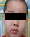
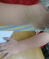
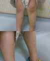
Examination revealed an otherwise healthy, active child. Multiple pink to red-brown papules and vesicles, coalescing into plaques with secondary excoriations were noted on his face, extensor surfaces of upper and lower extremities and buttocks (Figs. 1-3). Individual lesions measured 2 to 5 mm in diameter and some were flat-topped. Some ruptured vesicles were weepy and crusted suggestive of secondary bacterial infection. Other than a few redbrown papules on the back, the patient's trunk was spared. There were also no lesions on the patient's palms and soles. The rash appeared to affect the exposed areas of the body more severely. No hepatosplenomegaly or lymphadenopathy was detected.
The patient was treated symptomatically with Atarax (hydroxyzine), chlorhexidine lotion and moisturizing lotion, and advised to go swimming in chlorinated water to help lower the risk of secondary bacterial infections.
DISCUSSION
GCS is a sporadic dermatosis that commonly affects children three months to 15 years of age, with more than 90% younger than four years. However, it has been reported in adults where exclusively women are affected, suggesting that hormonal factors play a role [2].
It is characterized by the acute onset of multiple monomorphous, pink to red-brown papules or papulovesicles that may become confluent. The papules are distributed symmetrically on the face, extensor surfaces of the extremities, and buttocks. The rash may be pruritic and is typically nonhyperkeratotic; spares the trunk, palms of the hands, and soles of the feet. The eruptions can persist longer than 6 weeks in more than 50% of patients and complete resolution typically takes more than 2 months. The cutaneous manifestations can be preceded by an upper respiratory tract infection or diarrhea. The rash may be accompanied by a low-grade fever, hepatosplenomegaly, or cervical, inguinal or axillary lymphadenopathy. Koebner's phenomenon may also be present [2-4].
Although the exact underlying pathophysiologic mechanisms remain unclear, GCS is considered a cutaneous response to various immunologic triggers, with viral infections being the key factor. The most likely process is a local type IV hypersensitivity reaction to the offending antigen [2]. However, immunohistochemistry and electron microscopy findings are nonspecific and fail to demonstrate the presence of viral particles or antigens in skin lesions, thus suggesting that lesion development does not involve a direct local interaction between viral antigens and immune cells in the skin [5]. The syndrome was first described by Gianotti in association with hepatitis B virus. But other viruses such as EBV, cytomegalovirus, coxsackievirus, hepatitis A virus, parainfluenza virus, respiratory syncytial virus, rotavirus, mumps, parvovirus and molluscum contagiosum have been reported to be associated with this distinctive exanthema, EBV being the most common etiologic factor worldwide [2]. Studies have also shown that recent immunization and rarely, bacterial infections such as Bartonella henselae, Mycoplasma pneumoniae, and group A streptococci may trigger GCS [2, 6, 7]. In addition, a study has shown a higher prevalence of atopic dermatitis, family history of atopy and elevated IgE (total and specific) in GCS cases than controls [8], thus indicating that atopy is significantly associated with GCS.
GCS is diagnosed clinically by the presence of a characteristic rash and its distribution, with differential diagnoses including erythema multiforme, lichen planus, papular urticaria, scabies and atopic dermatitis [2, 3]. Being primarily a clinical diagnosis, no investigations were performed in this patient to determine the causative agent since he was well with no localizing signs of infection. Also, the diagnosis of GCS requires clinical suspicion, lest it be mistaken for one of its differential diagnoses such as atopic dermatitis as in this case. This is because the lesions in atypical, early or resolving cases can resemble that of atopic dermatitis. The presence of specific IgE on RAST testing and positive family history of atopy in this patient is consistent with findings of Ricci et el. [8] and could make distinguishing between atopic dermatitis difficult.
As GCS is generally benign and self-limiting condition, no definitive treatment is required. However, oral antihistamines and topical anti-pruritics can be prescribed as needed for pruritis. The effectiveness of topical steroids has not been established in a controlled setting [2]. Anti-virals such ribavirin have been reported to induce dramatic symptomatic remission in prolonged and disabling cases, but further studies are also required to confirm its efficacy in GCS [9]. Patients' parents should be reassured that the rash usually resolves without scarring within 10 to 60 days. Recurrences are rare.
 XML Download