Journal List > Restor Dent Endod > v.40(4) > 1090137

Kim, Hong, Choi, and Park: The effect of saliva decontamination procedures on dentin bond strength after universal adhesive curing

Abstract

Objectives

The purpose of this study was to investigate the effectiveness of multiple decontamination procedures for salivary contamination after curing of a universal adhesive on dentin bond strength according to its etch modes.

Materials and Methods

Forty-two extracted bovine incisors were trimmed by exposing the labial dentin surfaces and embedded in cylindrical molds. A universal adhesive (All-Bond Universal, Bisco) was used. The teeth were randomly divided into groups according to etch mode and decontamination procedure. The adhesive was applied according to the manufacturer's instructions for a given etch mode. With the exception of the control groups, the cured adhesive was contaminated with saliva for 20 sec. In the self-etch group, the teeth were divided into three groups: control, decontamination with rinsing and drying, and decontamination with rinsing, drying, and adhesive. In the etch-and-rinse group, the teeth were divided into four groups: control, decontamination with rinsing and drying, decontamination with rinsing, drying, and adhesive, and decontamination with rinsing, drying, re-etching, and reapplication of adhesive. A composite resin (Filtek Z350XT, 3M ESPE) was used for filling and was cured on the treated surfaces. Shear bond strength was measured, and failure modes were evaluated. The data were subjected to one-way analysis of variation and Tukey's HSD test.

Results

The etch-and-rinse subgroup that was decontaminated by rinse, drying, re-etching, and reapplication of adhesive showed a significantly higher bond strength.

Conclusions

When salivary contamination occurs after curing of the universal adhesive, additional etching improves the bond strength to dentin.

Introduction

Isolation is one of the most important factors for ensuring the adhesion of composite resin to dentin. Saliva contamination is more likely to occur when the operative site is near or at the gingival margin. Contamination can also occur as a result of uncooperative patients, malpositioned teeth, or cervical lesions. Saliva can cause several problems, such as increasing microleakage and reducing the bonding strength of the composite resin.1234
Clinicians must consider the effects of oral fluids on bond strength during the clinical application of bonding systems. However, the reduction of bond strength is related to the type of adhesive system being used and to the stage of the bonding process when contaminations occur. As a result, previous studies have shown conflicting results. Some studies have shown that contamination with saliva reduces the bond strength of the dental bonding agents to dentin, whereas others have reported the opposite.123456
Recently, a new type of adhesive system, known as universal or multi-mode adhesives, has been introduced. These can be applied with either the etch-and-rinse technique or the self-etch technique.7 The literature contains little information regarding this new class of universal adhesives, and the effect of salivary contamination on the performance of the universal adhesives has not yet been evaluated.78 The purpose of this study was to investigate the effects of decontamination procedures for salivary contamination after curing of a universal adhesive on dentin bond strength according to its etch modes.

Materials and Methods

A single variety of commercially available universal adhesive was used in this study. All-Bond Universal (Bisco, Schaumburg, IL, USA) was applied using the self-etch technique or the etch-and-rinse technique. All teeth were restored with Filtek Z350XT (3M ESPE, St. Paul, MN, USA). The materials used in this study are presented in Table 1.
Forty-two extracted bovine incisors were cleaned of tissue remnants and stored in saline until they were used (less than one month after extraction). The teeth were sectioned at the cementodentinal junction, and the labial surfaces of the teeth were trimmed to create flat dentin surfaces. The coronal part of the teeth was embedded in cylindrical molds using a self-curing acrylic resin, with the labial surface facing outwards and parallel to the base of the molds. The labial surfaces were divided into two parts mesiodistally, and the bonding procedure was performed separately on each part.
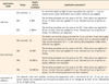
The teeth were randomly divided into seven groups of 6 teeth (12 surfaces) each. In the self-etch group, the teeth were divided into three groups: control (SE1), decontamination with rinsing and drying (SE2), and decontamination with rinsing, drying, and adhesive (SE3). In the etch-and-rinse group, the teeth were divided into four groups: control (ER1), decontamination with rinsing and drying (ER2), decontamination with rinsing, drying, and adhesive (ER3), and decontamination with rinsing, drying, re-etching, and reapplication of adhesive (ER4). The prepared teeth were stored in 100% relative humidity until preparation of the specimens for bonding. Immediately prior to bonding, fresh whole saliva was collected from an experimenter. The dentin surfaces were polished with 600 grit silicon carbide abrasive paper for 30 seconds under wet conditions to create a uniform surface and smear layer. The surfaces were then rinsed with air-water spray for 15 seconds and gently air-dried before application of the adhesive.
The universal adhesive was applied either in self-etch or etch-and-rinse mode and cured according to the manufacturer's instructions. Except for the control groups (SE1, ER1), fresh whole saliva was applied to the surface of the teeth with a disposable brush for 20 seconds. Decontamination procedures were performed on the dentin surfaces according to the experimental protocol for each group, as listed in Table 2.
A cylindrical plastic tube 3 mm long and with an inner diameter of 3.5 mm was placed on the surfaces. The tube was filled in two increments with composite resin and placed on the pretreated dentin surfaces. Each increment was cured with an LED curing light (Elipar S10, 3M ESPE) for 40 seconds at a minimum of 1,000 mW/cm2. Excess composite was carefully removed from the periphery of the matrix using an explorer. The prepared specimens were subsequently stored in 100% relative humidity for 24 hours.
The shear bond strength (SBS) was tested using a universal testing machine (Zwick Z020, Zwick GmbH, Ulm, Germany) with a crosshead speed of 0.5 mm/min. Failure modes were evaluated using an optical microscope and classified as adhesive failure, mixed failure, dentin cohesive failure, or resin cohesive failure. The SBS data were analyzed using one-way analysis of variation and Tukey's honest significant difference test to compare the groups, and the level for statistical significance was set at p < 0.05. Statistical analysis was performed using SPSS for Windows version 18.0 (SPSS Inc., Chicago, IL, USA).

Results

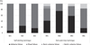
The results of the SBS test are shown in Table 3. The ER4 group (etch-and-rinse method, decontaminated by rinsing, drying, re-etching, and reapplication of adhesive) showed a significantly higher bond strength (16.22 ± 3.54 MPa) than the other groups (p < 0.05). The SE2 and ER2 groups (decontaminated by rinsing and drying only) showed no significant differences compared to the control group. No significant difference was found between the SE and ER groups overall.
The failure modes of all groups are shown in Figure 1. Most of the specimens showed adhesive or mixed failure. The SE2 and ER2 groups showed a greater frequency of adhesive failure than the control group, but had a similar SBS to that of the control group.

Discussion

Sal iva is mostly composed of water (99.4%), macromolecules, such as proteins, enzymes, mucins, immunoglobulins, and nitrogenous products, electrolytes, such as calcium, sodium, and chloride, and organic particles, such as urea, amino acids, fatty acids, and free glucose.910 The water in saliva can reduce the bond strength of dentin adhesives.11 Salivary glycoproteins may also interfere with proper adhesion.11121314 Many studies have shown that whole healthy human saliva functions as a contaminating medium.23111516 For this reason, fresh whole saliva from a single donor who had not eaten for one to two hours before saliva collection was used in this study. To our knowledge, no studies have yet evaluated the effect of contamination with saliva after curing of a universal adhesive on the bonding strength. Using other adhesive systems, several studies have shown that salivary contamination diminishes bond strength to dentin.1234 Still further studies have found that rinsing and drying of the contaminated surfaces alone, without reapplication of the bonding system, cannot restore the bonding strength to dentin in three-step etch-and-rinse, two-step etch-and-rinse, or one-step self-etching systems.1718 Glycoproteins may adsorb to the poorly polymerized adhesive surface and act as a barrier, thereby decreasing the wettability of the composite resin and preventing adequate copolymerization.19 Furthermore, water incorporated within the partially cured resin may interfere with the copolymerization of the subsequent resin increment.20
The SE2 and ER2 groups (decontaminated by rinsing and drying) in this study were not significantly different from their respective control groups. All-Bond Universal was resistant to salivary contamination that occurred after curing of the adhesive.32122 Although rinsing and drying are an accepted treatment for restoring the SBS, adhesive failure occurred more often in Groups SE2 and ER2 than the corresponding negative control groups (SE1 and ER1), especially in the ER groups.
Simple rinse and reapplication of the adhesive to the contaminated surface can restore the bond strength to dentin in two-step self-etching systems and one-step self-etching systems.42324 Salivary proteins can be removed by rinsing and reapplication of the self-etching primer.24 In a study by Farideh et al., when contamination of bonding surface with saliva took place after the curing of Single Bond, rebonding followed by water rinsing and drying was sufficient. In the present study, the SE3 and ER3 groups showed recovery of the SBS.25 Due to the acidity of the All-Bond Universal adhesive (pH 3.2), it is likely to have removed the salivary proteins without difficulty.
Group ER4 in the present study showed a significantly increased SBS. Two possible explanations exist for this result. The first potential explanation is that re-etching can increase the bond strength of adhesives. In 1992, Kanca recommended an additional 10 seconds of acid etching. Several studies have also shown that similar techniques can improve SBS.2627 However, another study showed that re-etching was not necessary because the bonding thickness was decreased after removing the oxygen-inhibited layer by acid etching and rinsing.25 In this study, the two-coat application of All-Bond Universal in the etch-and-rinse mode led to the formation of a thick hybrid layer with the sufficient bonding thickness to resist re-etching.
Second, two coats of bonding agent can improve the bond strength of single-step adhesives. Many studies have shown improvements in one-step self-etching adhesive systems when two coats of bonding agent were applied.282930 Indeed, cured one-step self-etching adhesives act like a permeable membrane, and dentinal fluid therefore transudates across the polymerized adhesive.31 This is especially true in the case of 2-hydroxyethyl-methacrylate (HEMA)-containing adhesives.32 In order to prevent phase separation between the hydrophilic and hydrophobic components, most self-etching primers contain HEMA.333435 The newly developed universal adhesive used in this study also contains HEMA (5 - 15% by weight). Although the manufacturer insists that the adhesive layer becomes hydrophobic after curing, water sorption may have occurred. Therefore, a second application of the universal adhesive may block water sorption and significantly improve SBS to the dentin surface.
Miguel et al. conducted an experiment in which a significant reduction in nanoleakage was observed in All-Bond Universal in the etch-and-rinse mode when a hydrophobic resin coating (Heliobond, Ivoclar vivadent, Schaan, Liechtenstein) was applied.29 This additional layer of hydrophobic resin adhesive adds unsolvated hydrophobic monomers to the bonded surface. Consequently, the relative concentration of retained solvents and unreacted monomers in the adhesive layer is decreased.36 Since the hybrid layer is more densely packed, the adhesive can resist the tensile forces during the microtensile bond strength test, and has less tendency to degrade over time.373839 Failure mode analysis also showed less adhesive failure in the group that underwent additional etching and application of the adhesive (ER4).
Lee et al. and Ahn et al. observed statistical differences in bond strength depending on the application mode of All-Bond Universal.4041 The lack of active brushing was suggested as a possible cause for the low bond strength in the self-etch mode for All-Bond Universal. However, in the present study, All-Bond Universal was applied along with scrubbing the dentin surface, which could explain the similar SBS findings in the self-etch and etch-and-rinse modes. The bond strength did not show significant differences in SBS according to the application mode. The presence of 10-methacryloyloxydecyl dihydrogen phosphate monomer in the composition of universal adhesives may well explain their good performance regardless of the application mode.

Conclusions

Within the limitations of this in vitro study, it can be concluded that when salivary contamination occurs after a universal adhesive is cured, simply rinsing and drying can restore the SBS to dentin regardless of the application mode. Re-etching and additional adhesive application improved the bond strength and affected the failure mode. Further long-term studies are necessary to evaluate the clinical performance of these techniques.

Figures and Tables

Figure 1

Percentage of failure modes of the tested groups.

rde-40-299-g001
Table 1

Materials used in this study

rde-40-299-i001
Material Manufacturer Lot number Composition
Ultra-Etch Ultradent, South Jordan, UT, USA ET463137, ET436237 35% phosphoric acid, cobalt aluminate blue spinel, cobalt zinc aluminate blue spinel
All-Bond Universal Bisco, Schaumburg, IL, USA 1400004366 MDP, bis-GMA, HEMA, ethanol, water
Filtek Z350XT 3M ESPE, St. Paul, MN, USA N497426 bis-GMA, UDMA, TEGDMA, bis-EMA, PEGDMA, silica filler, zirconia filler, zirconia/silica (aggregated)

MDP, Methacryloyloxydecyl dihydrogen phosphate; bis-GMA, Bisphenol A glycidyl methacrylate; HEMA, Hydroxyethylmethacrylate; UDMA, urethane dimethacrylate; TEGDMA, triethyleneglycol dimethacrylate; bis-EMA, Ethoxylated bisphenol A dimethacrylate; PEGDMA, poly(ethylene glycol) dimethacrylate.

Table 2

Dentin surface treatment and application procedures

rde-40-299-i002
Application mode Group Dentin surface treatment Application procedure*
Self-etch SE1 (control) A An absorbent pellet or high volume evacuation was used for 1 - 2 sec to remove excess water. Desiccation was avoided. Adhesive was applied.
SE2 A SRD The bonding procedure was the same as for SE1. Fresh saliva was applied for 20 sec. A water rinse was applied for 5 sec, followed by 5 sec of gentle air-drying.
SE3 A SRD A The bonding procedure was the same as for SE1. Fresh saliva was applied for 20 sec. A water rinse was applied for 5 sec, followed by 5 sec of gentle air-drying. The adhesive was reapplied.
Etch-and-rinse ER1 (control) EA The dentin was etched using an etchant for 15 sec and then rinsed thoroughly. Excess water was removed by blotting the surface with an absorbent pellet or high volume evacuation for 1 - 2 sec, leaving the preparation visibly moist. Adhesive was applied.
ER2 EA SRD The bonding procedure was the same as for ER1. Fresh saliva was applied for 20 sec. A water rinse was applied for 5 sec, followed by 5 sec of gentle air-drying.
ER3 EA SRD A The bonding procedure was the same as for ER1. Fresh saliva was applied for 20 sec. A water rinse was applied for 5 sec, followed by 5 sec of gentle air-drying. Adhesive was reapplied.
ER4 EA SRD EA The bonding procedure was the same as for ER1. Fresh saliva was applied for 20 sec. A water rinse was applied for 5 sec, followed by 5 sec of gentle air-drying. The ER1 bonding procedure was then repeated.
Application of adhesive Two separate coats were applied, scrubbing the preparation with a microbrush for 10 - 15 sec per coat. Excess solvent was evaporated by thoroughly air-drying with an air syringe for at least 10 sec, followed by 10 sec of light-curing.

*According to the manufacturer's instructions.

A, adhesive (applied according to the manufacturer's instructions); S, salivary contamination (scrubbing with a microbrush for 20 sec); R, rinsing (5 sec); D, drying (5 sec); E, etching.

Table 3

Shear bond strength values (MPa, n = 12)

rde-40-299-i003
Application mode Group Dentin surface treatment SBS
Self-etch SE1 A 10.61 ± 2.64
SE2 A SRD 8.84 ± 1.67
SE3 A SRD A 11.43 ± 2.65
Etch-and-rinse ER1 EA 10.61 ± 2.62
ER2 EA SRD 9.30 ± 3.56
ER3 EA SRD A 10.80 ± 3.83
ER4 EA SRD EA 16.22 ± 3.54*

The asterisk indicates a statistically significant difference (p < 0.05).

SBS, shear bond strength; A, adhesive; S, salivary contamination; R, rinsing; D, drying; E, etching.

Acknowledgment

This paper was supported by Wonkwang University in 2013.

Notes

Conflict of Interest No potential conflict of interest relevant to this article was reported.

References

1. Hiraishi N, Kitasako Y, Nikaido T, Nomura S, Burrow MF, Tagami J. Effect of artificial saliva contamination on pH value change and dentin bond strength. Dent Mater. 2003; 19:429–434.
crossref
2. Park JW, Lee KC. The influence of salivary contamination on shear bond strength of dentin adhesive systems. Oper Dent. 2004; 29:437–442.
3. Yoo HM, Oh TS, Pereira PN. Effect of saliva contamination on the microshear bond strength of one-step self-etching adhesive systems to dentin. Oper Dent. 2006; 31:127–134.
crossref
4. Sattabanasuk V, Shimada Y, Tagami J. Effects of saliva contamination on dentin bond strength using all-in-one adhesives. J Adhes Dent. 2006; 8:311–318.
5. Johnson ME, Burgess JO, Hermesch CB, Buikema DJ. Saliva contamination of dentin bonding agents. Oper Dent. 1994; 19:205–210.
6. Taskonak B, Sertgöz A. Shear bond strengths of saliva contaminated 'one-bottle' adhesives. J Oral Rehabil. 2002; 29:559–564.
crossref
7. Hanabusa M, Mine A, Kuboki T, Momoi Y, Van Ende A, Van Meerbeek B, De Munck J. Bonding effectiveness of a new 'multi-mode' adhesive to enamel and dentine. J Dent. 2012; 40:475–484.
crossref
8. Perdigão J, Sezinando A, Monteiro PC. Laboratory bonding ability of a multi-purpose dentin adhesive. Am J Dent. 2012; 25:153–158.
9. Eiriksson SO, Pereira PN, Swift EJ Jr, Heymann HO, Sigurdsson A. Effects of saliva contamination on resin-resin bond strength. Dent Mater. 2004; 20:37–44.
crossref
10. Humphrey SP, Williamson RT. A review of saliva: normal composition, flow, and function. J Prosthet Dent. 2001; 85:162–169.
crossref
11. Xie J, Powers JM, McGuckin RS. In vitro bond strength of two adhesives to enamel and dentin under normal and contaminated conditions. Dent Mater. 1993; 9:295–299.
crossref
12. el-Kalla IH, Garcia-Godoy F. Saliva contamination and bond strength of single-bottle adhesives to enamel and dentin. Am J Dent. 1997; 10:83–87.
13. Fritz UB, Finger WJ, Stean H. Salivary contamination during bonding procedures with a one-bottle adhesive system. Quintessence Int. 1998; 29:567–572.
14. Townsend RD, Dunn WJ. The effect of saliva contamination on enamel and dentin using a self-etching adhesive. J Am Dent Assoc. 2004; 135:895–901.
crossref
15. Yazici AR, Tuncer D, Dayangaç B, Ozgünaltay G, Onen A. The effect of saliva contamination on microleakage of an etch-and-rinse and a self-etching adhesive. J Adhes Dent. 2007; 9:305–309.
16. Jiang Q, Pan H, Liang B, Fu B, Hannig M. Effect of saliva contamination and decontamination on bovine enamel bond strength of four self-etching adhesives. Oper Dent. 2010; 35:194–202.
crossref
17. Patil SB, Shivakumar AT, Shah S. Effect of salivary contamination on shear bond strength of two adhesives: An in vitro study. Dent Hypotheses. 2014; 5:115–120.
crossref
18. Guerriero LN, Vieira SN, Scaramucci T, Kawaguchi FA, Sobral MAP, Matos AB. Effect of saliva contamination on the bond strength of an etch-and-rinse adhesive system to dentin. Rev Odonto Ciênc. 2009; 24:410–413.
19. Perdigão J, Lambrechts P, van Meerbeek B, Tomé AR, Vanherle G, Lopes AB. Morphological field emission-SEM study of the effect of six phosphoric acid etching agents on human dentin. Dent Mater. 1996; 12:262–271.
crossref
20. Abdalla AI, Davidson CL. Bonding efficiency and interfacial morphology of one-bottle adhesives to contaminated dentin surfaces. Am J Dent. 1998; 11:281–285.
21. Koppolu M, Gogala D, Mathew VB, Thangala V, Deepthi M, Sasidhar N. Effect of saliva and blood contamination on the bond strength of self-etching adhesive system-An in vitro study. J Conserv Dent. 2012; 15:270–273.
crossref
22. Pinzon LM, Oguri M, O'Keefe K, Dusevish V, Spencer P, Powers JM, Marshall GW. Bond strength of adhesives to dentin contaminated with smoker's saliva. Odontology. 2010; 98:37–43.
crossref
23. Cobanoglu N, Unlu N, Ozer FF, Blatz MB. Bond strength of self-etch adhesives after saliva contamination at different application steps. Oper Dent. 2013; 38:505–511.
crossref
24. Ari H, Dõnmez N, Belli S. Effect of artificial saliva contamination on bond strength to pulp chamber dentin. Eur J Dent. 2008; 2:86–90.
crossref
25. Darabi F, Tavangar M, Davalloo R. Effect of different decontamination procedures from a saliva-contaminated cured bonding system (Single Bond). Dent Res J (Isfahan). 2012; 9:399–403.
26. Justin RM, Paranthaman H, Rajesh AG, Varghese RP, Ranganath LM. Effect of salivary contamination on the bond strength of total-etch and self-etch adhesive systems: an in vitro study. J Contemp Dent Pract. 2012; 13:655–660.
27. Suryakumari NB, Reddy PS, Surender LR, Kiran R. In vitro evaluation of influence of salivary contamination on the dentin bond strength of one-bottle adhesive systems. Contemp Clin Dent. 2011; 2:160–164.
crossref
28. Reis A, Albuquerque M, Pegoraro M, Mattei G, Bauer JR, Grande RH, Klein-Junior CA, Baumhardt-Neto R, Loguercio AD. Can the durability of one-step self-etch adhesives be improved by double application or by an extra layer of hydrophobic resin? J Dent. 2008; 36:309–315.
crossref
29. Perdigão J, Muñoz MA, Sezinando A, Luque-Martinez IV, Staichak R, Reis A, Loguercio AD. Immediate Adhesive Properties to Dentin and Enamel of a Universal Adhesive Associated With a Hydrophobic Resin Coat. Oper Dent. 2014; 39:489–499.
crossref
30. Muñoz MA, Sezinando A, Luque-Martinez I, Szesz AL, Reis A, Loguercio AD, Bombarda NH, Perdigão J. Influence of a hydrophobic resin coating on the bonding efficacy of three universal adhesives. J Dent. 2014; 42:595–602.
crossref
31. Salz U, Zimmermann J, Zeuner F, Mozner N. Hydrolytic stability of self-etching adhesive systems. J Adhes Dent. 2005; 7:107–116.
32. Hosaka K, Nakajima M, Takahashi M, Itoh S, Ikeda M, Tagami J, Pashley DH. Relationship between mechanical properties of one-step self-etch adhesives and water sorption. Dent Mater. 2010; 26:360–367.
crossref
33. Van Landuyt KL, De Munck J, Snauwaert J, Coutinho E, Poitevin A, Yoshida Y, Inoue S, Peumans M, Suzuki K, Lambrechts P, Van Meerbeek B. Monomer-solvent phase separation in one-step self-etch adhesives. J Dent Res. 2005; 84:183–188.
crossref
34. Moszner N, Salz U, Zimmermann J. Chemical aspects of self-etching enamel-dentin adhesives: a systematic review. Dent Mater. 2005; 21:895–910.
crossref
35. Van Landuyt KL, Snauwaert J, Peumans M, De Munck J, Lambrechts P, Van Meerbeek B. The role of HEMA in one-step self-etch adhesives. Dent Mater. 2008; 24:1412–1419.
crossref
36. Breschi L, Mazzoni A, Ruggeri A, Cadenaro M, Di Lenarda R, De Stefano Dorigo E. Dental adhesion review: aging and stability of the bonded interface. Dent Mater. 2008; 24:90–101.
crossref
37. de Andrade e Silva SM, Carrilho MR, Marquezini Junior L, Garcia FC, Manso AP, Alves MC, de Carvalho RM. Effect of an additional hydrophilic versus hydrophobic coat on the quality of dentinal sealing provided by two-step etch-and-rinse adhesives. J Appl Oral Sci. 2009; 17:184–189.
crossref
38. Reis A, Grande RH, Oliveira GM, Lopes GC, Loguercio AD. A 2-year evaluation of moisture on microtensile bond strength and nanoleakage. Dent Mater. 2007; 23:862–870.
crossref
39. Reis A, de Carvalho Cardoso P, Vieira LC, Baratieri LN, Grande RH, Loguercio AD. Effect of prolonged application times on the durability of resin-dentin bonds. Dent Mater. 2008; 24:639–644.
crossref
40. Lee IS, Son SA, Hur B, Kwon YH, Park JK. The effect of additional etching and curing mechanism of composite resin on the dentin bond strength. J Adv Prosthodont. 2013; 5:479–484.
crossref
41. Ahn J, Jung KH, Son SA, Hur B, Kwon YH, Park JK. Effect of additional etching and ethanol-wet bonding on the dentin bond strength of one-step self-etch adhesives. Restor Dent Endod. 2015; 40:68–74.
crossref
TOOLS
ORCID iDs

Sujung Park
https://orcid.org/http://orcid.org/0000-0003-3457-1242

Similar articles