

 PDF
PDF ePub
ePub Citation
 Print
Print


Introduction
The modern exploration of regenerative dentistry has added impetus onto the field of molecular biology. Assuming the present-day situation, it can be categorically documented as an archetype stint in the therapeutic armamentarium for dental disease. Regenerative endodontic procedures are defined as biologically based procedures designed to replace damaged structures, including dentin and root structures, as well as cells of the pulp-dentin complex.1 The harmonized spurs of biology and mechanical regulators promoting cellular activities have critically enhanced the acceptance of regenerative therapy for dental tissues.
The regenerative potential of platelets has been deliberated. The platelets release growth factors that are trapped inside the fibrin matrix following activation. These are considered to be the stimulant for mitogenic response in the periosteum and are responsible for bone repair during normal wound healing.2 The superior understanding of physiologic properties of platelets in wound healing has led to their augmented therapeutic applications.3 Nevertheless, there is still concern linked to the procedures for production of autologous fibrin adhesives.4,5 Besides, legal restrictions on blood handling with concentrated platelet rich plasma have coexisted. In an effort to overcome these problems, it was contemplated to develop a new family of platelet concentrates, which came to be recognized as the platelet rich fibrin (PRF).5
Choukroun's PRF is a fibrin matrix where platelet cytokines and cells are wedged.6 They serve as a resorbable membrane following their release after a certain time.7 PRF was considered to be a healing biomaterial and was initially used in oral implantology.8 Presently, several investigations have shown its application in diverse disciplines of dentistry.9-14 This paper is intended to add light on the various prospects of PRF and clinical insights to regenerative endodontic therapy. The appraisal focused on original research articles and case reports entailing the use of PRF in endodontics. The search terminologies used on PubMed database were 'platelet rich fibrin (PRF)'. The criteria were further specified by use of Boolean operators (AND, OR, NOT) and permutation of precise keywords as 'platelet rich fibrin dental' and 'platelet rich fibrin endodontics'.
Review
What is PRF?
PRF is often named as Choukroun's PRF after its inventor.5,6 It is a second-generation platelet concentrate. The PRF constitutes components of blood sample that are beneficial to improve wound healing and immunity. Ross et al. were amongst the pioneers who first described a growth factor from platelets.15
Preparation
The procedure involves drawing of blood that is collected into test tubes without an anticoagulant and needs to be centrifuged instantaneously. A tabletop centrifuge can be used for this purpose for 2 minutes at 2,700 rpm.6 The resultant product consists of the three layers.16
The blood coagulation starts instantaneously as it comes in contact with the glass surface due to the lack of anticoagulant. If the time necessary to collect blood and launch centrifugation is exceedingly prolonged, the fibrin will polymerize in a diffuse way in the tube and only a small blood clot without consistency will be obtained.5 Consequently, blood collection should be prompt and instant centrifugation is a prerequisite in the production protocol for PRF.5
Recently, PRF box (Process, Nice, France) has been announced.2 It is formulated to produce homogenously thickened hydrated membrane and an exudate rich in platelets, leukocytes, vitronectin and fibronectin expressed from the fibrin clots.17 It has improved the issues regarding the handling of the PRF clot.18
Biological properties of PRF
PRF can be considered as an immune concentrate with specific composition and a three dimensional architecture.17,19,20 It contains multitude of growth factors like platelet derived growth factor (PDGF), transforming growth factor β1 (TGF β1), insulin like growth factor (IGF), etc., exhibiting varied potent local properties such as cell migration, cell attachment, cell proliferation, and cell differentiation.19,21 It has been shown as an ideal biomaterial for pulp-dentin complex regeneration.22 PRF is both a healing and interpositional biomaterial.23 It accelerates wound closure and mucosal healing due to fibrin bandage and growth factor release. In addition, it prevents the early invagination of undesired cells, and thus acts as a viable barrier between desired and undesired cells.19,23
Biochemical analysis of PRF
PRF consists of an intimate assembly of cytokines, glycan chains, structural glycoproteins enmeshed within a slowly polymerized fibrin network.17 These biochemical components have well known synergistic effects on healing processes.24 Fibrin is the natural guide of angiogenesis. Fibrin constitutes a natural support to immunity.20
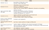
Proposed mechanism of action
In vitro release of growth factors from PRF and the results of in vivo studies have now put forward a proposal to optimize the clinical application of PRF.25 PRF is a concentrated suspension of the growth factors found in platelets (Table 1).18,26,27 These growth factors are involved in wound healing and are postulated as promoters of tissue regeneration.17,20
PRF as a tissue-engineering scaffold for endodontics
In any tissue engineering procedure, the cell growth and differentiation are related to an apposite scaffold.28-31 Furthermore, the differentiation of stem cells is controlled by extracellular matrix molecules.32 In this regard, it is anticipated that a suitable scaffold that contains growth factors might be promising tool to enrich the rate of tissue differentiation as it would selectively bind and localize cells and undergo biodegradation over time.33 PRF can be considered as an appropriate scaffold for regenerative endodontics as it fulfills all the properties as mentioned above. As a potential scaffold for regenerative endodontic therapy, PRF entails prospective research.
Review of regenerative endodontic applications of PRF
Following is a brief literature review on the various applications of PRF in endodontic therapy. Shivashankar et al. described a case report highlighting the combined use of graft material (PRF and hydroxyapatite [HA]) and barrier membrane in the treatment of large periapical lesion.23 The authors hypothesized that the use of PRF in conjunction with HA crystals accelerates the resorption of the graft crystals and induces rapid rate of bone formation. Likewise, Sculean et al. in their study concluded that the combination of barrier membrane and grafting materials might result in histological evidence of periodontal regeneration, predominantly bone repair.34 Gassling et al. demonstrated that PRF membranes are suitable for cultivation of periosteal cells for bone tissue engineering.35 Pradeep et al. concluded that when HA is combined with PRF, it increases the regenerative effects observed with PRF in the treatment of human three wall intrabony defects.36
Jayalakshmi et al. used PRF in combination with beta tricalcium phosphate (β-TCP) bone graft in the treatment of periapical cyst.37 The authors reported progressive, significant, and predictable clinical and radiographic bone regeneration/healing with the use of PRF. The authors suggested that the combined use of PRF and β-TCP for bone augmentation in treatment of periapical defects is a potential treatment alternative for faster healing than using biomaterials alone. Similar results were reported by Kim et al. using combination therapy of PRF with β-TCP.38
Keswani et al. reported that PRF might serve as a potentially ideal scaffold in revascularization of immature permanent teeth with necrotic pulps as it is rich in growth factors, enhances cellular proliferation and differentiation, and acts as a matrix for tissue ingrowth.39 In this context, Simonpieri et al. reviewed advantages of the use of PRF as it acts as a stabilizing sheath and offers mechanical sustenance.40 Also, the authors proposed that PRF fragments act as biological connectors and the cellular migration is accelerated which is critical for neo-angiogenesis and vascularization. Furthermore, the platelet cytokines (PDGF, TGF-alpha, IGF-1) are gradually released as the fibrin matrix is resorbed, thus creating a perpetual process of healing.
Bains et al. reported the applicability of PRF for the management of an iatrogenic perforation of pulpal floor in the furcation region of mandibular first molar.13 According to the authors, the autologous and biocompatible nature of PRF and mineral trioxide aggregate (MTA) appeared to be favorable for the long-term clinical results. Shivashankar et al. reported a case of revitalization of tooth with necrotic pulp and open apex using PRF.22 They described evidence of continued thickening of the dentinal walls, root lengthening, regression of the periapical lesion and apical closure with use of PRF. The authors considered PRF to be an excellent biomaterial for pulp-dentin complex regeneration. Analogously, Rudagi et al. also reported a case demonstrating the successful healing and apexification with combined use of MTA as an apical barrier, and autologus platelet rich fibrin membrane as an internal matrix.41
Huang et al. conducted an investigation into the biological effects of PRF on human dental pulp cells.42 PRF was found to increase dental pulp cell proliferation as well as osteoprotegerin (OPG) expression in a time-dependent manner. Alkaline phosphatase (ALP) activity was also significantly up-regulated by PRF. These findings might serve as a basis for preclinical studies that address the role of PRF in reparative dentin formation.
In regard to regenerative endodontic procedures with PRF, it is assumed that some amounts of human dental pulp cells present in the apical papilla usually remain vital even in case of a large periapical lesion. After the regression of the inflammation and under the influence of Hertwigs epithelial root sheath, these dental pulp cells differentiate into odontoblasts like cells. OPG and ALP expression are generally regarded as markers of odontoblastic differentiation. The only disadvantage associated with PRF is its manipulation to place inside the canal. Clinical trials are crucial to equate the effect of PRF in the revitalization of tooth with necrotic pulp and open apex on a long term basis.
Hiremath et al. reported affirmative results with pulpotomy using PRF.43 However, the authors suggested that long term trials with larger sample sizes are required to justify the use of PRF for treatment of pulpitis. It was concluded that pulpotomy with PRF could be a substitute treatment to MTA or other materials.
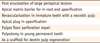
It is apparent persuasively that PRF, when used as a scaffolding material in an infected necrotic immature tooth for pulpal regeneration and tooth revitalization, satisfies many criteria of an ideal physical scaffold. Another advantage of using PRF as a scaffold is that it has a trimolecular or equilateral fibrin branch junction that makes its architecture flexible and can support cytokine enmeshment and cellular migration.5 However, long-term exploration still needs to be focused for therapeutic benefits of PRF. We present a summary of the potential regenerative endodontic applications of PRF as reported by various clinical studies (Table 2).
Conclusions
With the present knowledge, it can be confirmed that PRF can be considered a therapeutic biomaterial. However, despite the evident regenerative benefits of PRF, substantiation of its clinical applications is still limited. Consequently, there is a constraint for the rationalization of its use. Additional randomized, controlled clinical trials are defensible to test the long-term benefits and ultimate outcomes associated with PRF.
 XML Download