

 PDF
PDF ePub
ePub Citation
 Print
Print


INTRODUCTION
Direct anterior approach (DAA) of the hip has recently gained popularity as an alternative way to access the hip joint for the hip arthroplasty operation.
The procedure is performed with patient in supine position and is approached through the intra-muscular interval between tensor fascia lata (TFL) and sartorius free from disruption of the surrounding muscles. For this reason, number of studies suggested that hip arthroplasty done in DAA may provide number of advantages compare to that done in posterolateral approach (PLA) in terms of stability and early rehabilitation123). Also, as the patient is in supine position, it is easier to orient the native pelvis anatomy and therefore, theoretically, implantation of the acetabular cup can be done more accurately. However, due to the unfamiliarity, number of complication can also be a problem. Several studies showed that at least in the early phase of learning period, the operation time is significantly longer and there are number of potential problems including damage of the surrounding muscle structures and the fracture of the greater trochanter45). Nevertheless, the number of hip arthroplasty done with DAA is globally increasing.
However, there is limited description of DAA in the Korean literature. Thus in this study, we report our early experience of DAA and report its pros and cons by comparing it with the conventional PLA. More specifically, we assessed the intraoperative problems and perioperative assessments of the surgical factors between the two approaches to validate the advantages and disadvantages of DAA.
MATERIALS AND METHODS
Between October 2016 and February 2017, 25 consecutive hip arthroplasties were performed using DAA in our institution. This composed of 12 total hip arthroplasties (THA) and 13 bipolar hemiarthroplasties (BHA). All operations were done by a single surgeon who had hip arthroplasty fellowship in both the PLA and the DAA. This was the first 25 cases of hip arthroplasty done by the operator since the fellowship was completed. The cohort was compared with the 25 consecutive patients who had hip arthroplasty by the same surgeon between December 2015 to April 2016 with PLA which composed of 13 primary THA and 12 BHA. Retrospective chart review was done on this patient cohort.
All operations were primary arthroplasties. For DAA, the mean age of the patients were 65.4 years (range, 25 to 92 years) while that for PLA was 68.4 years (range, 37 to 98 years). In DAA cohort, the reason for operation was femoral neck fracture in 15, avascular necrosis in 8 and osteoarthritis in 2 while the composition was 16, 6 and 3 respectively for PLA. There was no contraindication for selecting PLA but the patient with severe spine deformity and the patient who were non ambulatory prior to surgery were exempt from using DAA. This study was approved by institutional review board of Chosun University Hospital (IRB File No. 2016-07-010-004).
1. Operation Methods
1) Direct anterior approach
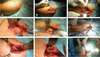
Conventional DAA was performed using previously described method by Matta et al6). Breifely, the patient was positioned in supine on a regular operation table and the skin incision was made starting at 2 cm lateral and 1cm distal to the anterior superior iliac spine. This incision was made approximately 7 to 10 cm in length along the course of the TFL 2 to 3 cm anterior to the greater trochanter. After the skin incision, TFL was identified and the fascia of the TFL was incised along the muscle fibers which was then followed by blunt dissection of the muscle from the fascia. The muscle belly was retracted laterally and ascending branch of the lateral circumflex artery was identified which was then ligated to prevent unexpected bleeding. To approach the hip joint, anterior portion of the hip capsule was resected. On exposure of the femoral head, osteotomy of the femoral neck was done at subcapital region and the femoral head was removed. Acetabulum was exposed and the cup and liner was inserted after routine reaming (Fig. 1).
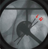
Our goal was to insert the cup at 40° of inclination and at 15° of anteversion. We used two methods to enhance the accuracy of the cup position. For the inclination, we developed a custom made guide which indicates 50° and 30° inclination when viewed by fluoroscopy (Fig. 2). For anteversion, the cup was mobilized until it made perfect hemisphere in 15° tilted c-arm image (Fig. 3).
For the stem insertion, the operation bed was extended approximately 45° at the hip joint. The superior and the inferior capsule was gradually resected until hip joint was in sufficient extension for femoral broach insertion. The hip was then externally rotated to make a figure of 4 position. The neutral rotation of femur can be marked and this can be used as a reference to insert the broach at 15° of anteversion. Routine broaching of the femoral canal was done using the offset broach handle designed for DAA. The prosthesis was inserted after the sufficient broaching was performed. The final size and the alignment of the broach were confirmed with the fluoroscopy.
2) Intraoperative assessments
To compare the time consumed for each approach, the operation and the anesthetic time was recorded. Operation time was defined as the time between the skin incision and skin closure while the anesthetic time was recorded according to the anesthesiologist's note. The amount of bleeding was also measured. Any unexpected complications or events that occurred during the operation were recorded.
3) Immediate postoperative assessments
The cup inclination and anteversion was measured using Infinitt Pacs system (Seoul, Korea). Pradhan's method was used to measure anteversion which has been validated previously to have high accuracy7). This method uses an equation arcsin (P/0.4D) where D represents maximum distance across the long axis of the ellipse of the cup and P represents a perpendicular distance measured from 1/5th of the distance along the D to the rim of the cup. Leg length discrepancy (LLD) was compared using the anteroposterior pelvis radiograph by measuring the distance between the anterior superior iliac spine and the lesser trochanter. The alignment and the size adequacy of the stem was done by performing postoperative templating. If there was more than 5° malalignment or if the templating of the bigger size stem was possible, we determined it to be inadequate.
2. Statistics
Descriptive analysis was done using JMP software (SAS Institute, Cary, NC, USA). The following variables were compared between PLA and DAA patients using Fisher's exact test and Student's t-test while non-normal non-parametric Mann-Whitney test was utilized when the distribution was non-normal; Operation time, anesthetic time, bleeding amount, percentage of acetabular cup within in safe zone (inclination between 30° and 50°, anteversion between 5° and 25°)8), the difference of the inclination and anteversion from the target value, adequacy of femur prosthesis and LLD. The significance was set at P-value of less than 0.05.
RESULTS
The mean operation time for THA was 110±66 minutes using DAA and 88±67 minutes for PLA and for BHA was 74±29 minutes and 55±28 minutes respectively. While the DAA took approximately 20 minutes more both for THA and for BHA, we found no significant difference between the two approaches. Also, while the anesthetic time was longer in DAA, no significance was found between the two groups. No difference was found for bleeding amount (Table 1).
For unexpected events or complications, 9 patients in DAA had TFL partial rupture at the muscle belly. Debridement was done on the disrupted muscle. One trochanteric fracture was presented. This was treated conservatively. While our initial plan was to not release any additional structure other than described previously, quadriceps tendons were released in 4 cases and piriformis in 3 cases so that femur could be further extended for femoral broach insertion. For the PLA, one case had unexpected bleeding at the contyloid fossa where Homan retracter was inserted. The bleeding stopped after the packing for approximately 5 minutes.
For the cup position, all the cups in the DAA were found to be in the safe zone while only 83.3% and 75.0% were in safe zone respectively for inclination and anteversion in the PLA. The difference from the targeted 40° inclination was 5°±2.4° in DAA and 8°±4.8° in PLA which was statistically insignificant. However, the difference from the targeted 15° anteversion showed significantly better result in DAA compare to PLA (4°±3.6° vs. 8°±4.2°, P<0.0001).
For the femur prosthesis adequacy, all prosthesis were adequate in size and alignment in DAA. For the PLA, 3 were found to be inadequate; 3 were undersized of which 2 of them being more than 5° varus aligned.
LLD showed mean of 3±1.4 mm in DAA and 5±2.4 mm in PLA. While there was no significant difference between the two groups, 2 patients in PLA had more than 1 cm difference while the maximum LLD was 6.5 mm in DAA (Table 2).
DISCUSSION
THA has been shown to provide significant pain relief and functional improvement in diseased hip and has been recognized as treated of choice for unsalvageable hip joint9). To access the hip joint during THA procedure, number of surgical approaches has been introduced which includes anterior, anterolateral, direct lateral, transtrochanteric, and posterior approach10).
While most of the previous approaches has shown high satisfaction rate both for the surgeon and for the patients, there are growing interest to the DAA and this has now being widely used5). Unlike other approaches described, DAA enable hip joint access by inter-muscular interval and theoretically it could provide less pain, faster recovery and improved hip stability leading to less dislocation compared to the common approaches which typically involves muscle detaching or splitting11121314). Number of studies reported significantly better outcome at least during the immediate postoperative period which includes less postoperative pain1213151617), better short term functional outcome313141819) and less length of hospital stay1620). The current study aims to look at the perioperative assessment of DAA compare to that of the PLA and we are unable to report the postoperative functional assessments at this point. Nevertheless, our result shows that the two approaches showed comparable operation time and anesthetic time and although statistically insignificant, we found DAA to provide advantage in implant positioning and selecting appropriate size. However, the result also shows high intraoperative complication rate when DAA was done by a novice surgeon.
Our result on operation and anesthetic time is consistent with the previous studies which reported longer operation time with DAA13182021). In our experience, the most time consuming part was accessing the femoral canal. As the patient is in supine position, the femur needs to be hyperextended and externally rotated to have access to the canal. In order to do this, we sequentially released the following structures in order; superior capsule and soft tissue (saddle), inferior capsule, piriformis tendon. When the canal was still not unapproachable, partial release of the quadriceps femoris tendon was performed. At release of the each structure, femur was extended and attempt to insert the broach into the canal was done which was considerably time consuming. However, we believe the operation time would be significantly decreased once the surgeon is more familiar with the approach. Also, if the special table designed for DAA could be used, that is table enabling distraction, external rotation and extension of the femur, the operation time could be shortened. Number of previous reports suggest that the increased operation time would be associated with the learning curve and once the sufficient number of operation is done, the operation time would be similar to the conventional approaches4121318).
With the small number of operation performed in our study, we are unable to provide the number of operation required to safely perform the operation. However, a recent study from de Steiger et al.22) reviewed revision rate among different THA approaches using Australian joint replacement registry and concluded that 50 or more procedure should be performed per year to have the comparable outcome between the approaches.
The potential complication of DAA includes lateral femoral cutaneous nerve damage, bleeding from circumflex vessel, muscle damage, femoral perforation, greater trochanter fracture and neuropraxia23). We were able to stay away from most of the complications through careful approach but the damage to the TFL was the most common complication. In our experience this occurred during the femoral head extraction. The cork screw femoral head extractor is used for femoral head extraction and often the extractor disrupted TFL by levering it during the extraction process.
Our result indicates that DAA may provide better implant positioning. As the patient is in supine position, use of c-arm was easier and the image of pelvis could be taken more accurately compare to when the patient was in lateral decubitus position. The previous report from Barrett et al.13) reported significantly better anteversion (% within safe zone) with DAA and the study from Rathod et al.24) reported decreased variability of cup anteversion with use of fluoroscopy. It should be noted that both study emphasized on the importance of learning curve. We believe high accuracy of cup alignment in the current study was due to the use of fluoroscopy and using custom made guide. The canal filling of the stem was also easily viewed with c-arm and this has made our decision easier to alter size of the femur implant. In the other hand, a study from Abe et al.25) reported no difference in stem anteversion when DAA was compared with PLA.
While we have found several advantages of the DAA in the early experience, it should be noted that currently there are lack of clear evidence that long term outcome of DAA is superior. A recent systematic review by Meermans et al.4) reviewed 42 studies comparing DAA to conventional approaches. They found conflict result among the different approaches and concluded that there is little evidence DAA can achieve better long-term outcome. Another systematic review by Connolly and Kamath5) reported that although DAA can provide number of advantages, all other standard approaches to the hip has also shown to be safe and effective. Also, recent study by Mohan et al.26) suggest that major search engines provide information that DAA is better than other approaches while only 22% of this information was in fact provided by the hospital and university.
We acknowledge there are number of limitations to the current study. The number of patients are small and this may have potentially effected the statistics. Also, the operation was done by novice surgeon and the result maybe different with more experience. As with the previous literatures, we agree that there will be less intraoperative complications and shorter operation time with sufficient experience. Nonetheless, this study has shown the early result of the DAA and this could provide guideline for what to be cautious of in the early experience of DAA.
CONCLUSION
The current study indicates that with use of DAA, cup and stem could be in more accurate position and in adequate size which is likely due to the accurate use of fluoroscopy. Also, although significant was not reached, there was trend toward more operation time and anesthetic time when DAA was used. Sufficient experience is required to decrease the incidence of complications.


 XML Download