

 PDF
PDF ePub
ePub Citation
 Print
Print


INTRODUCTION
The number of patients with osteoporosis and osteoporotic fractures is increasing annually, and these conditions commonly occur in elderly postmenopausal women. Osteoporosis causes microfractures by making bones more fragile, and fractures and pain are commonly experienced in the pelvis, spine, wrist, and other parts of the body12). Along with the recent increase in the number of elderly patients with severe osteoporosis, cases of pelvic insufficiency fracture (PIF) at the sacrum and pubis have been increasingly reported. Insufficiency fractures are caused by normal daily forces in bones of reduced internal strength and typically occur in the spine, pelvis, femoral neck and femur3). Of these, pelvic bone insufficiency fracture was first described by Lourie4) in 1982. This condition is clinically characterized by pain in the inguinal and gluteal regions and commonly occurs in patients with no trauma or minor trauma resulting from a fall5).
Since the prevalence of osteoporosis continues to increase dramatically along with the recent aging of populations, the incidence of PIF is projected to grow. However, studies on PIF have been rarely reported in Korea. Therefore, the authors of this study aimed to investigate clinical features and the effect of parathyroid hormone on treatment outcomes of PIF.
MATERIALS AND METHODS
Patients diagnosed with PIF at Chosun University Hospital in Gwangju, Korea between April 2011 to March 2014 were included in this study. A total of 15 patients met inclusion criteria (14 females and 1 male; mean age of 76.5 years [range, 67-83 years]). These patients visited our hospital due to difficulty in ambulation caused by pelvic pain or discomfort despite experiencing no or minor trauma resulting from a fall. In-depth interviews of patients and their family members revealed that 7 cases had pain in the absence of trauma and the remaining 8 cases had experienced a minor trauma resulting from a fall. The average duration of onset of symptoms to hospital admission was 4.46 weeks (range, 3-8 weeks).
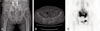
After simple radiography of the pelvis and spine, pelvic computed tomography (CT) was performed in all cases to confirm the diagnosis and fractured area, and PIF was diagnosed upon observing cortical destruction. For 3 patients, additional tests were not conducted at the initial visit because a fracture was not suspected; however, they were all diagnosed with insufficiency fractures following CT scans 2 weeks after their initial visits. The final diagnosis was made based on radiologist's interpretation of CT scans. Additional magnetic resonance imaging (MRI) of the hip joints were conducted in 2 of the 3 cases which were initially inaccurately diagnosed and the final diagnosis in these cases was made based on the presence of intramedullary edema. Bone scans were used to diagnose microfractures of the ribs and compression fractures of the spine in 6 cases, and increased uptake in a Honda-shaped pattern (H-sign) was observed in all 6 cases.
All patients had no history of steroid use or radiation therapy. There was also no case with underlying diseases including rheumatoid arthritis, a history of replacement surgery and secondary hyperparathyroidism which are suggested to be risk factors of insufficiency fracture. Before hospital admission, 8 patients received medication and injection therapy (oral bisphosphonates, 5 cases; intravenous ibandronic acid, 3 cases) at an average of 5.7 years (range, 6 months-7.6 years). Bone mineral density (BMD) was assessed in all patients with dual energy X-ray absorptiometry (DEXA) at the time of admission and the presence of osteoporosis was determined by measuring the mean BMD in lumbar spine vertebra (from L1 to L4) without compression fractures. All patients had osteoporosis and the mean lumbar T score was −3.9 (range, −3.1 to −6.4).
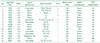
Conservative treatment with non-steroid anti-inflammatory drugs was the first line of therapy for all patients. Ten out of the 15 patients received outpatient treatment, to help control pain, the remaining 5 patients were treated in hospital for an average of 5 weeks (range, 3-6 weeks). Those experiencing severe pain were put on bed rest, and then wheelchair and partial weight-bearing ambulation was permitted within an allowable range of pain until bone union was confirmed. After confirmation of bone union, full weight bearing was started. PTH treatment was additionally advised for all patients, but only 5 patients consented and were administered PTH treatment (Forteo®; Eli Lilly, Indianapolis, IN, USA) until time of fracture healing. The mean follow-up period was 14.3 months (range, 12-18 months) and radiological follow-ups were carried out every 4 weeks from initiation of treatment to 6 months and every 3 months thereafter. Bone union was determined by a single orthopedic surgeon who treated patients during the entire follow-up period. The presence of tenderness was examined through simple radiography of the pelvis. In 9 cases with persistent pain at the fractured area and indistinct bone healing on simple radiographs, bone union was determined based on the continuity of the cortical bone by performing a CT scan of the pelvis. Improvement in clinical symptoms was measured using visual analogue scale (VAS) score (Table 1).
Statistical analyses were performed using the IBM SPSS Statistics version 21.0 (IBM Co., Armonk, NY, USA). Time to bone union and VAS scores were compared using t-tests between PTH therapy and no PTH therapy groups. P-values of less than 0.05 were considered statistically significant.
RESULTS
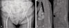
In regards to fracture location, an isolated sacral fracture was present in 4 cases (26.7%), an isolated pubic fracture was detected in 2 cases (13.3%), and both sacral and pubic insufficiency fractures were observed in 9 cases (60.0%). According to the Denis classification, sacral insufficiency fractures were most commonly located within zone 1 of the sacral body (11 cases) or within zone 2 (2 cases). With respect to fracture type, a vertical fracture line was observed in the sacrum parallel to the sacroiliac joint in all cases, and an additional fracture line parallel to the vertical fracture line was detected in 2 cases. Table 2 summarizes the 15 patients fragility fractures of pelvis6) classification; fracture types were Ia (2 cases), IIa (4 cases), IIb (5 cases), IIc (3 cases) and IIIc (1 case).
Follow-up radiographs revealed the presence of vertebral compression fractures in 14 out of 15 patients (93.3%). Vertebral compression fractures in more than two sites were detected in 11 cases, and 3.6 compression fractures were present on average. As shown in Fig. 1, vertebral compression fractures were more common in the lumbar spine than the thoracic spine (mean 2.4 vs 1.1). Compression fractures were old in 10 patients; these patients were put under follow-up observation. Conservative therapy (i.e., wearable assistive devices) was implemented in 4 cases complaining of tenderness in the fracture site. Additional treatment was not required since progression of compression fracture and neurological deficit were not detected upon radiological follow-up.
One case with no use of PTH showed pain aggravation and fracture displacement on simple radiographs at 4 week follow-up and the patient was treated with percutaneous sacro-iliac fixation using cannulated screws. Fracture healing was observed 6 months later.
The mean duration of bone union was 21.6 weeks (range, 18-27 weeks) in the group treated with PTH and 30.0 weeks (range, 22-34 weeks) in those receiving conservative therapy. The mean duration of bone union was significantly shorter in the PTH group than those receiving conservative therapy (P<0.05). VAS scores improved from 7.2 (range, 6-8) at the time of hospital admission to 3.2 (range, 2-4) at the final follow-up in the PTH treatment group, and improved from 7.44 (range, 6-9) to 3.67 (range, 2-5) in the conservative treatment group. VAS scores were lower in the PTH treatment group at the latest follow-up but did not reach statistical significance (Table 1).
DISCUSSION
PIF has been rarely reported. However, Weber et al.7) determined a prevalence rate of 1.8% in their prospective study of patients aged 55 years and older and Melton et al.8) suggested that the incidence of PIF significantly increased in those over the age of 75 years based on retrospective data analysis.
PIF occurs either as an isolated fracture or complex fractures of the sacrum and pubis, and insufficiency fractures predominantly affect elderly women with osteoporosis, rheumatoid arthritis, steroid use, radiation therapy or other clinical history. Risk factors for PIF are identified to be vitamin D deficiency and hypocalcemia9). In the present study, patients had no other risk factors except osteoporosis; however, serum vitamin D and calcium levels were not measured.
Insufficiency fractures are difficult to diagnose using simple radiography and are best diagnosed with bone scan, CT, MRI, and other imaging techniques. According to Grangier10), diagnosis is often delayed as the duration from pain onset to diagnosis takes about 40-55 days. Bone scans are a relatively sensitive diagnostic method characterized by H-sign which is a H-shaped area of increased tracer uptake, but it lacks specificity (Fig. 2)11). CT scan helps determine fracture patterns and assists in the discrimination between infection and tumor. MRI is highly sensitive for revealing intramedullary edema, but it doesn't easily differentiate between infection and tumor11). In our study, the initial diagnosis was made using simple radiography and CT. In cases of gait disturbance and persistent pain, the final diagnosis was made by excluding tumors and infections after identifying the H-sign on bone scan and bone marrow edema on MRI.
Insufficiency fractures that commonly occur in elderly patients usually extend parallel to the sacroiliac joint, vertically in the sacral ala or lateral to the margins of the lumbar spine12). This distribution suggests that the weight of the body transmitted through the spine may be partially responsible for sacral insufficiency fractures, and pubic insufficiency fractures are frequently associated with these fractures713). In this study, sacral insufficiency fractures occurred majorly in a vertical pattern, parallel to the sacroiliac joint and were commonly associated with pubic fractures (13 out of 15 cases)6).
Sacral insufficiency fractures more commonly occur when the deflection angle of the pelvic bone increases. The deflection angle of the pelvis is higher in women than men by 2.25°. This explains why osteoporosis and sacral insufficiency fractures are more common in women14). In the current study, the mean anteversion was 15.5° (11.2° to 20.4°) in women and 11.4° in the male patient. Since this study only includes only one male patient, the results from this study cannot be generalized.
Conservative treatment, sacroplasty, screw fixation, PTH therapy and others have been used in treatment of PIFs, but the optimal treatment of PIF has not yet been established. Conservative treatment consists of bed rest and restricting activity to within an allowable range of pain. Early weight-bearing exercises and activities are impossible in cases of delayed fracture healing in elderly patients with PIF, and the incidence of complications (e.g., generalized weakness, weakness of cardiopulmonary function, decubitus ulcer, pneumonia and others) is high due to prolonged bed rest1516). Sacroplasty has gained attention as a surgical treatment for sacral insufficiency fractures1117), but this technique requires long-term studies because surgical procedures remain challenging due to: i) complex anatomical structures of the sacrum and ii) complications that may be associated with bone cement leakage-induced nerve injury and cortical perforation18). Screw fixation of the sacroiliac joint has been recently attempted to assist in pain relief and early ambulation by providing stability for direct healing of sacral fractures. Since sacral screw fixation is associated with increased risk of neurological or vascular injuries, and most patients with severe osteoporosis have poor bone quality, there is the burden of anesthetic risk for screw loosening and old age1920). Rommens et al.21) implemented conservative treatment in cases without fracture displacement, despite the presence of lesions in the anterior pubis or sacrum, but recommended open reduction and internal fixation in cases of complex fractures of the sacrum and pubis with displacement. In our study, only one patient required screw fixation, but a single case was insufficient to conclude whether screw fixation was warranted as a preventive measure, despite the presence of concurrent sacral and pubic fractures.
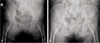
PTH stimulates osteoblast activities by acting directly on osteoblasts, and increases osteoclast formation by upregulating receptor activator of nuclear factor kappa-B ligand (RANKL) synthesized in osteoblasts. Matured osteoclasts facilitate bone resorption. In healthy individuals, serum PTH concentrations differ during the day and at night. Serum levels of PTH increase during inactive night time and decrease during active day time, and this intermittent PTH action promotes bone formation in osteoblasts and inhibits apoptosis22). As described in the above, elderly patients with PIF are mostly associated with multiple spinal fractures, and have high complication rates due to prolonged bed rest resulting from delayed fracture healing. Therefore, PTH treatment helps improve clinical outcomes including ambulation and symptoms such as pain by accelerating fracture healing in patients with PIF23). This study has demonstrated that conservative treatment is a favorable treatment option by improving bone union and clinical symptoms such as pain. The use of PTH shortens the treatment period and improves clinical symptoms, even if it is statistically insignificant (Fig. 3). Treatment with PTH can be considered as a favorable option for patients with PIFs.
This study was limited by the relatively small sample size (5 patients in PTH treatment group and 10 who received only conservative treatment) and the retrospective nature of the analysis. Other limitations are that PTH was not administered in all cases, and patient's nutritional status and bone turnover were not evaluated. Taking into consideration the fact that PIFs occur rarely, multicenter studies are warranted to clearly prove the efficacy of PTH therapy.
CONCLUSION
PIF should be considered in patients confirmed to have osteoporosis (using BMD test), those experiencing multiple vertebral fractures, and those with pain in pelvic area in the absence of major trauma. A thorough radiographic examination (e.g., CT or MRI) is required for those patients for whom PIF is suspected. Since PIF is frequently associated with compression fractures of the the thoracic and lumbar spine, assessment of these fractures is recommended. Our study shows that PTH therapy shortens treatment period for patients with PIF and could be a favorable treatment option.

 XML Download