

 PDF
PDF ePub
ePub Citation
 Print
Print


Introduction
1. Background, purpose, and scope of the clinical practice guidelines for gastric cancer
Recently, the cancer incidence rate has increased in South Korea. According to data from the National Cancer Information Center, under the Ministry of Health and Welfare, the rate has significantly increased by 3.3% annually during 1999~2008.1,2 According to 2008 data, 1 in 3 Koreans might have cancer, with probabilities of 37.2% for men, assuming a mean life expectancy of 77 years, or 30.5% for women, assuming a mean life expectancy of 83 years. The effective treatment has increased the survival rate of gastric cancer patients. However, the prognosis remains relatively poor, with a 5-year survival rate of 63.1% during 2004~2008.1 According to the data on the cause of mortality, 28.6% of the 246,942 total cancer-related deaths in 2009 were caused by malignant neoplasms, among which gastric cancer was the leading cause of cancer-related deaths.2 Economic burden related with gastric cancer is considerably high. In 2007, the Health Insurance Review and Evaluation Service indicated that the total medical costs incurred by gastrointestinal diseases (excluding meals, selective treatment fees, and medication costs from the total expenses paid for Health Insurance Reimbursement) were approximately 3.65 trillion wons. The medical costs incurred by malignant gastrointestinal diseases accounted for 36.6% of the above amount, and gastric cancer accounted for 10.9% (390 billion wons) of all gastrointestinal diseases, the highest percentage for a single disease (unpublished data).
The present clinical practice guideline is intended for use in both male and female adult patients with gastric cancer. This guideline, which is based on domestic and overseas evidences, have been developed to suit Korea's current medical practices and to ensure their widespread adoption in clinical practice. It was intended to help all medical staffs at the primary, secondary, and tertiary care medical institutions including physicians, surgeons, radiologists, pathologists, family doctors, and general practitioners. Additionally, it was designed to allow patients and populations to find optimum care by providing adequate medical information. Furthermore, it was intended for widespread adoption in order to increase the standard of gastric cancer treatment, thereby contributing to improvement in the patients' quality of life, as well as in the national health care. It will also provide information on clinical practices and principles for medical residents and other medical staffs.
The present guideline is specific and comprehensive for gastric cancer diagnosis and treatment; however, it does not address issues related to prevention, screening, and care of pediatric patients. In addition, it does not address controversial issues with inadequate evidence. However, the nominal group technique was applied to include consensus of the clinically important issues with weak evidences.
2. Composition and progress of guideline development group
The present guideline was prepared as a designated project assignment (no. 1020440) under the Research and Development Program for Cancer Control, conducted by the Ministry of Health and Welfare, South Korea. This guideline was prepared in an integrated and comprehensive manner through an interdisciplinary approach that included the Korean Academy of Medical Sciences, the Korean Association of Internal Medicine, the Korean Society for Radiation Oncology, the Korean Society of Pathologists, the Korean College of Helicobacter and Upper Gastrointestinal Research, the Korean Society of Gastrointestinal Endoscopy, the Korean Society of Gastroenterology, the Korean Cancer Association, the Korean Society of Radiology, the Korean Gastric Cancer Association, and the Korean Society of Nuclear Medicine, along with the participation of experts in the guideline development methodology. To develop this guideline, the Organizing Committee for Clinical Practice Guidelines for Gastrointestinal Cancer, the Development Committee for Standard Clinical Practice Guidelines for Gastric Cancer, and the Review Committee for Standard Clinical Practice Guidelines for Gastric Cancer were established, and the members were recommended by each participant associations and societies (http://www.guideline.or.kr).
3. Literature search, evaluation, and preparation of recommendations
The fundamental and important issues concerning gastric cancer diagnosis and treatment were selected as key questions according to the patient, intervention, comparator, and outcome (PICO). Search terms for each respective key question were selected according to the medical subject headings (MeSH) terms of National Library of Medicine. For each key question, the inclusion/exclusion criteria were determined and the search words were properly combined to conduct the literature search. PubMed, MEDLINE and the Cochrane Library were used for the international literature search, while KoreaMed was used for the purpose of domestic literature search. The literature search was conducted only for the documents published from 1980~2011 in either English or Korean.
To evaluate the validity of the documents selected as evidence, a systematic and consistent evaluation method was adopted. In order to apply different evaluation methods, the documents were classified according to the study design.3 To evaluate randomized controlled studies, the risk of bias (ROB) method of Cochrane Collaboration was adopted.4 The Review Manager (RevMan) 5 (The Nordic Cochrane Centre, The Cochrane Collaboration, 2012) and GRADEpro program (Jan Broeck, Andrew Oxman, Holger Schünemann, 2008) were used to arrange the evidence and evidence summary table.5 To evaluate non-randomized controlled studies, the Newcastle-Ottawa evaluation scale was applied. To evaluate diagnostic studies, the Quality Assessment of Diagnostic Accuracy Studies (QUADAS) tool was used;6 accordingly, the evaluations were classified as 'yes', 'no', or 'not clear' for 11 assessable items among the 14 items.
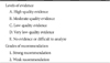
The Grading of Recommendations, Assessment, Development, and Evaluation (GRADE) method was used for the evidence summary.7,8 Levels of evidence of studies were ranked according to the type of research, with a high level evidence for randomized clinical trials and a low level evidence for observational studies. Next, the qualitative level of the corresponding study was increased or decreased after considering the factors that influenced the quality of each study. The levels of evidence were classified as high-quality, A; moderate-quality, B; low-quality, C; and very low-quality, D. For cases with no evidence or difficult to analyze, a fifth classification (no evidence or difficult to analyze, E) was added and used (Table 1).
For the grading of recommendations, (1) a balance of desirable and undesirable effects, (2) evidence quality, and (3) values and preferences were taken into account, according to the GRADE method. Any area with difficult-to-determine recommendations is not mentioned in the present guideline, but will be reviewed during the next guideline development. The grades of recommendations are divided into (1) strong recommendations and (2) weak recommendations (Table 1). A strong recommendation indicates that it is strongly recommended for most patients because compliance with this recommendation for a specific intervention will result in a desirable rather than an undesirable effect, along with a high quality of evidence and a better value and preference, compared to other interventions. A weak recommendation indicates that it is better for many patients to comply with this recommendation because despite rather weak evidence, a desirable effect has been shown. With a weak recommendation, some patients or medical staff might select different interventions according to the values or preferences.
4. Review and approval process
Based on the present guideline as developed by the Development Committee and reviewed by the Review Committee, a public hearing was held on October 29, 2011. The concerned experts, patients and other interested people participated the hearing. Revisions that reflected the opinions expressed in the public hearing were made. This guideline was then approved by the Korean Association of Internal Medicine, the Korean Cancer Association, the Korean Society of Pathologists, the Korean College of Helicobacter and Upper Gastrointestinal Research, the Korean Society of Gastroenterology, the Korean Society of Gastrointestinal Endoscopy, the Korean Society of Radiology, the Korean Surgical Society, the Korean Gastric Cancer Association, the Korean Society for Radiation Oncology, and the Korean Society of Nuclear Medicine.
5. Renewal procedure and monitoring
The present guideline for gastric cancer will be available free of cost at the web sites of the Korean Academy of Medical Sciences (http://www.kams.or.kr/), the Korean Association of Internal Medicine (http://www.kaim.or.kr/), the Korean Society for Radiation Oncology (http://www.kosro.or.kr/), the Korean Society of Pathologists (http://www.pathology.or.kr/), the Korean Society of Helicobacter and Upper Gastrointestinal Research (http://hpylori.or.kr/), the Korean Society of Gastrointestinal Endoscopy (http://www.gie.or.kr/), the Korean Society of Gastroenterology (http://www.gastrokorea.org/), the Korean Cancer Association (http://www.cancer.or.kr/), the Korean Society of Radiology (http://www.radiology.or.kr/), the Korean Gastric Cancer Association (http://www.kgca-i.or.kr/), and the Korean Society of Nuclear Medicine (http://www.ksnm.or.kr/), as well as through Facebook and Twitter, where monitoring can be processed and all opinions regarding this guideline are welcome. The guidelines will be renewed in 3 to 5 years based on the accumulated clinical evidence.
6. Absence of conflicts of interest
The present guideline was prepared as a designated project assignment, entitled the "Development of clinical practice guidelines methods for gastrointestinal cancer and clinical practice guidelines for gastric cancer/colon cancer" (no. 1020440), under R&D Program for Cancer Control, conducted by the Ministry of Health and Welfare. The members comprised of a principal investigator, Hyeong Sik Ahn (Department of Preventive Medicine, Korea University College of Medicine, Seoul), a principal investigator for gastric cancer guideline, Jae Gyu Kim (Department of Medicine, Chung-Ang University College of Medicine, Seoul), and a principal investigator for colon cancer guideline, Jun Won Uhm (Department of Surgery, Korea University College of Medicine, Seoul). The contents of the present guideline were not influenced by the opinions of financially sponsoring entities. All of the members who participated in the development of the present guideline have submitted a confirmatory document with their signatures to assure that there were no conflicts of interest. None of the participants were involved in issues related to conflicts of interest.
7. Limitations of the present guideline
One of the major limitations of the present guideline is that there is not enough evidences in Korea. Data from studies in other countries may be slightly different in terms of epidemiological and clinical aspects. Considering the established therapeutic efficacy of current treatment methods, it is difficult to conduct new randomized controlled studies in some areas because of the ethical issues.
Endoscopic diagnosis
1. Conventional endoscopy
1) Upper gastrointestinal endoscopy and biopsy
Upper gastrointestinal endoscopy is the primary tool for the diagnosis of gastric cancer. By using the proper technique such as air infusion and removal of mucus, it is possible to observe the entire stomach clearly. One of the major advantages of endoscopy is that tissue sampling can be performed immediately by using biopsy forceps. The recently introduced magnifying endoscopy may help to observe the gastric lesion.
To achieve the best results, endoscopy should be performed by properly trained endoscopists using appropriate endoscopic devices. Biopsy is necessary because there are limitations to the sensitivity and specificity of direct observation for diagnosing gastric cancer.9 Normally, acquisition of more than 4 tissue samples is recommended in order to increase the diagnostic accuracy. However, a fewer number of samples might be enough in patients considering endoscopic resection. Biopsy has a low sensitivity for detecting Borrmann type IV advanced gastric cancers. Endoscopic clips or dye injection can be used to determine the resection margin during the surgery.10 If it is difficult to localize the cancer lesion, as in rare cases, an intraoperative endoscopy can be performed.11
2) Chromoendoscopy
Chromoendoscopy is easy and simple method that allows better visualization of the lesions with unclear color changes or minute surface irregularities. It is also useful in determining the lateral borders during endoscopic submucosal dissection (ESD). Dyes include methylene blue, indigocarmine, acetic acid, and crystal violet, and among them, indigo carmine is the most commonly used.12 After indigocarmine spraying, the dye fills the depressed mucosal sites, thus highlighting the surface irregularities. Such characteristics can help to estimate the depth of invasion of early gastric cancers (EGCs) and to determine the range of resection for endoscopic therapies.
2. Endoscopic ultrasonography
Endoscopic ultrasonography (EUS) can be used to evaluate the depth of invasion of gastric cancers and the presence of local lymph node metastases. Because of the widespread use of laparoscopic surgery for EGC, evaluation of the stage of gastric cancer before therapy has become more important. A recent meta-analysis13 of 54 studies, including 5,601 gastric cancer patients, showed that EUS had a relatively good sensitivity and specificity for differentiating between T1 to 2 and T3 to 4 lesions (0.86 and 0.91, respectively). However, the sensitivity and specificity for detecting lymph node metastases were lower (0.69 and 0.84, respectively).13 According to a recent Korean study,14 which compared the accuracies of white light endoscopy and EUS, the accuracy of white light endoscopy for predicting the depth of invasion of EGCs was 73.7%, while that of EUS was only 67.4%. Therefore, the usefulness of endoscopic ultrasonography before endoscopic therapy for gastric cancer remains controversial.
Radiology
1. Upper gastrointestinal series
The upper gastrointestinal series is a radiologic test that is widely used for the diagnosis of gastric cancer as it is safe and non-invasive without the need for specific preparation. It is useful as a preoperative test because of its high sensitivity and the ability to visualize the lesion location accurately and objectively.15,16 To perform an upper gastrointestinal series, it is important to obtain appropriate mucosal coating and gastrointestinal distention. For this purpose, a 240% weight per volume (w/v) high-density barium solution is generally used in gastrointestinal series. To ensure the accuracy of the upper gastrointestinal series, a single-contrast study including the compression and mucosal relief views should be combined with a double-contrast study that uses a high concentration of barium to coat the mucosa via air distention.17
2. Computed tomography
From the late 1970s, computed tomography (CT) is a diagnostic and preoperative test for detecting gastrointestinal tumors including gastric cancer. To date, special CT techniques for evaluating the stomach have been widely used to detect and diagnose gastric cancers, to determine the optimal treatment method via accurate staging, and to identify therapeutic effects after surgery or anticancer treatments. After the multidetector row CT (MDCT) was introduced, the diagnostic accuracy has increased and the detection of small lesions, including EGCs, has improved.
1) Multidetector row computed tomography
After the introduction of MDCT, the z-axis resolution improved, and thus the ambiguity of lesions due to partial volume averaging, which was considered as the disadvantage of conventional-type single-channel helical CT, was decreased.
When using MDCT to diagnose gastric cancer, according to related literatures, at least a 4-channel MDCT is required, with a detector collimation ≤2.5 mm, an imaging section thickness ≤5 mm, and the administration of approximately 500 ml of water or an effervescent agent; the patient is then instructed to change position until each part of the stomach is properly distended.18,19,20 Additionally, it is better to perform dynamic CT after contrast enhancement because arterial-phase imaging allows easy detection of enhanced mucosal lesions of the stomach, while portal venous-phase imaging provides more accurate information including the depth of invasion of gastric cancer and the involvement of adjacent organs and facilitates the evaluation of lymph node metastases. In addition, delayed-phase imaging might also be useful because in some cases there is enhanced fibrosis surrounding the gastric cancer, thus allowing a more accurate evaluation of tumor infiltration into the gastric wall. The reconstruction of arterial-phase and portal venous-phase images will permit CT angiography, which provides preoperative evaluation of perigastric blood vessels.
Upon examining the reports of preoperative tumor node metastasis (TNM) staging via MDCT, the accuracies ranged from 67.9% to 90.9% for T (median value, 82.1%) and from 56.9% to 86% for N (median value, 69.5%). In particular, the specificity for T4, which determines whether surgery is indicated, ranged from 81.8% to 99.4% (median value, 96.5%).18,20,21,22,23,24 Nevertheless, CT allows clinicians to diagnose advanced gastric cancer that has spread to the peritoneum or shows distant metastases, thereby preventing unnecessary surgeries.
CT is also useful for evaluating the therapeutic effects after anticancer therapy. The Response Evaluation Criteria in Solid Tumors (RECIST), which is used to evaluate response to treatment in solid cancers, states that CT is among the most useful modalities, along with magnetic resonance (MR) for evaluating the sizes of solid tumors.25
2) Three-dimensional computed tomography gastrography; virtual gastroscopy
This technique utilizes the 3-dimensional reconstruction of CT images to provide similar imaging to the upper gastrointestinal series or endoscopy. Its advantages include 3-dimensional representation of the anatomical location of gastric cancers, facilitation of surgical planning, and the ability to detect depressed areas or changes in gastric mucosal folds that are difficult to observe in cross-sectional CT images; thus helping in identifying small lesions on the gastric mucosal surface in EGC.19
3. Magnetic resonance imaging Magnetic resonance imaging facilitates the
Magnetic resonance imaging facilitates the diagnosis of extra-serosal invasion or invasion into adjacent organs and the identification of distant metastasis, and it is therefore useful in preoperative staging of gastric cancer. In particular, it has been widely used for the evaluation of liver metastases. Recently, liver-specific MR contrast agents have been developed and this allows us to achieve more accurate diagnoses of hepatic metastases in the future.26
Nuclear imaging
1. Role of [18F]-fluorodeoxyglucose positron emission tomography/computed tomography in the diagnosis of gastric cancer
Although [18F]-fluorodeoxyglucose (FDG) positron emission tomography/computed tomography (PET/CT) is increasingly being adopted for the diagnosis of cancer and determination of therapeutic effects and staging, its role in detection of primary lesions is limited in some cases. According to the existing literature, the PET detection rate of EGC is less than 50%, and therefore PET alone is not recommended for gastric cancer screening in asymptomatic patients.27 In a study on the diagnosis of advanced gastric cancer, the success rate of PET in establishing the diagnosis ranged from 62% to 98% because FDG uptake varies according to the characteristics of gastric cancer.28,29,30,31 For example, PET/CT shows strong FDG uptake in the intestinal type gastric cancer and demonstrates high sensitivity, while a relatively low sensitivity was reported in diffuse-type tumors with a low level of FDG uptake.32 Considering the above findings, endoscopy and barium upper gastrointestinal series are still considered to be more effective for diagnosing EGC.
2. Staging and prediction of prognosis
FDG PET/CT has a limited role in the detection of early primary gastric cancers. However, this is the same as that with contrast-enhanced CT, because it is difficult to evaluate primary tumors accurately due to the characteristics of the stomach. Additionally, according to the pathologic evaluation of biopsies, the cell densities and the degree of malignancies are not uniform;32,33 and therefore although PET/CT allows evaluation of the degree of metabolic activity and targets the highly malignant areas, its role is still limited with regard to accurate measurement of the depth of invasion into the gastric wall.
Next, when evaluating invasion into adjacent lymph nodes, the ability of PET to evaluate glucose metabolism is expected to allow more accurate detection of metastases to the local lymph nodes. According to Mochiki et al.,31 the diagnostic performance of PET for N1 lymph nodes is not satisfactory, but this might have little clinical significance because these lymph nodes will be resected during gastric cancer surgery. On the other hand, invasion of lymph nodes in the N3 group is classified as distant metastasis, and it is very important to determine distant metastasis accurately. Yun et al.34 reported that PET might provide useful information on distant lymph node invasion.
In a study by Hillner et al.35 of 3,025 gastric cancer patients, distant metastases to other internal organs was evaluated, and patient management was altered significantly after the PET results in 37% of cases and PET showed better results than the conventional tests in the detection of liver metastases.36 In terms of skeletal evaluation, bone scanning still plays an important role and PET scanning can play a compensatory role in the detection of osteolytic bone metastasis.29
In PET scanning, the degree of FDG uptake in primary gastric cancer is expected to be an additional predictive factor along with the conventional anatomical imaging-adapted TNM staging.32,33 Additionally, peritoneal metastasis is also one of the important prognostic factors of gastric cancer and PET has a higher specificity (99%), lower sensitivity (35%), and equal accuracy when compared to CT for detection of peritoneal metastasis.37
3. Evaluation of recurrence
Postoperative evaluation of recurrence is very important in the management of gastric cancer. Sites of gastric cancer recurrence include adjacent organs, lymphatic system, blood circulation, and peritoneum. Consequently, recurrence sites include the local area, liver, lungs, skeletal system, peritoneum, and many other sites. Changes in the anatomical structure after gastric surgery can make it difficult to diagnose recurrence accurately in many cases. Some studies have reported that PET is better than conventional CT in terms of specificity and positive predictive value.33,38 Several studies reported that PET scanning after gastric distension due to water intake is useful for determining postoperative recurrence.39 According to the recent studies that assessed PET/CT, PET generally provides better results for the evaluation of gastric cancer recurrence compared to contrast-enhanced CT40 and this might be due to the combination of anatomic and functional data.
4. Determination of therapeutic effects
During gastric cancer therapy, each individual patient presents with a different gastric cancer cell type, degree of differentiation, malignant potential of tumors, and depth of invasion, and therefore, a careful approach is essential in selecting anticancer drugs. Ott et al.39 performed baseline PET scans and additional PET scans after 2 to 3 cycles of chemotherapy, and they proposed that the therapeutic effects of anticancer drugs can be determined by comparison of these two scans.39 Recently, many reports have suggested that the efficacy of therapeutic options can be determined by evaluating not only changes in size, but also changes in lesion metabolism. Therefore, the role of FDG PET/CT in management of gastric cancer will continue to expand in the future.
Surgery
1. Principles of gastric cancer surgery
1) Gastric resection
The standard surgical procedures for gastric cancer are distal subtotal gastrectomy (gastric resection of two-thirds) for cancers in the lower or middle third of the stomach and total gastrectomy for cancers in the upper or middle third of the stomach, as well as radical lymphadenectomies. Limited or function-preserving surgeries include pylorus-preserving gastrectomy, local resection, segmental resection, and proximal gastrectomy. Before performing limited surgeries, factors such as the location of the lesion, the extent of lymphadenectomy, and resection margins should be considered. Proximal gastrectomy is mainly performed for EGCs in the upper stomach, but care should be taken to prevent the risk of developing reflux esophagitis.41,42,43,44
2) Lymphadenectomy
Perigastric lymphadenectomy can be performed in patients with EGC. Here, lymph node dissections are performed for group 1 lymph nodes plus LN7, 8, 9, [+11p].45,46 Peri- and extragastric lymphadenectomy is performed in patients with advanced gastric cancer (AGC) and patients with EGC who have suspected lymph node metastasis. Lymphadenectomy beyond perigastric lymph nodes is not currently accepted as a standard therapy, but it is considered to be an extensive operation. The effects of para-aortic lymph node dissection have not been reported.47
3) Combined resection
Combined resection of involved organs can be performed in cases showing direct invasion into the adjacent organs,48,49,50,51,52,53,54,55,56,57,58 gastric cancers in the greater curvature with muscularis propria invasion, suspected splenic hilar lymph node involvement,59,60,61,62 suspected distant metastasis,63,64,65,66 and in patients undergoing palliative surgery.
4) Reconstruction
Only a few studies have compared the differences between postoperative reconstruction methods. A few studies have compared Billroth-I and Billroth-II and have reported that the 2 types of anastomosis did not show significant differences with regard to therapeutic performance or difficulty in ingestion.67,68 Additionally, Roux-en-Y gastric bypass might be superior to Billroth-I anastomosis in terms of postoperative bilious reflux.69
2. Surgery for early gastric cancers
1) Surgical indications in early gastric cancer
EGC refers to gastric cancers limited to the mucosal and submucosal layers regardless of lymph node metastasis; the frequency of lymph node metastasis is approximately 5% in mucosal cancers and approximately 20% in submucosal cancers.70,71 Any EGC without distant metastasis can be a candidate for surgery which is composed with gastric resection and lymph node dissection.72,73
3. Surgery for advanced gastric cancer
1) Surgical indications in advanced gastric cancer
AGC is defined as gastric cancers with invasion into the proper muscle and/or deeper layers. In cases with perigastric organ invasion, combined radical resection can be performed. In cases with distant metastasis, resection limited to the primary tumor can be performed to prevent or treat cancer-related symptoms such as bleeding and obstruction, and to improve the quality of life.87,88,89 In unresectable cases, palliative surgeries such as gastrojejunal bypass can be performed.90
Endoscopic therapy
1. Absolute indications
Traditionally, the standard treatment for gastric cancer has been surgery. Endoscopic mucosal resection, which was first introduced in Japan in 1984, and the more recently introduced ESD can replace standard surgery if applied to limited stages of EGCs. Endoscopic therapy can minimize surgical complications, and the quality of life is only slightly affected. In multiple retrospective studies, survival after endoscopic treatment is comparable to that after surgery.91,92,93,94,95,96 However, according to a Cochrane review, there are no randomized controlled trials comparing endoscopic therapy and surgery.97
Theoretically, the indication for endoscopic therapy is EGC without risk of lymph node metastasis. However, it is impossible to diagnose lymph node metastases accurately before treatment. Therefore, indications for endoscopic treatment were defined based on the analysis of surgical date of lymph node metastasis.98,99,100,101,102 Currently, the absolute indications of endoscopic therapy for EGC include (1) lesions limited to the mucosal layer, (2) well and/or moderately differentiated adenocarcinomas, (3) tumors ≤2 cm in length, (4) absence of ulcer or ulcer-scar tissue, and (5) tumors without lymphovascular involvement.100 Because it is impossible to clearly determine the depth of invasion or lymphovascular involvement, additional treatment is sometimes required based on the pathological results.
2. Expanded indications
With the development of ESD techniques, there have been attempts to extend the indications of endoscopic resection.96,103,104 Expanded indications include (1) well or moderately differentiated adenocarcinoma in the mucosal layer without an ulcer regardless of the size, (2) well or moderately differentiated adenocarcinoma measuring less than 3 cm in the mucosal layer with ulcer, (3) small (less than 2 cm) intramucosal cancer with undifferentiated histology, and (4) well or moderately differentiated adenocarcinoma with minute submucosal invasion (≤500 µm, SM1).
3. Follow-up
The National Comprehensive Cancer Network guidelines recommend that follow-up after gastric cancer treatment should be conducted every 3 to 6 months for the first three years after R0 resection. For 3 to 5 years, the follow-up interval is 6 months, and there it is yearly. The guideline states that complete blood count, biochemical tests, radiologic studies, and endoscopy can be performed if they are clinically necessary.105 The recurrence rate after endoscopic therapy for EGC is 3.3% to 14.0%. It is recommended that patients should undergo an endoscopic follow-up at least annually because of the risk of missing multiple synchronous cancers.
Chemotherapy
1. Postoperative adjuvant chemotherapy for gastric cancer
Gastric cancer recurs in 22% to 45% of patients after curative resection.106,107 In this regard, there have been many studies that evaluated the effect of adjuvant chemotherapy. In meta-analyses of clinical trials between 1980 and 2000, adjuvant chemotherapy increased the rate of survival.108,109 In 2010, the Global Advanced/Adjuvant Stomach Tumor Research International Collaboration (GASTRIC) group performed a meta-analysis of 17 clinical trials, which showed that adjuvant chemotherapy increased the survival duration and that fluoropyrimidine-containing therapy lowered the risk of death.110
In a western study, administration of epirubicin+cisplatin+5-fluorouracil ([5-FU]: ECF) chemotherapy after surgery significantly increased the overall survival (hazard ratio [HR], 0.75) and disease-free survival (HR, 0.66).111 It is difficult to accept this regimen as a standard treatment in Korea because only 42.5% of the patients underwent D2 lymph node dissection. In this context, clinical studies in Korea and Japan have attempted to determine the effect of adjuvant chemotherapy after D2 lymph node dissection. Among the 1,059 patients with stage II disease, a comparison was made between an S-1 (tegafur+gimeracil+oteracil)-treated group and an untreated group after D2 dissection.112 The S-1-treated group had a significantly higher 5-year survival rate, compared to the surgery-alone group (71.7% vs. 61.1%; HR, 0.669).113 A clinical trial (CLASSIC trial) conducted by Korean researchers among 1,035 patients with stage II, IIIa, and IIIb (T3N2) diseases showed that the group treated with capecitabine and oxaliplatin combination therapy had a significantly higher 3-year survival rate compared to the untreated group (74% vs. 60%).114
In conclusion, S-1 monotherapy and capecitabine+oxaliplatin therapy can be standard adjuvant treatments for stage II and III diseases after D2 lymph node dissection. In the future, well-designed clinical trials are needed for the development of more effective postoperative adjuvant therapies.
2. First-line palliative chemotherapy for recurrent and/or metastatic gastric cancer
The main goals of chemotherapy for recurrent and metastatic gastric cancers are to prolong the survival and to improve the quality of life by providing symptom palliation. Since 1990s, 4 small randomized Phase III clinical trials that were published115 have consistently shown survival benefit (approximately 3 to 7 months) and improved quality of life in the patients receiving palliative chemotherapy, compared to the patients receiving best supportive care alone.116,117,118
Recommendation: First-line palliative chemotherapy is recommended for recurrent and/or metastatic gastric cancer patients in the context of performance status, medical comorbidity and toxicity profile (Recommendation Grade 1, Evidence Level B).
Among the gastrointestinal cancers, gastric cancer is known to respond relatively well to chemotherapy. Chemotherapeutic agents for gastric cancer including 5-FU, mitomycin C, cisplatin, and etoposide have shown response rates of at least 10% when administered as monotherapies. Other newer agents including irinotecan, oral etoposide, paclitaxel, docetaxel, and pegylated doxorubicin have shown response rates ranging from 10% to 20%. Generally, the response rates are low with very short response duration (within 3~4 months) when anticancer drugs are administered alone. Therefore, combination chemotherapies have been attempted in order to increase the response rates and prolong the survival time; in studies in which combination chemotherapy was administered, response rates of 25% to 50% and median survival of 6 to 12 months have been reported.119,120,121,122
A recent meta-analysis of randomized trials showed that combination chemotherapy significantly improves the survival, compared to single chemotherapy or best supportive care alone.115 First-line palliative chemotherapy with a two-drug combination of fluoropyrimidines and platinum is preferred for patients with advanced or metastatic disease. Three-drug combination chemotherapy with docetaxel or epirubicin should be reserved for medically fit patients with a good performance status.
Recently, molecular targeted agents such as trastuzumab, bevacizumab, cetuximab, and lapatinib have been tested with the standard chemotherapy for recurrent and metastatic gastric cancers. Based on the results of the ToGA study, which showed a significant improvement in the median overall survival (13.5 vs. 11.1 months) with the addition of trastuzumab to chemotherapy (5-FU or capecitabine and cisplatin), trastuzumab in combination with chemotherapy is recommended for patients having HER2 over-expression or amplification (HER2 IHC 3+ or HER2 IHC 2+ with FISH/SISH+).123 However, the addition of either bevacizumab,124 cetuximab,125 or lapatinib126 to chemotherapy failed to show survival benefit in recent phase III clinical trials.
Recommendation: First-line palliative chemotherapy regimens include fluoropyrimidines (5-FU, capecitabine, S-1), platinums (cisplatin, oxaliplatin), taxanes (paclitaxel, docetaxel), irinotecan and anthracyclines (doxorubicin, epirubicin). Single or combination (two or three drugs) therapy can be given (Recommendation Grade 1-2, Evidence Level B-C).
Preferred regimens for first-line chemotherapy include DCF (docetaxel, cisplatin, and 5-FU) and its modification (1B), ECF and its modification (1B), fluoropyrimidines (5-FU, capecitabine, or S-1)+cisplatin (1B), fluoropyrimidines (5-FU or capecitabine)+oxaliplatin (2B), fluoropyrimidines (5-FU)+irinotecan (2C), taxanes (docetaxel or paclitaxel)+cisplatin (2C), and trastuzumab with fluoropyrimidines (5-FU or capecitabine)+cisplatin for HER2-overexpressing adenocarcinomas (1B). Single agent chemotherapy using fluoropyrimidines (5-FU, capecitabine, or S-1; 2C) can be given for patients who are medically unfit for receiving combination chemotherapy.
3. Second-line palliative chemotherapy for recurrent and/or metastatic gastric cancer
A complete cure cannot be expected in recurrent/metastatic gastric cancer patients. Instead, an extended survival period and alleviation of symptoms can be expected with chemotherapy. However, there are no established results for second-line palliative chemotherapy, although progressive diseases have been detected in many patients who received first-line palliative chemotherapy.
Recently, 2 randomized phase III clinical trials reported significantly extended survival durations after second-line palliative chemotherapy compared to best supportive care in advanced patients who had received first-line palliative chemotherapy for metastatic gastric cancer.127,128 Among these patients (Eastern Cooperative Oncology Group performance statuses: mostly 0 to 2), the group that received irinotecan or docetaxel as a second-line palliative chemotherapy agent showed a significant increase in the overall survival duration compared to the group that received best supportive care. A meta-analysis of the results from these 2 studies showed that second-line chemotherapy had a significant effect on survival duration when compared to best supportive care (HR, 0.52; 95% confidence interval, 0.30~0.90).
Recommendation: In cases of recurrent and/or metastatic gastric cancer after first-line palliative chemotherapy, second-line palliative chemotherapy is recommended if the patient's performance status is good (Recommendation Grade 1, Evidence Level B).
Although a standard second-line palliative chemotherapy has not been established for advanced cancer, the type, dosage, and administration method of second-line chemotherapy should be determined after consideration of the toxicity of the intended agent, differences between patients, the first-line chemotherapy type, performance status, accompanying diseases, available agents, and economic aspects. For second-line palliative chemotherapy, regimens based on existing phase II clinical trial results can be adopted or regimens from well-designed clinical trials can be used. The recommended second-line palliative chemotherapies, based on clinical trial results, include paclitaxel-based chemotherapies (paclitaxel; paclitaxel with doxifluridine, capecitabine, or 5-FU and leucovorin; and paclitaxel with cisplatin or carboplatin), docetaxel-based chemotherapies (docetaxel; docetaxel with cisplatin or oxaliplatin; docetaxel with 5-FU or capecitabine; docetaxel with etoposide; docetaxel with epirubicin; and DCF if not used as a first-line therapy), irinotecan-based chemotherapies (irinotecan; irinotecan with cisplatin; irinotecan with 5-FU and leucovorin or capecitabine; and irinotecan with mitomycin), platinum-based chemotherapies (5-FU or capecitabine with cisplatin; 5-FU or capecitabine with cisplatin and trastuzumab for Her2-neu overexpressing adenocarcinomas if not used as a first-line therapy; fluoropyrimidine [capecitabine or 5-FU and leucovorin] with oxaliplatin; pegylated liposomal doxorubicin with oxaliplatin; and epirubicin, cisplatin and 5-FU [ECF] or epirubicin, cisplatin and capecitabine [ECX] if not used as a first line therapy), fluoropyrimidine-based chemotherapies (fluoropyrimidine [S-1, capecitabine, or 5-FU] and leucovorin); fluoropyrimidine [S-1 or 5-FU and leucovorin] with mitomycin; 5-FU with methotrexate; and capecitabine with doxorubicin).
Radiation therapy
1. Neoadjuvant radiation therapy
Neoadjuvant radiation therapy can be performed before surgery to increase the possibility of performing radical resection in patients with locally advanced gastric cancer. To date, there have been three randomized controlled trials regarding neoadjuvant radiation therapy. Zhang et al.129 reported a significant increases in the 5- and 10-year overall survival and resection rates in neoadjuvant radiation therapy group compared to surgery alone group, when 370 patients with gastric adenocarcinomas located in the gastric cardia were compared. Skoropad et al.130,131 reported that neoadjuvant radiation therapy tended to increase survival rates among patients with preoperatively positive lymph node metastases or clinical T3 or higher stage.
2. Adjuvant radiation therapy
Adjuvant radiation therapy can be performed alone or in combination with chemotherapy if there is a possibility of recurrence after curative resection. Postoperative recurrences are mainly divided into local recurrences, regional recurrences, and distant metastases. Among these, local and regional recurrences can be reduced with adjuvant radiation therapy and eventually the cure rate can be increased. There have been several studies which have compared surgery alone with surgery plus postoperative adjuvant chemoradiotherapy in gastric cancer patients who underwent curative resection.
According to the result of a randomized controlled trial in 556 patients, conducted by Macdonald et al.,132 the adjuvant chemoradiotherapy group showed increased survival duration compared to surgery alone group. However, lymph node dissection was either not performed at all or was only partially performed in 90% of the patients, and this limitation made it difficult to apply the findings of this study to the patients in Korea, where D2 lymph node dissection is considered a standard treatment. Therefore, this study showed that adjuvant chemoradiotherapy would increase the survival period for gastric cancer patients without extended lymph node dissection.
An observation study of 990 patients who underwent D2 lymph node dissection, conducted by Kim et al.,133 showed that adjuvant chemoradiotherapy resulted in a reduced recurrence rate and a better survival rate compared to that in surgery alone group. Therefore, adjuvant chemoradiotherapy can be considered as a postoperative adjuvant therapy for gastric cancer patients, including those who have undergone D2 lymph node dissection. Meanwhile, according the results of an observational study conducted by Dikken et al.,134 adjuvant chemoradiotherapy reduced the local recurrence rate after D1 lymph node dissection, but no difference was observed after D2 lymph node dissection. Hence, a randomized phase 3 clinical study is needed to determine the effects of adjuvant chemoradiotherapy after extended lymph node dissection.
Recommendation: Chemoradiotherapy may be considered as a postoperative adjuvant therapy for radically resected gastric cancer patients (Recommendation Grade 2, Evidence Level C).
Although it is not possible to obtain a curative effect, palliative radiation therapy can be used to alleviate patients' symptoms and to increase the quality of life of patients. Palliative radiation therapy can be applied to reduce severe bleeding or difficulty in swallowing caused by stomach cancers and severe pain due to metastasis to other organs (e.g., brain, bone, and abdomen).
Pathologic evaluation
1. Handling of gastric cancer specimens
Gastric cancer specimens are obtained by endoscopic biopsy, endoscopic resection including polypectomy, and surgical resection. In case of endoscopic biopsy, the biopsy sites and number of specimens should be clearly defined. For the pathologic evaluation of endoscopic mucosal resection (EMR) or ESD specimens, the samples should be spread on a plate without overstretching, fixed with a pin to prevent contraction, and placed in formalin. Mapping can be performed when the specimen is submitted properly. The guideline items of pathologic diagnosis are described only when mapping is performed. If mapping is not possible, only the histological classification and differentiation can be mentioned in the pathologic report. Surgically resected specimens should be opened along the greater or lesser curvature of stomach without causing damage to the lesion. The specimen should be stretched and pinned onto a plate to prevent contraction. The stomach should be immediately fixed in 10% neutral formalin for routine microscopic examination. The amount of fixative should be sufficient to completely submerge the specimens. At least 3 to 4 hours are required to fix biopsies and at least 8 hours are required to fix the resection samples. Fixed specimens should be cut into sections according to the recommendations of the Gastrointestinal Pathology Study Group of Korean Society of Pathologists.135 For frozen section or tissue banking, the use of a fresh unfixed tissue is recommended.
2. Pathologic diagnosis of gastric cancer
The World Health Organization classification136 is used for histological classification of gastric cancers, and Lauren classification137 can be added in it. If histological classification is difficult, immunohistochemical or histochemical staining will be helpful. Tubular adenocarcinomas should be classified according to the grade of differentiation, for which the 2-, 3-, or 4-tier grading system can be used. Generally, the 3-tier grading system is used.
Grade 1: Well differentiated; >95% gland forming
Grade 2: Moderately differentiated; 50%~95% gland forming
Grade 3: Poorly differentiated; 0%~49% gland forming
The following items should be included in the pathologic report, along with optional information according to the recommendations of the Gastrointestinal Pathology Study Group of Korean Society of Pathologists.138 For the following items in the pathological report, all processes should be performed properly including prescribing, tissue processing, and diagnosis. Additional reasonable prescriptions and tests should be added for providing more information. The pathologic staging for gastric cancer is based on the American Joint Committee on Cancer (AJCC) Cancer Staging Manual, 7th edition.139
Biopsy: Histological classification, differentiation
Endoscopic resection*: Histological classification, differentiation, tumor size, depth of invasion, presence of lymphatic vascular invasion, resection margin status
Surgery: Histological classification, differentiation, tumor size, depth of invasion, proximal and distal resection margins, number of resected regional lymph nodes, number of lymph nodes with tumor invasion
*The items can be described only when the pathologic interpretation is possible after mapping.
3. Pathologic evaluation of lymph node metastasis
Accurate pathologic evaluation of lymph nodes is mandatory for cancer staging. As many lymph nodes as possible should be microscopically examined in the radically resected specimens. The pN staging is determined using the conventional hematoxylin and eosin stain. Tumor staging is based on the AJCC staging system.139
4. Pathologic markers associated with targeted agents for gastric cancer
Some changes in the protein or gene expression in gastric cancers are recognized as important prognostic and therapeutic markers. Immunohistochemical staining is one of the useful methods for detecting the change in protein expression. For targeted therapy, Her2 protein expression tests or gene amplification tests are necessary for selecting gastric or gastroesophageal cancer patients. When the score of immunohistochemical staining for Her2 is 3+, targeted therapy is indicated. In case the score of immunohistochemical staining for Her2 is 2+, an additional FISH (or SISH) test is recommended. However, even when the score of immunohistochemical staining for Her2 is 0 or 1+, Her2 gene amplification is present in 2% to 11% of patients by FISH (or SISH). Therefore, the FISH (or SISH) test is not meaningless even when the immunohistochemical staining score is 0 or 1+.140,141,142,143,144
 XML Download