

 PDF
PDF ePub
ePub Citation
 Print
Print


INTRODUCTION
Familial Mediterranean fever (FMF) is an autosomal recessive, autoinflammatory disease characterized by periodic fever, synovitis, serositis and/or skin manifestations [1]. It was first described in 1997, and shown that mutations in the Mediterranean fever (MEFV) gene, which is found in the short arm of the 16th chromosome (16p13.3), is responsible for FMF. The disease is frequently seen in Turks, Jews, Armenians, Arabs, and Japanese [2]. More than 70 mutations have been identified in the MEFV gene and the most common mutations is M694V [3]. MEFV, regulates neutrophil activation and consists of 781 amino acids, is a gene responsible for the synthesis of pyrin protein, which has anti-inflammatory features. Pyrine plays a role in the natural immune response, and provides interleukin-1 release, leukocyte apoptosis, and blockage of nuclear factor-kappa B pathway. Excessive secretion of inflammatory cytokines occurs as a result of the failed pyrine protein synthesis due to MEFV mutation. Increased cytokine secretion may effected mucosal surface of the gastrointestinal tract, and patients may present with clinical semptoms due to mucosal erosions [14]. One of the main clinical finding of FMF is abdominal pain secondary to peritoneal inflammation, and more than 50% of these patients present to gastroenterology departments before the diagnosis [5]. Intestinal amyloidosis, which is one of the most serious and late complications of the disease develops as a result of deposits of amyloid in the intestinal lamina propria and submucosal veins. It may be asymptomatic as well as manifests as gastrointestinal system (GIS) symptoms such as abdominal pain, dysmotility, diarrhea, pseudo-obstruction, perforation, and malabsorption [6]. In addition, colchicine which is used in the treatment has many GIS side effects including chronic diarrhea, chronic abdominal pain and colitis [7].
The studies in the literature on the endoscopic findings of patients with FMF, which has so widespread gastrointestinal involvement, are not sufficient especially in children. Therefore, we aimed to analyze the clinical and endoscopic findings of FMF patients who underwent endoscopic intervention due to GIS symptoms in our pediatric gastroenterology outpatient clinic. Additionally, we aimed to analyze the diagnostic utility of endoscopic procedure in patients with FMF under colchicine treatment.
MATERIALS AND METHODS
Patients with FMF, that were underwent endoscopic procedures between January 2010 and December 2017 due to GIS symptoms before or after the diagnosis, were included into the study. Demographic characteristics, laboratory values (complete blood count, C-reactive protein [CRP], erythrocyte sedimentation rate [ESR]), medical treatment, endoscopic and histopathologic findings were recorded, retrospectively. The diagnosis of FMF was made according to the Tel Hashomer criteria by pediatric rheumatologist [8].
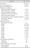
Thirty-nine (23.8%) of the 164 FMF patients were underwent 53 endoscopic procedures (21 esophagogastroscopy, 4 colonoscopy, and 14 both esophagogastroscopy and colonoscopy). Of all patients, 64.1% (n=25) were girls, the mean age at the diagnosis of FMF was 7.2±3.7 years, and the mean age at the time of endoscopy was 8.2±4 years. A total of 13 patients (15 endoscopic procedures; 9 esophagogastroscopies, 6 colonoscopies) underwent endoscopy due to GIS symptoms before the diagnosis of FMF. Other 26 patients, who underwent endoscopy after the diagnosis of FMF, were receiving medical therapy colchicine (Colchicum-Dispert; Recordati İlaç; Istanbul, Turkey; 0.03–0.05 µg/kg). The demographic characteristics of the patients are shown in Table 1.
Age and gender matched four patients who underwent endoscopic procedure in the pediatric gastroenterology unit were randomly selected as the control subjects for each endoscopic procedure (n=212, 62.1% female, median age of 8.4±3.9 years). Total number of patients who underwent endoscopic procedure between 2010 and 2017 were 6,435 in our pediatric endoscopy unit (after excluding the patients with FMF). Only inclusion criteria was age and gender matching, and exclusion criteria were used.
Detection of endoscopic appearance of reflux esophagitis (Los Angeles classification) and/or histopathological anomaly (papillary elongation, basal cell hyperplasia, increased intraepithelial neutrophil) on the esophagogastroscopic examination was considered as reflux esophagitis. In addition, detection of an abnormal number of eosinophil on the esophageal histopathological examination was considered as esophageal eosinophilia [9]. On the colonoscopic examination; more than 10 lymphoid nodules with a diameter greater than 2 mm were defined as lymphoid nodular hyperplasia (LNH) [10]. The diagnosis of inflammatory bowel disease (IBD) was made according to Porto criteria [11].
Informed verbal and written consent were obtained from the parents before the endoscopic prosedure in all patients. The study was retrospective and based on the file records (observational study), therefore no approval was made to the ethics committee, only written permission was received from the pathology department for to use the results of pathological examination. The study was made in accordonce to Helsinki Declaration.
Statistical analysis
All calculations in our study were performed using the IBM SPSS Statistics, ver. 23.0 (IBM Co, Armonk, NY, USA), and the continuous variables were expressed as mean±standard deviation and categorical variables as percentage (%). Comparison of the quantitative data between the groups was performed using Student t-test in the normally distributed variables, and Mann-Whitney test in the non normally distributed variables. Whereas qualitative data were compared using chi-square test. The p-values ≤0.05 were considered statistically significant.
RESULTS
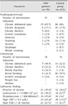
Indications for endoscopic procedures and laboratory findings of 39 patients (53 procedures) and control group are shown in Table 2. There was no significant difference between the groups in terms of esophagogastroscopy and colonoscopy indications. Pre-procedural anemia and elevated inflammatory markers (leukocytosis, thrombocytosis, CRP, and ESR elevation) were more common in FMF patients (p<0.05 for all).
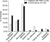
When the esophagogastroscopic examinations of the patients were compared with the control group, there was no significant difference in esophageal, gastric and duodenal findings (Fig. 1). Three of 13 patients without any abnormal finding on esophagogastroscopy were diagnosed FMF based on their clinical and laboratory findings on the follow up. Other patients (n=10) were diagnosed FMF based on recurrence of abdominal symptoms in addition to periodic fever despite the treatment of endoscopic pathology. Of the 26 FMF patients who underwent esophagogastroscopy after diagnosis, proton pump inhibitor therapy was administered in seven patients for nonspecific gastritis, and two for reflux esophagitis. Three patients with Helicobacter pylori gastritis were treated with tripple eradication regimen. Patients' symptoms improved during the follow-up, and endoscopic procedure was found to provide contribution by 46.2% (12/26 patients) in determining of the etiology of the additional symptoms.
Colonoscopic examination revealed that the frequency of IBD was higher in undiagnosed patients compared to both the control group (50% vs. 6.9%, p<0.05, OR:13.4 and 95% CI: 2.1–84.3) and the patients under colchicine treatment (50% vs. 8.3%, p< 0.05, OR: 11 and 95% CI: 0.8–147.8). No significant difference was found in terms of incidence of LNH and non-specific colitis both in undiagnosed and in patients under treatment compared to controls (Table 3). The diagnosis of IBD was made based on clinical, laboratory and histopathologic findings in three patients in undiagnosed FMF patients. All of them were early onset IBD (ages at the time of first symptoms; 10 days, 4 years, and 4.5 years old), and receiving immunosuppressive therapy, but they were unresponsive to immunosuppresive treatment on the follow-up. Examination for the monogenic disorders revealed homozygot mutation for MEFV in all of them. Immunosuppressive therapy was discontinued and colchicine treatment was initiated. Clinical and laboratuary remision were observed during the follow up in all patients. Other patient who was receiving colchicine for FMF, was admitted with bloody diarhea, and colonoscopic-histopathologic examination revealed ulcerative colitis. Mesalazine was added to the existing treatment. She was on remision during the follow up. Budesonide was initiated for the treatment of the patient with nonspecific colitis (n=1). Colonoscopic procedure that was made after the diagnosis was found to provide contribution by 16.7% (2/12 patients) in determining the etiology of the additional symptoms. The patients with LNH were followed up without any medical tretment.
DISCUSSION
In this study, we investigated the endoscopic findings of pediatric patients with FMF, we found that (i) anemia and elevation in the inflammatory markers were more common in patients with FMF than the control group; (ii) there was no significant difference in esophagogastroscopic findings; (iii) IBD is more common in undiagnosed patients with FMF; and (iv) endoscopic procedure was helpful in determining the additional pathologies and guiding treatment in patients on colchicine treatment.
Studies in the literature about GIS mucosal involvement of FMF in pediatric patients are scarce. Only Gurkan and Dalgic [12] reported the endoscopic findings of the patients with FMF who admitted to the pediatric gastroenterology outpatient clinic. They were reported that colitis and aphtous bulbitis were common findings in patients with FMF, and these findings were associated with uncontroled recurrent FMF attacks. Amyloid deposition was not found in any patient. In an another study buy the same authors, FMF was shown to be one of the most common causes of colonic LNH [13]. In an adult study, endoscopic findings of the patients on colchicine treatment was analyzed and histopathologic changes were found in jejunal biopsies that were compatible with colchicine side effects [7]. In another adult study, esophagogastroscopy, colonoscopy and small bowel capsule endoscopy were performed in patients under the colchicine treatment. Similar to our study, there was no significant difference in emdoscopy findings but an increased amount of mucosal lesions were found in small bowel capsule endoscopy. Approximately half of the patients had mucosal lesions (such as ulcer, erosion) on small bowel capsule endoscopy. Mitotic changes in the lesions were attributted to the colchicine effect rather than autoinflammatory conditions [4].
The association of FMF and IBD has been emphasized more markedly, especially in the studies conducted in recent years. In a study by Beşer et al. [14], IBD was found in 15.4% of 78 FMF patients that were receiving medical treatment. Particularly M694V and K695R mutations were shown to be more frequent in these patients. In studies with larger series, the incidence of IBD was found as 1.16% in FMF patients [15]. On the other hand, in studies conducted in patients with IBD, the incidence of FMF was found between 5% and 25% [1416]. Salah et al. [17] analyzed the frequency of MEFV mutations in children with IBD and they found that 88% of the IBD patients carried the one of the MEFV mutations and especially common in indeterminate colitis. FMF should be considered in differential diagnosis especially in cases of early-onset and treatment-resistant IBD. The frequency of IBD was found higher in undiagnosed patients than the patients under colchicine treatment in our study, it may be related with anti-inflammatory effect of colchicine that may suppress the IBD related symptoms.
Functional GIS disorders such as diarrhea, constipation, dyspepsia and chronic abdominal pain are commonly observed in patients with FMF. These are encountered both as secondary to medical treatment or complications of FMF [18]. Clinical conditions such as H. pylori gastritis, reflux esophagitis and nonspecific gastritis may also cause gastrointestinal symptoms in patients with FMF. Although accompanying fever may suggest a FMF episode, inflammatory diseases such as IBD may also cause fever. If the gastrointestinal symptoms are chronic and do not likely with FMF attacks, esophagogastroscopic and colonoscopic procedures should be performed to make the differential diagnosis of all these clinical conditions.
The limitations of our study were (i) detailed histopathological examination may be made, such as analyzing the mitotic activity in detail, may give additional about information colchicine effect; (ii) the lack of small bowel examination (small bowel capsule endoscopy); and (iii) small number of patients may cause type 2 mistakes during the statistical examinations.
In conclusion, we investigated the endoscopic findings of FMF patients, and we thought that FMF has very broad endoscopic findings. FMF should be considered in differential diagnosis in early onset (<5 years) and treatment resistant IBD patients, and endoscopic examination should be performed when patients have chronic gastrointestinal symptoms.

 XML Download