

 PDF
PDF ePub
ePub Citation
 Print
Print


INTRODUCTION
Eosinophilic gastroenteritis (EGE) is a type of gastrointestinal disease associated with eosinophilic infiltration, although its pathogenesis is not yet fully understood. Patients are diagnosed based on significant pathological findings and are classified into one of three disease subtypes: mucosal, muscular, and subserosal. Each subtype shows distinct gastrointestinal symptoms and clinical manifestations based on the layers that have been infiltrated by eosinophils [1]. Because histologic confirmation is essential for the diagnosis of EGE, most patients with suspected EGE undergo gastroduodenoscopy or colonoscopy for biopsy of the gastrointestinal tract. Some patients with ascites undergo diagnostic tapping, and others receive a surgical biopsy [2].
EGE patients display a variety of gastrointestinal symptoms, such as diarrhea, abdominal pain, vomiting, and ascites. Peripheral eosinophilia, hypoalbuminemia, anemia, and immunoglobulin E (IgE) elevation are often found in EGE [34], and EGE patients may also present with allergic diseases, including allergic rhinitis, atopic dermatitis, and food allergy [5].
EGE is a rare disease, and treatment outcomes in children have not been studied. Therefore, we investigated the clinical manifestations, treatments, and outcomes of EGE in pediatric patients.
MATERIALS AND METHODS
Patient
The medical records of patients from 1993 to 2014 at the Department of Pediatrics, Seoul National University Children's Hospital (Seoul, Korea) were reviewed. These patients had undergone at least one or more procedures to obtain specimens, including endoscopy and ascites tapping; pathological confirmations were also performed.
Five patients with gastrointestinal symptoms and peripheral eosinophilia were excluded because endoscopy was not performed and the eosinophil infiltration was not confirmed by histology. Three patients with eosinophilic infiltration in the gastrointestinal tract were classified as having other diseases and were also excluded from our study. One patient was diagnosed with lymphoma, and eosinophilic infiltrations in other organs were found in 2 patients who were diagnosed with hypereosinophilic syndrome.
A total of 24 patients were eventually enrolled in this study. Their clinical manifestations, treatments, and outcomes were investigated. This study was approved by the Seoul National University Hospital Institutional Review Board (IRB No. 1504-126-670).
Diagnosis
The diagnostic criteria for EGE in the present study were as follows:
1) The presence of gastrointestinal symptoms including nausea, vomiting, weight loss, bloating, abdominal discomfort, abdominal pain, diarrhea, ascites, edema, and hematochezia.
2) The pathological confirmation of eosinophilic infiltration into gastrointestinal tissues or ascites. However, there is no established value for the normal upper limit of eosinophils in the gastrointestinal tract, as the normal distribution of eosinophils differs depending on the site within the gastrointestinal tract. Usually, the stomach is known for having a slightly lower eosinophilic distribution than other sites, whereas higher densities (up to 30 eosinophils per high-power field [hpf]) are commonly found in the appendix, terminal ileum, cecum, and proximal colon [6]. However, many reports set the limits of a clinically significant eosinophil count by microscopy as more than 20/hpf [278]. In our study, the pathologists set the upper limit of moderate eosinophil infiltration as more than 30/hpf and severe eosinophil infiltration as more than 50/hpf. Moderate and severe eosinophil infiltrations were defined as clinically significant infiltrations. In addition, pathologic findings such as the infiltration of eosinophils into the submucosa, eosinophils within the epithelium of the crypts or villi, crypt hyperplasia, villous atrophy, large numbers of degranulating mast cells, and contiguous smooth muscle fibers were helpful for the evaluation of eosinophil infiltration.
3) The exclusion of other diseases with eosinophilic infiltration. There are many eosinophil-infiltrative gastrointestinal diseases, such as eosinophilic granuloma, polyarteritis nodosa, intestinal parasitosis, lymphoma, gastric cancer, Crohn's disease, allergic proctocolitis, and hypereosinophilic syndrome, and these conditions were excluded by an accurate pathologic evaluation or the involvement of other organs [9].
Subtype
Klein et al. [1] classified EGE into three disease subtypes according to the predominantly involved intestinal layers: mucosal, muscular, and subserosal. Patients with hypereosinophilia in ascites (more than 500 eosinophils per µL) were also classified as having subserosal disease. If patients with muscular disease or subserosal disease had mucosal eosinophil infiltration, they were not classified as having mucosal disease but muscular disease or subserosal disease instead. The involvement of 2 or more intestinal segments was defined as extensive disease [10].
Growth and outcome
To investigate the growth status of the patients, a Z-score was calculated using the LMS formula; Z=(measured value/M)^L-1/L*S. The LMS values were acquired from the database of the Korean Pediatric Society and the Korea Centers for Disease Control and Prevention. An underweight status was defined as a Z-score of weight <-1.96, and stunting was defined as a Z-score of height <-1.96. The poor outcome group was defined as the steroid-dependent or steroid-resistant patients.
RESULTS
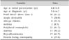
Twelve of the patients were boys, and 12 of the patients were girls. The mean age upon initial presentation was 4.4 years (range, 1.0-14.6 years), and the mean age at diagnosis was 5.3 years (range, 1.4-15.2 years). The mean duration of follow-up was 4.1 years (range, 1.2-5.4 years). Common symptoms included abdominal pain (45.8%), and diarrhea (54.2%). Some patients presented with vomiting (37.5%), weight loss (25.0%), hematochezia (25.0%), ascites (16.7%), and edema (29.2%).
In laboratory evaluations, 19 patients (79.2%) showed leukocytosis (above 10,000/µL), and 22 patients (91.7%) had peripheral eosinophilia (above 500/µL). The mean of the peak eosinophil count was 4,913/µL, and the mean of the peak eosinophil percentage among the leukocytes was 23.9%. Fifteen patients (62.5%) had hypoalbuminemia (below 3.3 g/dL). After currently active infectious states were ruled out, erythrocyte sedimentation rate elevation (above 20 mm/hr) and C-reactive protein elevation (above 0.5 mg/dL) were observed in 13% and 38% of the patients, respectively. Thirteen patients (54.2%) had anemia. An increased total IgE, defined by normal range of serum IgE standardized for age [11], was identified in 9 patients (37.5%). Protein-losing enteropathy was confirmed in 6 patients (25.0%) by an increased concentration of stool α-trypsin (Table 1).
A food multiple allergen simultaneous test (MAST) result above class II was defined as a positive result, which was displayed by 10 of the patients (41.7%). Allergic rhinitis (33.3%), atopic dermatitis (20.8%), and asthma (8.3%) were reported, and 13 patients (54.2%) had a history of allergy.
Nine patients underwent colonoscopy, 16 patients underwent esophagogastroduodenoscopy, and 2 patients received both procedures. Endoscopic examinations showed gastroduodenal ulcers in 3 patients (12.5%) and nonspecific findings, including gastritis and duodenitis, in 13 and 11 patients, respectively. Surgical biopsies were performed in three cases, and four patients underwent ascites tapping due to ascites with abdominal distension.
Eighteen patients were classified with mucosal layer disease by endoscopic biopsy. One patient had muscular disease and received therapeutic surgery for her gastric outlet obstruction. Five patients were classified with subserosal disease by surgical biopsy or ascites tapping. One patient received a subtotal gastrectomy for intractable ulcers, and the other patient received a full-thickness biopsy because of steroid resistance. The other 3 patients showed hypereosinophilia in their ascites.
Half of the patients had eosinophil infiltration in the stomach, and half of the patients showed infiltration in the duodenum. Some patients had infiltration into the colon (29.2%), ileum (16.7%), and esophagus (12.5%). Thirteen patients (54.2%) showed extensive disease. No difference was found based on disease subtype between the disease locations.
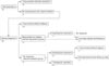
Nine patients had food allergy restrictions. Dietary restrictions were based on food allergy history and food-specific IgE tests. Five patients improved with food restrictions, and 4 patients required steroid treatment (Fig. 1).
Nineteen patients were treated with corticosteroids for 4 to 12 weeks. Steroids were effective in 78.9% of the patients (15/19). Eleven patients (57.9%) showed no relapses after discontinuing steroids. Montelukast, a selective leukotriene receptor antagonist, was administered to 5 patients to maintain remission. Three patients showed no relapse, but two patients relapsed and required steroid therapy repeatedly. A total of four patients (21.1%) had relapsing symptoms after stopping steroid treatment (steroid-dependent group). One steroid-dependent patient underwent azathioprine treatment for steroid sparing but did not show an improvement in gastrointestinal symptoms during those 4 months. The treatment strategy in this steroid-dependent group was changed to low-dose steroid maintenance.
The other 4 patients (21.1%) showed no response to steady steroid therapy (steroid-resistant group). Two of them received azathioprine treatment; 1 of these patients showed symptom relief and was taken off azathioprine after 6 months. The remaining 2 patients received therapeutic operations for gastric outlet obstruction and intractable ulcers.
Upon initial presentation, stunting was found in 2 children, and an underweight status was observed in two children. The Z-scores in weight at diagnosis and at the last follow-up were 0.08±1.01 and -0.20±1.04, respectively (p=0.602). The Z-scores in height at diagnosis and at the last follow-up were -0.06±0.85 and -0.044±0.927, respectively (p=0.516).
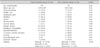
Symptoms, laboratory abnormalities, extensive disease, subtype, and history of allergy were not associated with poor outcomes. However, the presence of gastroduodenal ulcers was significantly associated with relapse and steroid resistance (p=0.01) (Table 2).
DISCUSSION
This study investigated the clinical manifestations, treatment outcomes and prognostic factors of 24 children with EGE. To our knowledge, this study is one of the largest studies on EGE in pediatric patients. Tien et al. [12] reported 14 Taiwanese children with EGE and did not evaluate the patients' Klein classifications. The epidemiology and characteristics of the patients in our study were similar to those reported in prior studies [41314]. Half of the patients were male, with other studies reporting 42.5% to 85% male patients. Most of the patients in our study had abdominal pain (45.8%), nausea/vomiting (37.5%), and diarrhea (54.2%). In a prior study, abdominal pain was the most frequent symptom (75% to 100%), while nausea (48% to 67%), vomiting (33% to 62.5%), and diarrhea (12% to 75%) were present in a wide frequency range.
Many studies have reported a predominance of mucosal layer disease (43% to 100%), and 18 patients (75.0%) were classified with mucosal layer disease in this study. Khan [15] suggested that mucosal disease is associated with nonspecific gastrointestinal symptoms. The incidence of muscular disease observed in this study was relatively low (4.2%) compared with other studies (13% to 70%). Our patient with muscular disease had vomiting and abdominal pain, which was consistent with other studies [1617]. If gastric outlet obstruction or intestinal obstruction is found, therapeutic surgery and pathologic evaluation should be aggressively considered. In most cases of subserosal disease in previous studies, the presence of ascites was typically found. Two of our patients had no ascites, and subserosal infiltration of eosinophils was confirmed by surgical biopsy. The presence of ascites is a typical symptom in the subserosal group, although eosinophils can involve the subserosal layer without ascites. Chen et al. [4] reported that one adult patient with subserosal disease underwent surgery for a refractory ulcer that had perforated.
Eosinophilic infiltration in the gastrointestinal tract is mediated by Th-2 cytokines (interleukin [IL]-3, IL-5, and IL-13) and eotaxin. Several inflammatory mediators and chemoattractants enhance eosinophil development, and peripheral eosinophilia can also be observed. IL-5 is important for upregulating eosinophilic differentiation and controlling peripheral circulating eosinophil levels [1819]. Pineton et al. [20] reported that peripheral eosinophilia is shown in 60% to 70% of EGE patients. In our study on pediatric patients, 91.7% of the patients had peripheral eosinophilia. Although IL-5 is important in regulating the circulating levels of eosinophils, their trafficking and accumulation in the gastrointestinal tract depends on a synergistic effect with eotaxin [19].
Food antigens are considered to play a role in the development of EGE [21]. In the present study, a food MAST result above class II was found in 10 patients (41.7%). Food restrictions were effective for some of these patients (50.0%) who showed positive food-specific IgE tests. Food restriction may be effective if a specific food allergy is identified. Khan et al. [16] reported the efficacy of exclusively elemental diets based on free amino acids, and Yamada et al. [22] suggested that the elimination of multiple food items, including the 6 most common allergenic foods (milk, soy, egg, wheat, peanuts/tree nuts, and shellfish/fish), as well as those specific foods to which the patient is allergic, can successfully treat EGE compared with an elemental diet. Recently, Lucendo et al. [23] announced a systematic review of dietary treatment in EGE patients; these authors argued that elemental diets and empirical elimination of allergy-associated foods could improve clinical symptoms but that their effect was questionable due to the lack of objective evaluation of clinical changes and the very limited assessment of histologic remission. Thus, an elemental diet and the elimination of multiple food items could be considered for patients who do not respond after the avoidance of allergenic food.
Steroid therapy and the re-application of steroids in cases of relapse are fundamental treatment strategies for EGE. Systemic steroids suppress eosinophil-induced inflammation in the gastrointestinal tract, and steroids are indicated for patients who are refractory to dietary therapy. Total of 78.9% patients improved after steroid treatment, although some patients showed dependency on steroids (21.1%) and others showed resistance to steroids (21.1%).
Relapses are frequently noted as steroids are tapered or discontinued [15]. In previous studies, high rates of relapse or resistance were reported in adult patients treated with steroids (38% to 57%) [420]. Total of 42.1% showed a relapse or steroid resistance in our study of pediatric patients, and Tien et al. [12] reported similar rates (33%) of steroid dependency in pediatric patients.
Systemic steroid therapy can induce growth failure, a cushingoid state, hyperglycemia, cataracts, and adrenal suppression. Thus, alternative treatments are needed for patients dependent on or resistant to steroids.
Montelukast, a leukotriene D4 receptor antagonist, is considered a maintenance treatment for EGE after steroid induction. Leukotrienes function as chemotactic factors for eosinophils and induce eosinophilic infiltration. It has been reported in various studies that leukotriene antagonists allow patients to taper off steroids and maintain remission [122425]. Among our five patients who received montelukast treatment, 3 showed no relapse. However, this drug's effect on the maintenance of remission was not clear. Thus, further prospective randomized studies are necessary to evaluate the effect of montelukast in children with EGE.
Azathioprine may be administered for steroid-dependent or steroid-resistant patients. Redondo-Cerezo et al. [13] reported that remission was induced by azathioprine. In our study, 1 patient showed improvement after azathioprine treatment, but the other 2 patients who received the treatment did not improve; thus, the effect of azathioprine as a steroid-sparing treatment was limited in this study.
Our study further demonstrated that gastroduodenal ulcers were a risk factor for steroid dependency or resistance. In one study, 13% of patients had shallow gastric or duodenal ulcers [4], and a similar proportion of patients in our study had gastroduodenal ulcers (13%). Pineton et al. [20] described the differences in prognosis according to EGE subtype, in which subserosal disease evolved in more than 50% of the cases as a single incidence without any recurrence, whereas more than 80% of the continuous courses (chronic form without remission) were of the mucosal disease type. We found no difference in treatment outcome between the subtypes.
Our study has some limitations. We could not evaluate the severity of symptoms because this study was retrospective. The relationship between severity of symptoms and eosinophilic infiltration in tissues could not be analyzed. The evaluation of treatment including steroid, montelukast, and azathioprine was limited. Controlled clinical trials are needed.
In conclusion, a high suspicion of EGE is necessary when children have nonspecific gastrointestinal symptoms and peripheral eosinophilia. In our study, most patients improved with food restrictions or steroid treatment, although one-third of patients experienced relapse or steroid resistance. Moreover, the presence of gastroduodenal ulcers was significantly associated with relapse and steroid resistance.
 XML Download