

 PDF
PDF ePub
ePub Citation
 Print
Print


INTRODUCTION
Iron is essential for iron-containing enzymes involved in brain-energy metabolism, neurotransmitter synthesis, and myelination, all of which are implicated in neurodevelopment during early infancy [1]. Fetal iron accretion usually occurs during the third trimester of gestation [2]. In preterm infants, fetal iron accumulation through transfer from the mother is interrupted, leading to an increased risk of iron deficiency at birth. Approximately 80% of very low birth weight (VLBW) and 95% of extremely low birth weight (ELBW) infants require erythrocyte transfusions [3]. Multiple erythrocyte transfusions can result in excessively high iron levels [4], leading to oxidative injury as the production of oxygen radicals increases [5]. Multiple erythrocyte transfusions are also associated with the incidence and severity of retinopathy of prematurity (ROP) and bronchopulmonary dysplasia (BPD) [678]. Although enteral iron supplementation has not been reported to be a cause of iron overload or oxidative injury, the initiation of enteral iron supplementation in preterm infants receiving multiple erythrocyte transfusions should be performed carefully.
We aimed to investigate the iron status of VLBW infants receiving multiple erythrocyte transfusions during hospitalization in the neonatal intensive care unit (NICU).
MATERIALS AND METHODS
Patients
Our study enrolled 46 VLBW infants who were admitted to NICU of the Kyungpook National University Hospital between January 2012 and December 2013. Infants with congenital anomalies, metabolic disorders, culture-positive sepsis, and intrauterine transfusions (that is, twin-to-twin transfusion) were excluded. We obtained approval from the institutional review board of Kyungpook National University Hospital for the present study (IRB No. 2013-12-042).
Assessment of iron status
We measured serum iron and ferritin concentrations as well as total iron binding capacity (TIBC) as markers of iron status on the infants' first day of life and weekly thereafter during the course of their hospitalization. Hemoglobin and hematocrit levels, mean corpuscular volume (MCV), and reticulocyte count were also obtained. Maternal hemoglobin, hematocrit, and serum ferritin levels were assessed before delivery. Maternal anemia was defined as hemoglobin levels <12 g/dL, and iron deficiency as serum ferritin levels <10 ng/mL.
Red blood cell parameters, including hemoglobin, hematocrit, MCV, and reticulocyte count were assessed by flow cytometry using an automated blood cell counter. Plasma iron levels and TIBC were measured using the nitro-PSAP test, and ferritin concentration was measured through a turbidimetric immunoassay using an automated method.
Iron supplementation
When the infants reached full enteral feeding (100 mL/kg), iron supplementation (2 mg/kg) was started. We withheld iron supplementation if serum ferritin levels were >350 ng/mL.
Predominantly breast milk-fed infants were supplemented with a human milk fortifier (HMF, Similac; Abbott Laboratories, Abbott Park, IL, USA). Formula milk (Absolute Babywell Preemie; Maeil, Seoul, Korea), breast milk, and HMF contained 0.0029 mg/mL, 0.00047 mg/mL [9], and 0.36 mg/pack iron, respectively.
Erythrocyte transfusions
The threshold for administering erythrocyte transfusions was a hematocrit of 36%, requiring oxygenation of >35%, or a mean airway pressure of >6 to 8 cmH2O by positive pressure ventilation, and a hematocrit of 31% with respiratory support, oxygen therapy, or recurrent apneic episodes of more than 9 times per 12 hours. Based on the clinical conditions of the infants, the volume of erythrocyte transfusions ranged from 10 to 15 mL/kg.
Association between erythrocyte transfusion and neonatal morbidity
We evaluated the association between the volume and number of erythrocyte transfusions and neonatal morbidity, including ROP and BPD. A diagnosis of BPD was made when infants born at <32 weeks' gestation required oxygen for the first 28 days at a postmenstrual age of 36 weeks or when infants born at ≥32 weeks and remained on oxygen supplementation for 56 days [10]. We included infants with ROP stage 2 or 3.
Statistical analyses
Statistical analyses were performed using IBM SPSS Statistics ver. 20 for Windows (IBM Co., Armonk, NY, USA). The results are reported as means±standard deviation. Comparisons of the mean values of the continuous variables were performed by the Student t-test and one-way analysis of variance. Association between gestational age and serum ferritin concentrations after controlling for maternal ferritin concentrations, and correlation among the volume of erythrocyte transfusion, serum ferritin levels, and the duration of positive ventilation after controlling for the amount of iron intake were analyzed using partial correlation. Statistical significance was defined as p<0.05.
RESULTS
Patient baseline characteristics
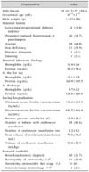
The baseline characteristics of the patients are presented in Table 1. One-fourth of the mothers were iron deficient, and over half were anemic; however, no infants had anemia or iron deficiency at birth. The mean serum concentration among the infants at discharge was 328.8±328.0 ng/mL (range: 33.8-1,510.0 ng/mL). No infants were iron-deficient during hospitalization in the NICU.
Thirty-eight of the VLBW infants received erythrocyte transfusions. The incidences of ROP, BPD, necrotizing enterocolitis, and intraventricular hemorrhage were 24%, 24%, 0%, and 2%, respectively.
Maternal factors related to neonatal iron status (measured on the first day of life)
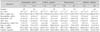
We found no statistically significant differences in red blood cell parameters and iron status between infants born to mothers with and without anemia or iron deficiency (Table 2). Moreover, no significant differences were observed for the red blood cell indices, plasma iron and ferritin levels, and TIBC between infants born to mothers with and without diabetes or hypertension (Table 2). The mean serum ferritin concentrations of infants born to mothers with hypertension were lower than those of infants born to mothers without hypertension; however, this difference was not statistically significant (p=0.085).
Red blood cell indices and iron status (measured on first day of life) by gestational age
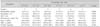
The infants were divided into eight groups, based on gestational age. No significant differences were found for red blood cell parameters and iron status (Table 3). Serum ferritin levels were not correlated with gestational age after controlling for maternal ferritin concentrations (r=-0.063, p=0.804).
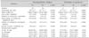
Volume of erythrocyte transfusions and serum ferritin concentrations
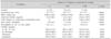
The infants were categorized as belonging to one of three groups, based on the volume of erythrocyte transfusion (calculated per body weight) (Table 4). Infants with a younger gestational age or lower birth weight required a higher volume of erythrocyte transfusions. The mean iron intake was statistically higher in non-transfused infants compared to infants receiving erythrocyte transfusions. Mean serum ferritin concentrations were not different on the first day and at discharge, but we observed differences for the minimum and maximum serum ferritin levels during hospitalization. Maximum serum ferritin levels were significantly higher in infants receiving ≥100 mL/kg erythrocyte transfusions, but the volume of erythrocyte transfusions (mL/kg) was not correlated with maximum serum ferritin levels after controlling for the amount of iron intake (r=0.164, p=0.338). Minimum and maximum serum ferritin levels of non-transfused infants were higher than those of infants receiving <100 mL/kg erythrocyte transfusions.
Neonatal morbidity and erythrocyte transfusions
The associations between erythrocyte transfusions and neonatal morbidity are presented in Table 5. A total of 10 and 11 VLBW infants were diagnosed as BPD and ROP, respectively. We found a significantly lower gestational age and birth weight among infants with morbidity. Infants with BPD or ROP received a higher volume and number of erythrocyte transfusions than infants without BPD or ROP. The total volume of erythrocyte transfusion was correlated with the duration of positive-pressure ventilation (r=0.676, p<0.001) but not with maximum serum ferritin levels (r=0.012, p=0.945) after controlling for the amount of iron intake. Infants with ROP required a longer duration of positive-pressure ventilation compared to infants without ROP. Minimum and maximum serum ferritin levels were not significantly different between infants with ROP and those without ROP. Mean iron intake was significantly higher in infants without morbidity than in those with morbidity.
DISCUSSION
In our study, no VLBW infants were iron-deficient, defined as serum ferritin levels <10 ng/mL, during their hospitalization in NICU. Their mean serum ferritin levels since 1 week of age were above the 95th percentile according to previous standards by Siddappa et al. [11]. The infants required a mean volume of 50.0±52.9 mL/kg for erythrocyte transfusions, and their iron status during hospitalization was considered as iron-replete or -retaining rather than iron deficient.
It has been reported that fetal iron status is independent of maternal iron indices [1213]. However, one previous study showed that lower maternal hemoglobin concentrations were correlated with lower fetal iron stores and lower cord hemoglobin concentrations [14]. Maternal anemia and iron deficiency did not affect neonatal red blood cell count and iron status in our study. Serum ferritin levels were measured after the infants were stable. About 50% of infants required oxygen and positive-pressure ventilation before blood sampling; thus, serum ferritin concentrations might have been affected by the oxygen exposure. Maternal hypertension causes inadequate placental circulation, which leads to a chronic hypoxic state, stimulating fetal erythropoiesis [15]. Infants born to mothers with hypertension are at risk for iron-deficiency due to the interrupted transport of maternal iron to the fetus and augmented erythropoiesis [11]. Infants born to mothers with gestational diabetes mellitus are also at risk for low serum ferritin levels [16]. In our study, we observed no significant differences in serum ferritin levels on the first day of life between infants born to mothers with and without hypertension or diabetes mellitus.
Excessive iron accumulation can occurs in preterm infants with multiple erythrocyte transfusions. The iron status of preterm infants undergoing multiple transfusions might be adequate without iron supplementation; in particular in transfused ELBW infants who have a high iron storage until 6 months of age [717]. Since the life span of transfused erythrocytes is shorter than that of erythrocytes in premature infants [18], the breakdown of erythrocytes and resulting damage during transfusion can result in an iron overload state [19]. High serum ferritin concentrations reflect not only excessive iron accumulation but also systemic inflammatory conditions and infections [20]. In our study, 38 VLBW infants required erythrocyte transfusions. The volumes (based on weight) of the erythrocyte transfusions were not correlated with serum ferritin concentrations. However, maximum serum ferritin levels were significantly higher in infants receiving ≥100 mL/kg erythrocyte transfusions compared to non-transfused infants and infants receiving <100 mL/kg erythrocyte transfusions. Although infants with sepsis were excluded from our study, the preterm infants had various inflammatory conditions, such as exposure to oxygen-free radicals. This might have affected serum ferritin levels.
A higher iron status, as measured by various biomarkers, in particular serum ferritin, might be mediated by iron-related oxidative stress though lipid oxidation [21]. In animal models, pharmacological iron supplementation significantly increased lipid oxidation in both normal and iron deficient states [22]. In a randomized controlled trial conducted in pregnant women during the third trimester, lipid peroxidation levels but not serum ferritin levels were significantly higher in women receiving iron supplementations than in controls [23]. In our study, non-transfused infants had a significantly higher oral iron intake than transfused infants. Although maximum serum ferritin levels were not associated with the amount of iron intake after controlling for the volume of erythrocyte transfusion, minimum and maximum serum ferritin concentrations of non-transfused infants were higher than in infants with receiving less 100 mL/kg of erythrocyte transfusion. In our study, infants without BPD or ROP had higher oral iron intake than those with BPD or ROP. It is still uncertain if low-dose iron supplementation increases lipid oxidation.
Preterm infants have an immature iron metabolism system and antioxidative activity [45], and excessive iron accumulation can cause and exacerbate major morbidities such as BPD and ROP, which are known to be associated with oxidative injury [78]. Higher serum ferritin concentrations have also been shown to be associated with the incidence and severity of ROP [24]. The volumes of erythrocyte transfusions were significantly higher in infants with BPD and ROP than in those without these conditions, but the maximum serum ferritin concentrations of infants with morbidity were not statistically different to those without morbidity. The duration of positive-pressure ventilation showed a strong positive correlation with the volume of erythrocyte transfusion. However, infants undergoing positive-pressure ventilation required more frequent erythrocyte transfusions to maintain adequate hematocrit levels. It was not clear whether the high total erythrocyte volumes resulted in or were caused by the longer duration of the positive-pressure ventilation.
Our study was conducted with a small number of VLBW infants; in particular, only a small number of infants did not receive erythrocyte transfusions. Serum ferritin concentrations were used as the primary marker for iron storage; however, serum ferritin levels might be affected by systemic inflammation. Although infants with sepsis were excluded from this study, the severity of individual inflammatory conditions was not controlled. Serum ferritin alone might not be representative of the iron status of VLBW infants under inflammatory conditions and receiving multiple erythrocyte transfusions.
The total volumes of erythrocyte transfusions were associated with neonatal morbidity, and the majority of VLBW infants were at risk for excessive iron stores during hospitalization. When serum ferritin levels were >350 ng/mL, iron supplementation was withheld. We did not assess if a change in serum ferritin concentrations through oral iron supplementation under an iron-overload state, which has a limitation to show the association between oral iron intake and oxidative stress.
In conclusion, VLBW infants receiving multiply erythrocyte transfusions had excessive iron stores during hospitalization in the NICU. Also non-transfused infants might had a risk of iron overload during hospitalization. This suggests that the iron status of VLBW infants should be assessed during hospitalization in the NICU and after discharge for an appropriate iron supplementation regimen.
 XML Download