

 PDF
PDF ePub
ePub Citation
 Print
Print


INTRODUCTION
Henoch-Schönlein purpura (HSP) is the most common systemic vasculitis affecting children. It typically involves the small vessels of skin, gastrointestinal (GI) tract, joints, and kidneys. HSP is usually diagnosed on the basis of clinical features, such as purpuric rash, colicky abdominal pain, arthralgia, and hematuria [1,2]. However, in approximately 10-40% of patients, GI and/or joint symptoms may precede the onset of skin purpura, which is a mandatory diagnostic criterion, by up to 2 weeks [2,3,4]. Therefore, it may be difficult to diagnose HSP when a patient exhibits an atypical presentation of HSP. Actually, the interval between symptom onset and diagnosis is reported to be 1-30 days [5,6].
Although the prognosis of HSP is generally good, GI tract involvement is the most serious potential complication of HSP; massive GI bleeding, bowel infarction, and perforation as well as intussusceptions may develop. In fact, surgical interventions associated with GI complications are performed in approximately 5-12% of patients with HSP, and the recurrence rate has been reported to be up to 30% [7]. However, abdominal symptoms are easily considered to be nonspecific in children. Furthermore, to date, no specific serologic marker that can aid in the diagnosis of HSP or distinguish it from other diseases with similar clinical manifestations has been identified. Therefore, there is a strong need in clinical practice to determine markers of GI tract involvement when the first or only presenting symptoms are GI-related to avoid invasive procedures, such as endoscopy and laparotomy.
HSP is an immune complex-mediated disease. Vascular deposition of immunoglobulin A (IgA)-dominant immune complexes usually induces endothelial cell damage via the actions of inflammatory cytokines and recruitment of inflammatory cells [8,9]. Theses endothelial cell alterations consequently lead to hemostatic alterations, such as fibrinolysis and the activation of coagulation [10]. A few studies have shown that biochemical markers reflecting endothelial cell damage or hemostatic alterations are elevated in several types of systemic vasculitis [11,12,13]. However, the correlations between these markers and disease severity, especially in HSP, are not completely clear.
Therefore, to determine clinically useful biochemical markers to support the clinical diagnosis of HSP, this study evaluated laboratory parameters of inflammation and activated coagulation during the course of the disease. Furthermore, we aimed to demonstrate potential biomarkers reflecting the disease activity of HSP and/or indicating GI involvement by comparing these parameters with a clinical scoring system that have been suggested for scoring the clinical severity of HSP [14,15].
MATERIALS AND METHODS
Patients
We retrospectively reviewed the clinical data and laboratory parameters of children diagnosed with HSP at the Seoul National University Bundang Hospital from January 2004 to February 2011. HSP was diagnosed on the basis of the criteria proposed by the American College of Rheumatology, which require individuals to meet at least 2 of the following criteria: younger than 20 years of age at the time of disease onset, palpable purpura on the skin, bowel angina manifesting as diffuse abdominal pain that worsens after meals or bloody diarrhea, and granulocytes in the walls of arterioles and venules detected in skin biopsy [1]. Patients were excluded if they had additional evidence of infection. Healthy children referred to our clinic because of abdominal pain and for whom thorough investigations did not reveal any organic diseases were recruited as controls. This study was approved by the institutional review board of the Seoul National University Bundang Hospital (IRB B-1107-132-114).
Assessment of HSP disease activity
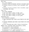
We evaluated the disease activity of HSP according to the previously described clinical scoring system [14,15]. Skin, joint, abdominal, and renal symptoms were scored as 0-3, and the sum of the 4 subscores was recorded as the total clinical score (0-12) for each patient (Table 1).
The scores were assessed by the same physician for both the acute and convalescent phases of the disease. The acute phase was defined as the period during which the patient presented with any clinical symptoms of HSP including purpura, abdominal pain, arthralgia, and hematuria. Meanwhile, the convalescent phase was defined as the period when all presented symptoms had resolved regardless of relapse.
Laboratory parameters
Blood samples were obtained from all patients with HSP during both the acute and convalescent phases. The following laboratory data indicating inflammation, standard coagulation, and activated coagulation were analyzed in the patients: hemoglobin, white blood cell (WBC) count, absolute neutrophil count (ANC), platelet count, erythrocyte sedimentation rate (ESR), C-reactive protein (CRP) level, prothrombin time (PT), activated partial thromboplastin time (aPTT), and fibrinogen, D-dimer, and fibrin degradation product (FDP) levels. Fibrinogen, D-dimer, and FDP levels were also measured in the controls and compared to those in the patients.
D-dimer levels were quantitatively analyzed by using an immunoturbidimetric method with a D-dimer assay kit (DiagnosticaStago, Asnieres, France), and the results were analyzed using an STA Compact CoagulationAnalyzer (DiagnosticaStago). FDPs were measured using a Pacific Hemostasis FDP Assay Kit (Fisher Diagnostics, Middletown, VA, USA).
Statistical analysis
The data were analyzed using PASW version 18.0 statistical software (IBM Co., Armonk, NY, USA). A linear mixed model was applied to evaluate differences in the factors between the acute and convalescent phases of HSP. Student's t-test, the Mann-Whitney U-test, and Spearman correlation coefficients were also used to analyze the data as appropriate. The acute- and convalescent-phase patients as well as the controls were compared using one-way ANOVA and the Kruskal-Wallis test. The level of statistical significance was set at p<0.05.
The statistical methods of this study were reviewed by Medical Research Collaborating Center at the Seoul National University Bundang Hospital.
RESULTS
Clinical features of patients
A total of 185 children (101 boys and 84 girls; mean age, 6.6±2.9 years) were enrolled in the patient group. An additional 130 healthy children (55 boys and 75 girls; mean age, 7.0±2.3 years) without HSP served as controls.
The clinical scores of the patients during the acute phase of HSP are shown in Table 2. A total of 132 patients (71.4%) had GI symptoms at disease onset, and renal manifestations were observed in 20 patients (10.8%) at the time of HSP diagnosis. Thirteen patients (7.0%) did not have skin lesions as an initial manifestation of the disease.
During the acute phase, 123 patients were initially treated with corticosteroids. HSP symptoms resolved spontaneously without any intervention in the other 62 patients. Among the 123 corticosteroid-treated patients, 29 required additional intravenous immunoglobulin administration for intractable HSP. Three of them who were refractory to immunoglobulin were additionally managed with high-dose methylprednisolone pulse therapy, and 2 who did not respond to methylprednisolone pulse therapy were ultimately treated with plasmapheresis. No patients underwent surgery for GI tract complications.
Laboratory parameters of HSP
The laboratory parameters of the patients in the acute and convalescent phases of HSP are presented in Table 3.
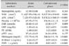
The WBC count, ANC, ESR, and CRP levels differed significantly between the acute and convalescent phases of the disease. However, there were no significant differences with respect to the hemoglobin level, platelet count, PT, or aPTT between phases. Moreover, the mean platelet level, PT, and aPTT were within normal ranges, even during the acute phase.
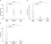
The fibrinogen, D-dimer, and FDP levels were significantly higher during the acute phase than during the convalescent phase. These markers were also significantly higher during the acute phase of the disease than the values in the control group (p<0.05) (Fig. 1).
Relationships between total clinical score and laboratory parameters during the acute phase of HSP
The total clinical scores were significantly correlated with the skin score (r=0.646, p<0.001), joint score (r=0.430, p<0.001), abdominal score (r=0.639, p<0.001), and kidney score (r=0.239, p=0.001) during the acute phase of HSP. The total scores were also significantly correlated with the WBC count (r=0.241, p=0.001), ANC (r=0.261, p<0.001), CRP level (r=0.260, p<0.001), D-dimer level (r=0.371, p<0.001), and FDP level (r=0.369, p<0.001).
Subgroup analysis according to the abdominal score of HSP
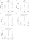
During the acute phase, the ANC, CRP, D-dimer, and FDP levels were significantly higher in the patients with GI symptoms (i.e., abdominal scores of 1-3) than in those without GI symptoms (i.e., abdominal score of 0) (p<0.05, Fig. 2).
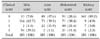
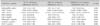
When the patients were categorized into 3 groups corresponding with the presence of no, mild, or significant GI symptoms according to their abdominal scores (abdominal scores of 0, 1, 2-3, respectively), there were significant differences in the WBC count, ANC, and CRP, D-dimer, and FDP levels among the subgroups of patients with GI symptoms (p<0.05, Table 4). In particular, the patients with significant GI symptoms (abdominal scores of 2-3) had higher WBC counts, ANC, CRP, D-dimer, and FDP levels than those without GI symptoms (abdominal score of 0) (p<0.05) (Fig. 2).
DISCUSSION
The results of the present study demonstrate that laboratory markers of activated coagulation significantly reflect the disease activity of HSP. In particular, fibrin-related markers, such as D-dimer and FDPs, were more strongly associated with clinical symptoms than inflammatory markers. Furthermore, D-dimer and FDP levels consistently reflected GI tract involvement during the acute phase of HSP.
Although the etiology of HSP remains unknown, infectious agents, vaccination, and medications have been suggested to trigger an immune response. In addition, IgA in serum and mucosal secretions plays a pivotal role in the pathogenesis of HSP [16]. Several studies have suggested that the abnormal glycosylation of IgA1 (1 of the 2 subclasses of IgA) aggregates into macromolecular complexes, which activate the alternative complement pathway and recruit inflammatory cells [17]. Moreover, recent studies have shown that a variety of IgA autoantibodies are associated with HSP. These autoantibodies have been reported to be cross-reactive with endothelial cells and to induce complement-dependent cell lysis [18].
It is understood that there is a bidirectional relationship between inflammation and coagulation systems. The activation of inflammation leads to the activation of the coagulation system, which also markedly affects inflammatory activity. This is considered important in the pathogeneses of vascular diseases [19]. Proinflammatory cytokines and chemokines, polymorphonuclear cells, platelets, endothelial cells, fibrinogen, and fibrin have been reported to be important mediators of the activation of inflammation and coagulation [19,20]. In fact, several studies have investigated the roles and patterns of these molecules in the pathogenesis of HSP [9,16,21].
To date, there have been some efforts to apply the molecules involved in endothelial cell damage and the consequent activation of coagulation as indicators of HSP disease severity. Factor XIII, a fibrin-stabilizing factor, is significantly decreased during the acute phase of HSP and has been suggested to be a prognostic indicator of this disease [22,23]. Another study has shown that only the level of von Willebrand factor, which is synthesized by endothelial cells and released into circulation upon endothelial stimulation, is correlated with disease activity, and there are no correlations between factor XIII and clinical symptoms [14]. In a small-scale study, factor XIII has been shown to decrease only in clinically severe cases [24]. However, none of these studies have simultaneously assessed both markers of inflammation and activated coagulation in patients with HSP.
The present study simultaneously analyzed the inflammatory cells recruited in association with endothelial cell activation and fibrin-related markers during the disease course. These inflammation and coagulation pathways were significantly activated in patients during the acute phase of HSP. Moreover, the results of standard coagulation assays show that PT and aPTT were within the normal ranges, even during the acute phase, and did not differ significantly between the acute and convalescent phases. These results are concordant with those of previous studies [14,24]. Furthermore, these results contribute to our understanding of the pathophysiology of HSP by adding to the existing evidence of the presence of an activated coagulation state secondary to the endothelial inflammation induced by IgA immune deposits in these patients.
In addition, fibrinogen, D-dimer, and FDP levels were significantly higher during the acute phase compared with the convalescent phase and also compared with the controls. The D-dimer and FDP levels were more strongly correlated with the total clinical score of HSP than the inflammatory markers, such as the WBC count, ANC, and CRP levels. Signs of hyperfibrinolysis have been observed in patients in a few small-scale studies. In particular, D-dimer has been found to be well correlated with the disease activity of HSP [15]. Therefore, the assessment of fibrin-related markers, such as D-dimer and FDPs, is expected to be clinically useful for the determination of disease activity along with the application of the previously suggested clinical scoring system for HSP.
The incidence of GI tract involvement has been reported to be between 50% and 75% [3,4,25]. In the present study, 71.4% (132/185) of the patients had GI symptoms during the acute phase of HSP; however, 7.6% (10/132) of them did not present with any skin lesions, which is a mandatory diagnostic criterion. Therefore, all of these patients underwent upper gastroendoscopy accompanied by biopsy to be diagnosed with HSP.
In the present study, D-dimer and FDP levels were more consistently and significantly elevated in the patients with abdominal symptoms compared with the inflammatory markers, such as the WBC count, ANC, ESR, and CRP levels. Although the patients with severe abdominal symptoms exhibited significant increases in the WBC count, ANC, and CRP levels as well as the D-dimer and FDP levels, these results suggest that D-dimer and FDPs may play important roles as indicators of GI tract involvement during the acute phase of HSP. Therefore, fibrin-related markers, such as D-dimer and FDPs, may enable clinicians to evaluate not only the effectiveness of a treatment but also disease relapse, especially in young children complaining of abdominal pain recurrence during the treatment of HSP.
To our knowledge, this is the first study to analyze the laboratory markers of both inflammation and activated coagulation and to demonstrate their relationships with disease activity and GI involvement in HSP using a clinical scoring system. Because the scoring system mainly relies on subjective descriptions, measuring D-dimer and FDP levels in combination with the use of a scoring system may aid in the diagnosis of HSP and help distinguish this disease from other conditions in children without purpuric rash complaining of abdominal pain. Nevertheless, additional studies are required before these fibrin-related markers may be utilized as indicators of disease activity, particularly with regard to the involvement of specific organs.
 XML Download