

 PDF
PDF ePub
ePub Citation
 Print
Print


INTRODUCTION
The endoscopic mucosal resection (EMR) technique has been widely used for the resection of colorectal neoplasms. Endoscopic piecemeal mucosal resection (EPMR) is used in up to 50% of cases for resection of large lesions of >20 mm in diameter.1234 The local recurrence rate is higher with EPMR than with en bloc resection. The local recurrence rate after EPMR has been reported to be up to 50%, compared with a rate of 0% to 17.8% after en bloc resection.156 Endoscopic submucosal dissection is an effective procedure for en bloc resection of large colorectal neoplasms. However, endoscopic submucosal dissection has not been widely used because it is technically difficult to perform and carries a considerable risk of perforation.578910 Therefore, EMR with circumferential precutting (precut EMR) has been introduced as an alternative method for resecting large polyps.11 Several studies demonstrated that precut EMR had higher en bloc resection rates and lower recurrence rates than conventional EMR.812 However, precut EMR also had a piecemeal resection rate with a range of 34.0% to 54.9% in large lesions.812 Despite this high piecemeal rate with precut EMR, the recurrence rate after precut EPMR (PEPMR) has not been extensively investigated compared with conventional EPMR (CEPMR).
There have been few studies of the clinical outcome of endoscopic treatment for recurrent lesions after EPMR.11314 A recent Australian study of colonic EMR reported that 135 of 145 recurrent/residual adenomas (93.1%) were successfully treated endoscopically.1 However, this study did not describe the detailed methods, such as EMR, avulsion using hot biopsy forceps, and argon plasma coagulation (APC). Thus, further studies are necessary to evaluate the performance and outcome of salvage treatment for recurrent lesions after EPMR.
The aims of our study were to compare the outcomes of CEPMR and PEPMR and to identify risk factors for local recurrence after EPMR. In addition, we aimed to investigate the performance and outcome of endoscopic treatment for recurrent lesions.
METHODS
1. Patients
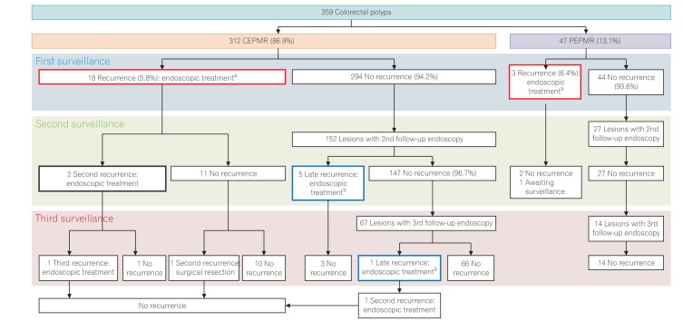
We retrospectively analyzed data regarding 359 lesions in 344 patients who underwent EPMR and who had at least 1 surveillance colonoscopy or sigmoidoscopy after the initial EPMR for a colorectal polyp of ≥10 mm diameter at the Asan Medical Center between January 2005 and December 2014 (Fig. 1). We searched for EPMR cases by searching for the term “piecemeal” in the colonoscopy report found with a search of the computerized database at our institution. After initial selection of EPMR cases by this system, we reviewed all the colonoscopy images of these patients and finally enrolled the EPMR cases in this study. We excluded patients with a positive deep resection margin. All procedures were performed by 8 experienced endoscopists and 15 trainee endoscopists using CEPMR or PEPMR methods. All of the 8 experienced endoscopists had >10 years of experience in diagnostic and therapeutic colonoscopies. They performed 94.9% of the CEPMR cases and 95.7% of the PEPMR cases. Cases in which the patient had undergone EPMR for subepithelial lesions, such as neuroendocrine tumor, leiomyoma, and granular cell tumor were excluded; the patients who had familial adenomatous polyposis or UC were also excluded. All patients provided written informed consent to undergo endoscopic resection of colorectal polyps. This study was approved by the Institutional Review Board of the Asan Medical Center, Seoul, Korea (IRB number: 2015-0954) and waived for informed consent.
2. EPMR Procedures
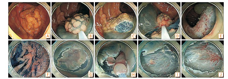
A single-channel endoscope was used to perform EPMR procedures. We used a mixed solution of normal saline with epinephrine (1:10,000) for submucosal injection. A small amount of indigo carmine was added to the solution to improve the visibility of the submucosal layer and polyp edges. For CEPMR, after submucosal injection, part of the polyp was snared and resected by electrical current. Repeat snaring and resection was performed until most of the lesion had been resected. If a small residual lesion was left, avulsion was performed with the use of hot biopsy forceps. If a tiny invisible residual was suspected because of multiple resections, APC or snare tip coagulation was used for fulguration. For PEPMR, a circumferential incision was made 2 to 4 mm from the edge of the polyp by electrical cutting with the snare tip or dual knife (Olympus Co., Tokyo, Japan) after submucosal injection. A snare was placed at the cut groove, and part of the polyp was grasped by squeezing the snare. Resection was then performed with the use of the electrosurgical current. Repeat snaring and resection were performed in a similar manner to CEPMR. Avulsion, APC, and snare tip coagulation were added in the same manner as CEPMR if they were needed. Fig. 2 shows representative figures of the CEPMR and PEPMR cases. ICC200 and VIO300D (ERBE, Tubingen, Germany) were used as electrosurgical units.
3. Surveillance Colonoscopy
All patients were advised to have periodic follow-up surveillance colonoscopies or sigmoidoscopies to evaluate local recurrence at the EPMR sites. The first surveillance colonoscopy or sigmoidoscopy was recommended to be at 3 to 12 months after the initial EPMR. The length of time to the second surveillance was determined based on the findings of the first surveillance endoscopy. If the first surveillance showed no recurrence, the second surveillance was recommended to be performed after 1 to 3 years at the discretion of the endoscopist, according to the number, size, and pathology of the polyps resected at the initial colonoscopy with EPMR. If the first surveillance showed local recurrence, endoscopic retreatment was attempted, and the next surveillance endoscopy was recommended again in 3 to 12 months.
4. Baseline Characteristics and Outcome Measurements
We investigated the baseline characteristics of the polyps, such as location, size, morphology, and presence or absence of previous biopsy before EPMR. The morphological types of colorectal polyps were classified as the laterally spreading tumor-granular type, the laterally spreading tumor-nongranular type, and the sessile/protruded type. The sessile/protruded type was a 0–Is polyp in the Paris classification. One of the complications, intraprocedural bleeding, was defined as bleeding that developed during the endoscopic resection, interrupted the endoscopic resection procedure, and finally required endoscopic hemostasis with the use of hemostatic forceps, APC, and/or clipping. Delayed bleeding was defined as hematochezia and/or melena that developed after the completion of the colonoscopy procedures and required endoscopic hemostasis. Delayed bleeding was divided into early (≤48 hours after EPMR) and late (>48 hours after EPMR) bleeding. Perforation was identified based on the endoscopic evidence of a definite mural defect with the visualization of intraperitoneal organs or periserosal fat tissue.
Resected specimens were retrieved and reconstructed to be as close to the original shape of the polyp as possible. They were then fixed in 10% formalin and microscopically evaluated. Histological diagnoses were made according to the Vienna classification.15 Histological complete resection was defined as the absence of tumor cells at the lateral and deep resection margins of the reconstructed specimen. The definition of a superficial submucosal cancer was a cancer with a depth of submucosal cancer invasion <1,000 µm from the muscularis mucosa.
During surveillance endoscopy, routine biopsy was not performed if the EPMR site showed a completely clear scar without any abnormality. Biopsy was performed if a visible recurrent lesion was detected at the scar or any suspicious abnormality was observed, even if it was subtle. Recurrence was defined as a histologically confirmed colorectal neoplasm that was detected on the scar of the previous EPMR site. Late recurrence was defined as local recurrence discovered at the subsequent colonoscopy approximately 12 months after the first surveillance colonoscopy in which the local recurrence was absent. If a recurrent lesion was detected, endoscopic treatment was attempted. The principal endoscopic treatment method for recurrences was EMR, and supplementary methods, such as cold biopsy, hot biopsy, and APC, were applied if necessary to completely remove all the recurrent lesions.
5. Statistical Analysis
Continuous variables were expressed as means with SD or medians with interquartile range (IQR), as appropriate. Continuous parameters were analyzed by the Student t-test or the Mann-Whitney U-test. Categorical variables were compared by the chi-square test or Fisher exact test, as appropriate. A logistic regression model was used for multivariate analysis of risk factors for recurrence. P-values of <0.05 were considered to indicate statistical significance. All statistical analyses were performed with SPSS version 21.0 for Windows (IBM Corp., Armonk, NY, USA).
RESULTS
1. Baseline Characteristics
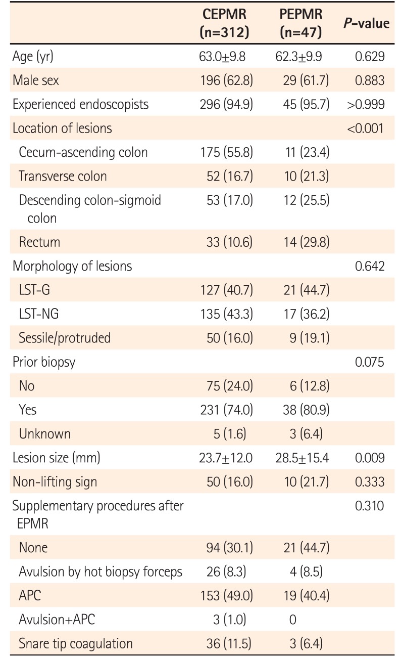
The baseline characteristics of the patients, polyps, and EPMR procedures are summarized in Table 1. Of 359 lesions in 344 patients, CEPMR was performed for 312 lesions (87%) and PEPMR was performed for 47 lesions (13%). The mean age of the patients was similar in the CEPMR and PEPMR groups. The morphological types of the lesions were not different in the 2 groups. However, the mean diameter of the tumors treated with PEPMR (28.5±15.4 mm) was larger than that of those treated with CEPMR (23.7±12.0 mm) (P=0.009).
2. Short-Term Outcomes after EPMR
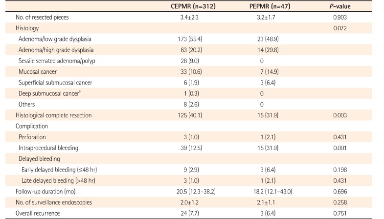
The details of the procedure-related variables and histological outcomes are described in Table 2. The average number of resected pieces per case was similar in the CEPMR and PEPMR groups. The distribution of histological diagnoses was also similar in both groups. Adenoma was the most common pathology in both groups. The histological complete resection rate was higher in the CEPMR group (40.1%) than in the PEPMR group (31.9%) (P=0.003). Intraprocedural bleeding was more frequent in the PEPMR group than in the CEPMR group (31.9% vs. 12.5%, P=0.001).
3. Long-Term Outcomes after EPMR
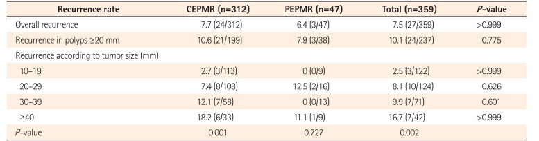
The median time to the first surveillance colonoscopy was 7.9 months (IQR, 4.9–12.8 months), and the overall duration of follow-up was 20.0 months (IQR, 12.3–38.3 months). The overall recurrence rate during the study period was 7.5% (27/359). The recurrence rates were not significantly different in the CEPMR and PEPMR groups (7.7% vs. 6.4%, P>0.751) (Table 2). Local recurrence at the first surveillance endoscopy was observed in 18 of 312 lesions (5.8%) after CEPMR and in 3 of 47 lesions (6.4%) after PEPMR. Among lesions that did not have a recurrence at the first surveillance colonoscopy, the rate of late recurrence on subsequent surveillance colonoscopy was 3.9% (6/152) after CEPMR and 0% after PEPMR (Fig. 1). The median time to late recurrence after CEPMR was 16.0 months (IQR, 14.5–20.6 months). Recurrence rates increased as lesion size increased in both groups (Table 3).
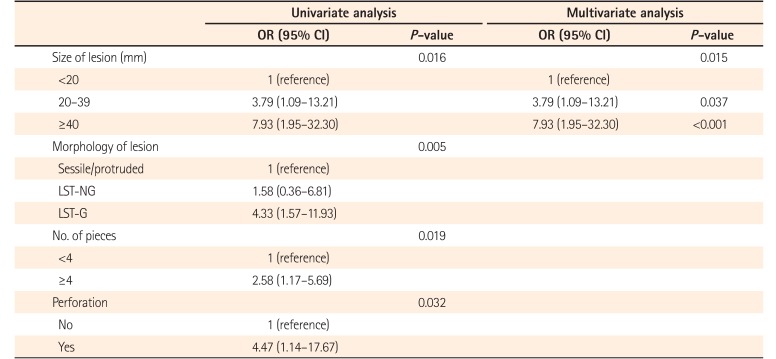
In the univariate analysis, local recurrence was associated with tumor size, tumor morphology, number of resected pieces, and perforation. However, multivariate analysis revealed that larger tumor size was the only independent risk factor for recurrence (Table 4).
4. Performance of Endoscopic Treatment for Recurrent Lesions
Among 27 recurrent tumors, 2 cases recurred as sessile serrated adenomas and the remaining 25 cases recurred as adenomas/villous adenomas. No case recurred as cancer after removal of an adenoma. Salvage endoscopic treatment was performed for all 21 recurrent lesions at the first surveillance endoscopy. Single endoscopic treatment methods, such as cold forceps polypectomy or EMR, were used in 3 recurrent lesions, and combination methods (EMR plus APC, EMR plus hot biopsy, EMR plus hot biopsy plus APC, cold forceps polypectomy plus APC, or hot biopsy plus APC) were used in 18 recurrent lesions. Of these 21 recurrent lesions, 15 underwent surveillance endoscopies after endoscopic treatment. Of the latter 15 lesions, 3 (20.0%) had a second recurrence at the subsequent surveillance endoscopy; 1 patient underwent surgical appendectomy because the recurrence was at the appendiceal orifice, and the other 2 underwent repeat endoscopic treatment.
Salvage endoscopic treatment was performed for all 6 late recurrences at the second or third surveillance endoscopy in cases without recurrence at the first surveillance endoscopy. A single endoscopic treatment modality was used in 5 late recurrences and a combination method in 1 late recurrence. Of these 6 patients with late recurrence, 5 underwent subsequent surveillance endoscopy and showed no further recurrence.
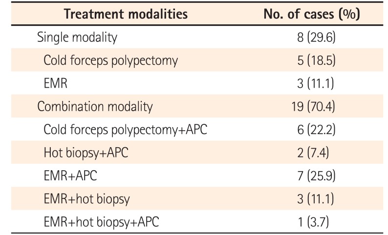
In summary, 21 lesions (5.8%) showed local recurrence at the first surveillance endoscopy, and 6 lesions (3.9%) showed late recurrence at subsequent surveillances, although they had not had recurrence at the first surveillance. All 27 recurrences were treated endoscopically. A single modality was used in 8 recurrences (29.6%), and combination methods were used in 19 recurrences (70.4%) (Table 5). Twenty of 27 recurrences were followed up by subsequent endoscopy. Of these, 1 (5.0%) was treated by surgical appendectomy because of re-recurrence at the appendiceal orifice, and the other 19 (95.0%) were cured by endoscopic treatment, although 3 of the 19 showed second or third recurrences, which were treated by repeat endoscopic treatment. A flowchart detailing recurrence and subsequent endoscopic treatment is presented in Fig. 1.
DISCUSSION
In our meticulous review, we found that the rates of recurrence after CEPMR and PEPMR were not different. Large tumor size increased the risk of local recurrence after EPMR. Recurrent lesions could be managed by endoscopic treatment in most cases, which was usually combination therapy rather than a single method.
Previous studies reported that the local recurrence rate after EPMR was 6.9% to 23.5%.1316171819 A recent meta-analysis showed that the recurrence rate after EPMR was 11% to 54% for lesions ≥20 mm and the pooled estimate of recurrence risk was 22%, compared with only 3% after en bloc EMR.18 In our study, the overall local recurrence rates of CEPMR and PEPMR were 7.7% (24/312) and 6.4% (3/47) for colorectal polyps ≥10 mm (P>0.999). For polyps ≥20 mm, the recurrence rates were 10.6% (21/199) after CEPMR and 7.9% (3/38) after PEPMR (P=0.775). The recurrence rates in our study are within the ranges of earlier reports. The rate of late recurrence at the second or third surveillance endoscopy without recurrence at the first surveillance was 3.9% (6/152) after CEPMR, whereas there was no late recurrence after PEPMR. The late recurrence rate after CEPMR in our study is also consistent with a previous study that showed a late recurrence rate of approximately 4%.12 An interesting point of our study is that there was no difference in recurrence rates after CEPMR and PEPMR. Although precut incision with a safety margin can guarantee a clear lateral resection margin and theoretically provide a higher complete resection rate than conventional EMR,1112 it cannot eliminate the risk of tumor remaining between the pieces after EPMR. The tumor remaining between the pieces may grow and lead to recurrent tumors detected at follow-up surveillance endoscopy. We suggest this may be one reason that the recurrence rates were not different between CEPMR and PEPMR. Another point worthy of note in our study is the lower histological complete resection rate (31.9%) after PEPMR compared with 40.1% after CEPMR. We expected a higher histological complete resection rate after PEPMR because of the precut incision with safety margin. However, the result was contrary to our hypothesis. We believe that the lower complete resection rate after PEPMR may be related to thermal injury of the resection margin during circumferential incision.11 Although the resection itself was complete, without involvement of the resection margins by tumor tissue, thermal injury may have made precise histological evaluation difficult and resulted in positive or indeterminate resection margins.
Larger tumor size (≥40 mm) was the only independent risk factor for local recurrence after EPMR, regardless of piecemeal resection methods. As the size of the polyp increases, the number of pieces may increase during EPMR. If the number of pieces increases, the risk of remnant tumor between pieces may increase, thereby increasing the risk of recurrence. Therefore, we suggest that adequate strategies to decrease the risk of recurrence should be considered after EPMR of large polyps, especially those of diameter ≥40 mm. Utilizing APC may be 1 option to decrease the risk of recurrence,2021 and routine application of APC may be considered after EPMR of polyps with ≥40 mm diameter. Avulsion of visible residual tumor with hot biopsy forceps may be another method to minimize the risk of recurrence after EPMR.22 In addition, we suggest that more careful endoscopic surveillance should be considered after EPMR of polyps with ≥40 mm diameter because of the higher risk of recurrence.
Although local recurrence rates were not low, endoscopic treatment ultimately provided a high cure rate of 95% (19/20), despite repeat recurrences in some cases. Only 1 case (5%) underwent surgical management because the recurrent lesion involved the appendiceal orifice and extended into the appendiceal lumen. Regarding the endoscopic treatment methods, 70.4% of all recurrences (19/27) were treated not by single endoscopic therapy but by combination therapy (EMR plus APC, EMR plus hot biopsy, EMR plus hot biopsy plus APC, cold forceps polypectomy plus APC, or hot biopsy plus APC). Because recurrent lesions after endoscopic resection, especially wide resection, such as EPMR of large polyps, may develop in the background of the scar tissue, the non-lifting sign may be present and endoscopic resection may be much more difficult than endoscopic resection of polyps with no previous attempt at resection.2123 Hence, a single endoscopic treatment method may not be sufficient for complete removal of the recurrent lesions on the scar.24 This may be a reason why combination therapy was used in most cases of recurrence. Even though a combination of methods was needed, endoscopic management was very effective for treatment of recurrent lesions after EPMR. Future studies should investigate the most useful endoscopic methods for treatment of recurrent lesions on the scar.
Complication rates after CEPMR and PEPMR were not different except for a higher rate of intraprocedural bleeding after PEPMR. Circumferential precutting by the snare tip or endo-knives may have been the reason for higher intraprocedural bleeding during PEPMR. Despite the absence of differences in delayed bleeding and other complications, considering the more frequent intraprocedural bleeding during PEPMR and the absence of difference in local recurrence rates between CEPMR and PEPMR, we suggest that PEPMR may not be recommended routinely for resection of large polyps and CEPMR should be the first choice. However, because of the theoretical advantage of minimal risk of slipping of the tumor out of the snare during closure of the snare, PEPMR may be indicated for resection of very flat tumors that are liable to slip out of the snare. Our study was a retrospective one, and we could not evaluate and analyze the usefulness of PEPMR from this viewpoint. Future studies are needed to clearly define the possible indications for PEPMR.
Our study had several limitations. First, because it was a retrospective study based on experience in a single center, selection bias related to the choice of PEPMR might have influenced the clinical outcomes and recurrence rates. Additionally, we searched for EPMR cases from the database of our institution by the computerized searching system using the term “piecemeal.” Because the searching system was not perfect, many EPMR cases may have been missed, which is another cause of selection bias of this study. The second limitation is the absence of a detailed analysis on the probable difference in the endoscopic skill between experienced and trainee endoscopists. Therefore, we do not know whether the expertise in endoscopic technique may have influenced the outcomes after EPMR. Third, because only half of the enrolled patients underwent a second surveillance, the local recurrence rates might be underestimated. Finally, the median follow-up period was <2 years. Despite these weaknesses, we believe that the present study is meaningful because, to the best of our knowledge, our investigation is the first study comparing clinical outcomes between CEPMR and PEPMR.
In conclusion, local recurrence rates were not different in CEPMR and PEPMR. Large tumor size was the only independent risk factor for local recurrence after EPMR. Endoscopic treatment for recurrent lesions resulted in a high cure rate, although the majority of recurrences required a combination of 2 or more endoscopic treatment methods.
 XML Download