

 PDF
PDF ePub
ePub Citation
 Print
Print


I. Introduction
Fracture of the zygomaticomaxillary complex (ZMC) is one of the most common facial injuries associated with mandibular fracture1,2. Treatment of ZMC injury has improved due to various reduction methods and development of miniplates and screws. The ZMC fracture treatment consists of the reduction and fixation of the dislocated bone fragments to their original location. Most ZMC bones are successfully repositioned with open reduction such as intraoral and transconjunctival approach or closed reduction such as Gillie's approach1.
Toriumi et al.3 have described complication of the ZMC injury such as asymmetry of zygoma, trismus, diplopia, exophthalmos, enophthalmos, limitation of eye movement and hypoesthesia due to injury of the infraorbital nerve. Reduction accuracy of fractured bone fragments were the major focus of most studies on facial asymmetry resulting from zygomatic asymmetry other than eye ball symptoms4,5. When bone fragments were precisely reduced, no asymmetry was reported1.
Ellis et al.6 reported that facial asymmetry which did not exceed 2 mm was difficult to perceive by experienced clinicians and most asymmetries after reduction of ZMC were acceptable. Unfortunately, the ZMC fracture pattern is diverse and soft tissue change after reduction surgery is difficult to predict. In addition, perceiving facial changes accurately is difficult due to the lack of information on patient's face before the injury. Gaziri et al.7 evaluated asymmetry of the hard tissue after ZMC fracture by comparing the affected with the unaffected side. Patients often complain of soft tissue asymmetry when comparing with the contralateral side of the face even when bone fragments were accurately reduced. Therefore, assessment of asymmetry should be based on the unaffected side of the face after surgery.
Regarding assessment of facial asymmetry, previous studies evaluated facial analysis methods using plain X-ray, 3-dimensional (3D) computed tomography (CT), cone-beam CT (CBCT) and laser scan8,9,10,11,12,13,14,15,16,17,18. However, most of the analyses were related with orthognathic surgery or craniofacial deformity. Evaluating asymmetry after reduction surgery where the distance of bone fragments was unplanned is difficult. Moreover, use of laser scan is limited due to high cost.
In this study, we assessed soft tissue asymmetry that occurred after open reduction of unilateral ZMC fractures. Soft tissue asymmetry of the affected side was quantitatively compared with the unaffected side using 3D CBCT scans. Tendency of soft tissue asymmetry after reduction was analyzed and usefulness of CBCT was also investigated. This study was authorized by the Wonkwang University Dental Hospital (WKDIRB 201404-01).
II. Materials and Methods
1. Subjects
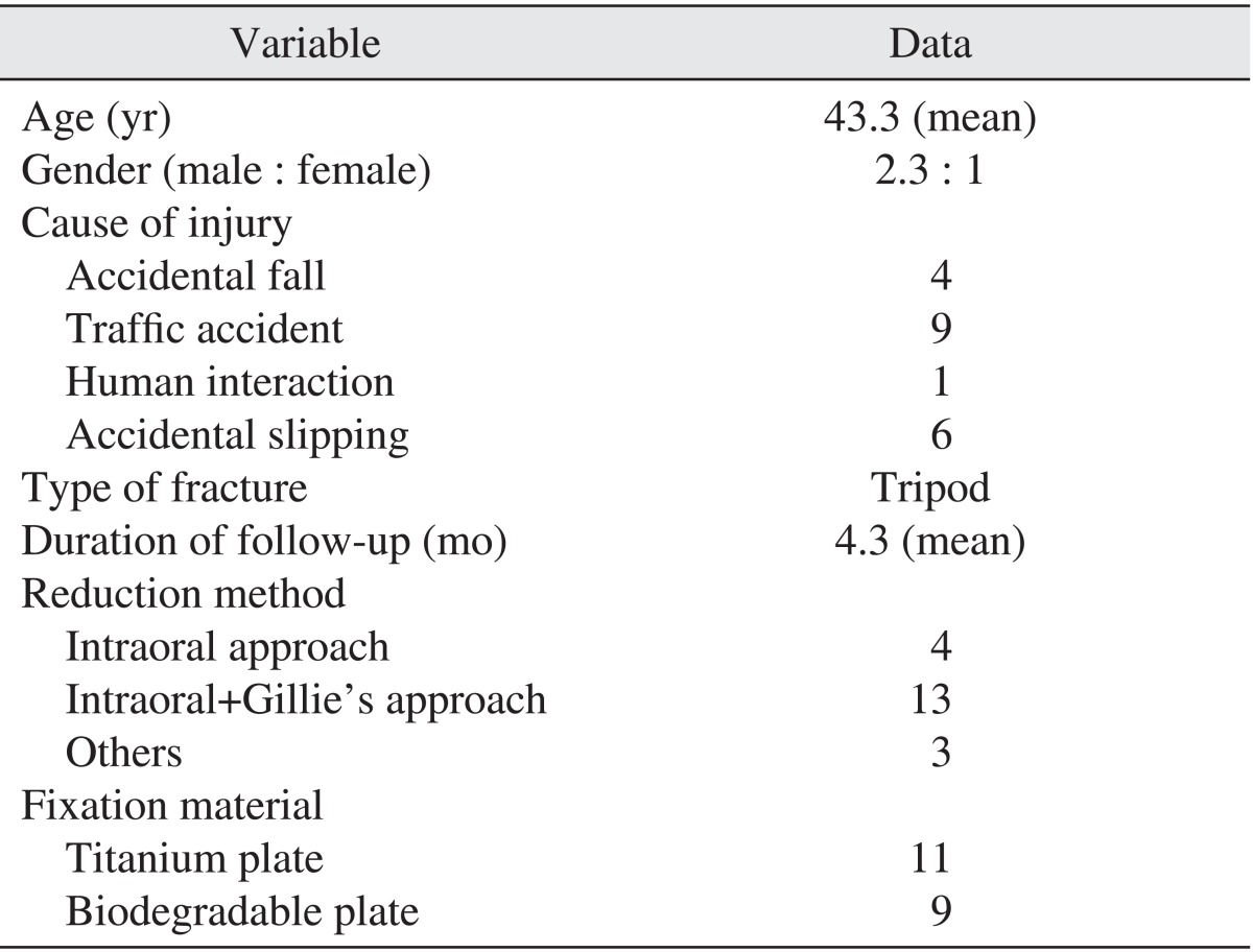
A total of 60 adults were chosen to participate in this study. The patient group consisted of 20 subjects who had open reduction surgery after being admitted to the emergency room of Wonkwang University Dental Hospital or medical hospital due to ZMC fracture. The control group comprised 40 healthy adults without any facial asymmetry. Subjects in the patient group had received a CBCT at least 3 months after 104the reduction surgery. Inclusion criteria for the patient group were lack of previous ZMC or maxilla fracture, the patient did not complain of asymmetry before the injury, diagnosis of unilateral ZMC fracture, and patient had open reduction surgery after the injury.
Inclusion criteria for the control group were absence of previous hard tissue surgery in the head and neck region and absence of facial asymmetry.
Intraoral, Gillie's and transconjunctival approaches were used for open reduction. Bone fragments were fixated with titanium and biodegradable plates. Fixation locations were zygomaticomaxillary buttress, frontozygomatic buttress and infraorbital rim.(Table 1)
2. Materials
1) Image acquisition
In this study, we used images obtained using the CBCT (The Alphad VEGA; Asahi Roentgen, Kyoto, Japan) system at Wonkwang University Dental Hospital. Images were taken under the following settings: field of view, 200×179 mm; 80 kV; 5.00 mA; exposure time, 17 seconds, voxel size, 0.39 mm; slice thickness, 1.00 mm.
2) Construction of 3D planes
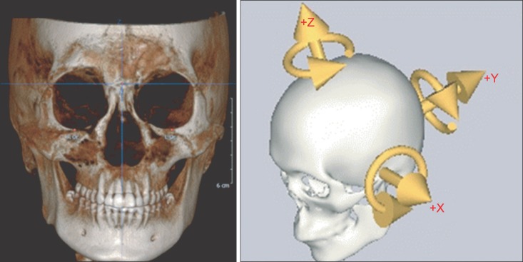
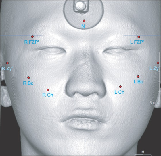
CT images were analyzed using 3D software (OnDemand3D; Cybermed Inc., Seoul, Korea) from DICOM format data. 3D coordinates were set up for each subject and cephalometric landmarks were designated. Nasofrontozygomatic (NFZ) plane was used as a reference plane of the skull base. NFZ plane was composed of the right and left frontozygomatic points and nasion. The coordinate origin (0, 0, 0) was set on N. Based on the origin, coordinates were constructed as x, y, z planes. X-axis (transverse axis) was a line parallel to the frontozygomatic (FZ) line. Y-axis (anteroposterior axis) was a line perpendicular to the FZ line and parallel to the right Frankfort horizontal (R FH) plane. The Z-axis was perpendicular to both FZ line and R FH plane. Using this coordinate system, three planes were defined. Midsagittal plane was defined as a plane perpendicular to R FH plane and NFZ line while passing through the origin. Horizontal plane (Frankfort plane) was defined as a plane passing through right porion (R Po), right orbitale (R Or), and left orbitale (L Or). Coronal plane was defined as a plane perpendicular to horizontal and midsagittal planes and passing through the origin.(Fig. 1)
3. Methods
To compare the amount of asymmetry between healthy adults and patients who underwent ZMC reduction, distance from landmarks on the right and left soft tissues to midsagittal and coronal planes were measured in both groups. The degree of asymmetry after ZMC surgery was assessed by comparing measurements between the two groups. Soft and hard tissues were analyzed with landmarks used in previous studies on midfacial asymmetry10,11,15. Next, factors affecting soft tissue convexity such as reduction techniques, presence of comminuted fractures, amount of existing deviation and fixation materials were evaluated. Relationship between subjective perception and actual amount of asymmetry was statistically examined.
1) Hard tissue landmarks
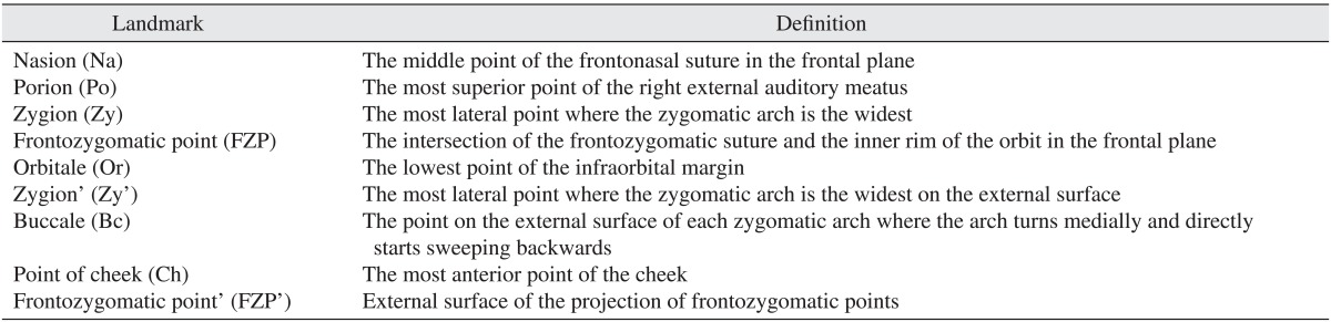
Nasion (Na), Or, Po, zygion (Zy) and frontozygomatic point (FZP) were used as hard tissue landmarks.(Table 2)
3) Soft tissue distance measuring
Zygomatic width was measured from midsagittal plane to Zy'. Zygoma and cheek projection were measured from the coronal plane to Bc, Ch and FZP'. In the control group, difference between right and left measurements was used. In the patient group, measurement differences between the unaffected and affected areas were used.
4) Hard tissue reduction accuracy
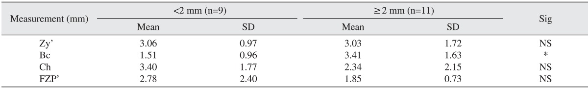
Reduction amounts of bone fragments were measured at four buttresses where the ZMC fracture occurred (zygomaticofrontal suture deviation, anterior wall of maxilla, zygomatic arch and infraorbital rim). The patient group was divided based on the amount of deviation (2 mm or more, less than 2 mm). Less than 2 mm of deviation was considered as the gold standard for hard tissue reduction.
5) Statistical analysis
Preparation and measurement of all coordinates were repeated again at 2 weeks by the same investigator to prevent intra-observer error. Intra-observer error between the 2 measurements was verified with paired t-test and no significant difference was found (P>0.05). The first measurements were used in this study.
Independent t-test and analysis of variance (ANOVA) were performed to assess significant difference between the measurements. Statistical analysis was conducted using SPSS software (SPSS version 12.0; SPSS Inc., Chicago, IL, USA) with a 95% reliability.
III. Results
1. Amount of asymmetry in control and patient groups
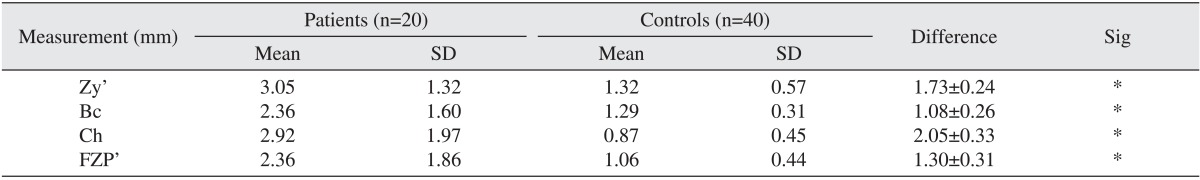
The average amount of asymmetry in the control group was less than 2 mm (Zy': 1.31±0.57 mm, Bc: 1.28±0.30 mm), Ch: 0.87±0.45 mm, FZP: 1.06±0.44 mm). Asymmetry greater than 2 mm was observed in the patient group (Zy': 3.05±1.32 mm, Bc: 2.36±1.60 mm, Ch: 2.92±1.97 mm, FZP': 2.36±1.86 mm). The measured symmetry amounts in the patient group were statistically significantly different from the control group (P<0.05).(Table 3)
2. Comparison among ZMC reduction patients
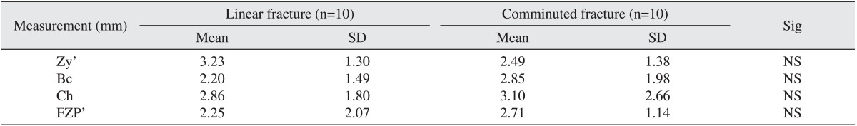
1) Effect of comminuted fractures
When a comminuted fracture of the maxilla was present, the amount of asymmetry was increased when compared with a linear fracture.(Table 4) However, soft tissue asymmetry was not statistically significantly different regarding the presence or absence of comminuted fracture (P<0.05).
2) Effect of the amount of deviation after reduction
When hard tissue deviation was 2 mm or greater, soft tissue depression was observed on Bc (3.41±1.63 mm; Table 5) and the depression was statistically significant (P<0.05).
3) Effect of fixation materials (titanium or biodegradable)
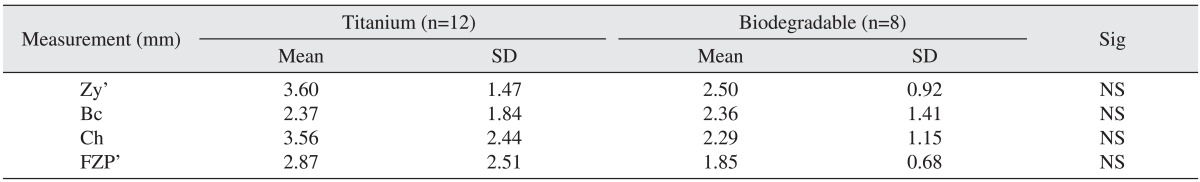
Titanium and biodegradable plates were used in 10 patients, separately. Soft tissue depression was more pronounced at Zy' and Ch' in the titanium plate group (Table 6); however, no statistically significant difference was found (P<0.05).
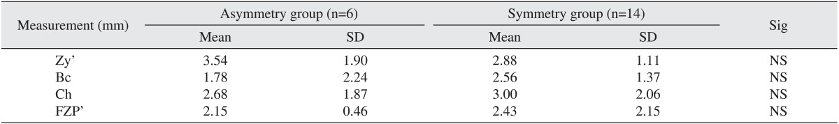
4) Effect of asymmetry perception after reduction surgery
Soft tissue depression was observed irrespective of asymmetry perception after reduction surgery.(Table 7)
IV. Discussion
Various analysis methods using 3D CT scans have been introduced to evaluate soft tissue change after surgery8,9,10,11,12,13,14,15,16,17,18 and are useful despite possible image errors19,20. Most of these studies focused on assessment of soft tissue change after orthognathic surgery or surgery for correction of craniofacial deformity. Evaluation of soft tissue change after ZMC fracture reduction surgery employing the same principles used in those studies is difficult. Recently, laser scans are gaining popularity to evaluate facial soft tissue. However, the high cost of laser scans can be a limiting factor. In this study, soft tissue was assessed using CBCT images with OnDemand3D software and relatively accurate results were obtained.
Furst and colleagues reported no statistically significant difference between ZMC CT images and dry skull21. Gwilliam et al.22 reported that cephalometric landmarks on the soft tissue were statistically reproducible. The present study also revealed no statistically significant difference when the measurements were repeated after 2 weeks to avoid intra-observer errors (P<0.05). 3D planes for image analysis were constructed using the same method described by Cho23.
CBCT images used in this study were taken after at least 3 months of follow-up. Modabber et al.24 investigated swelling after ZMC fracture reduction according to two different cooling therapy methods. They reported that swelling was the most severe 3 days after the surgery and no statistically significant difference was found between the two groups 28 days after the surgery. Generally soft tissue assessed at 6 months after surgery in orthognathic surgery is considered a relapse. This study evaluated CBCT images taken 3 months after surgery to minimize the effects of postoperative swelling and relapse was not considered because rigid fixation was used.
Hwang et al.25 evaluated asymmetry in adults with normal occlusion and reported a difference in distance from the midsagittal plane to right and left bilateral landmarks such as Bc, Ch and Zy' (Bc: 1.43±1.06 mm, Ch: 0.85±0.69 mm); similar results were obtained in our study for normal adults (Zy': 1.32±0.57 mm, Bc: 1.29±0.31 mm, Ch: 0.87±0.45 mm, FZP': 1.06±0.44 mm).(Table 3)
A variety of studies have investigated the accuracy of ZMC reduction surgery on the hard tissue and the majority reported satisfactory results26,27. However, patients perceive facial asymmetry in their soft tissue. In the present study, asymmetry was assessed on the soft tissue and the patient group showed statistically significantly more pronounced facial asymmetry than the control group (P<0.05). Soft tissue asymmetry is affected by soft tissue change after trauma or surgery, inappropriate reduction of bony fragments, and stability of bony fragments after the surgery.
Presence of comminuted maxilla fracture did not affect the amount of asymmetry on each landmark in a statistically significant manner. According to a study by Klotch and Gilliland28, facial expression muscles were detached from hard tissue and fibrosis occurred at an abnormal location if open reduction was not performed when a maxillary comminuted fracture occurred leading to inappropriate soft tissue contour. They recommended immediate reconstruction of hard tissue and best results were obtained when open reduction surgery was performed within 48 hours. Their recommendation is in contrast to conventional treatment of closed reduction to prevent demineralization of bone tissue when mandibular comminuted fracture occurs29. In the present study, open reduction was immediately performed when the comminuted fracture occurred. Slight soft tissue depression was observed compared with the control group. However, no significant difference was found when compared with cases where a comminuted fracture did not occur. Immediate open reduction is required in the maxilla for esthetic soft tissue contour because the maxilla has ample blood supply.
When soft tissue asymmetry was compared according to the amount of deviation after the reduction surgery, soft tissue was more conspicuous on the landmark Bc when hard tissue deviation exceeded 2 mm after surgery (Bc: 3.41±1.63 mm). Statistically significant difference was found (P<0.05). Soft tissue depression around zygoma would be more noticeable if reduction was not accurate. The majority of previous studies reported that hard tissue asymmetry less than 2 mm was not perceivable30,31; in the present study, patients with less than 2 mm deviation did not complain of asymmetry. Location of the landmarks in patients with less than 2 mm of deviation was not considerably different from patients with 2 mm or greater deviation. The amount of deviation in the patient group where deviation was 2 mm or greater ranged from 2 mm to 3.78 mm indicating that approximately 3 mm of soft tissue asymmetry can be expected if reduction is not precisely performed.
With respect to plate materials, titanium and biodegradable plates were used for 10 patients, separately. Soft tissue depression was more noticeable on Zy' and Ch when titanium was used in comparison to biodegradable plates (P<0.05) because titanium plates were used when the ZMC fracture dislocation amount was larger. Previous studies investigated tissue response to fixation materials. Wittwer et al.32 reported that a biodegradable plate achieved satisfactory fixation effects due to improvements in physical properties and reported induced inflammatory reaction or tissue response against soft tissue. Langford and Frame33 stated no evidence was found indicating titanium induced soft tissue inflammation. Accordingly, asymmetry on Ch was less prominent when a biodegradable plate was used because soft tissue response caused swelling and increased soft tissue volume when compared with titanium.
Irrespective of perception of asymmetry, soft tissue depression was observed after surgery. The average location of right and left landmarks was not statistically significantly different according to asymmetry complaint (P<0.05). In the present study, we evaluated the effects of surgical techniques, presence of comminuted fracture, fixation materials and accuracy of soft tissue contour reduction after ZMC fracture reduction surgery. In addition to those factors, presence of existing asymmetry and masseter muscle and facial expression muscles attached to the zygoma can also influence soft tissue contour34. In this study, five patients complained of asymmetry and their average deviation on Zy' was significantly different from patients who did not complain, indicating asymmetry caused by lateral zygoma protrusion was more easily perceived than asymmetry caused by posteroanterior zygoma protrusion. A study by Choi et al.35 and Zheng et al.36 also reported that people perceived reduced lateral zygoma protrusion as more esthetic.
Asymmetry in the surgery group was generally more pronounced than in the control group. Postoperative soft tissue asymmetry was observed even when reduction was less than 2 mm. The amount of asymmetry was not considerably different regardless if the patient complained. These findings can be used in clinics when informing patients that asymmetry is inevitable irrespective of reduction accuracy.
V. Conclusion
The amount of soft tissue asymmetry after open reduction of ZMC fracture was evaluated with CBCT scans taken 3-6 months postoperatively to exclude effects of postoperative swelling. OnDemand3D software was used and the following conclusions were reached;
Asymmetry was increased in the patient group with statistical significance (P<0.05) when compared with the control group.
Amount of protrusion on Bc was influenced by the amount of hard tissue dislocation after surgery.
Soft tissue contour was not affected by fixation materials.
Degree of asymmetry was not statistically significantly different (P<0.05) regardless of whether patients complained of asymmetry.
Soft tissue asymmetry occurs after open reduction surgery and various factors are associated. Asymmetry of ZMC fracture patients after surgery was evaluated using CBCT.
 XML Download