Journal List > J Korean Assoc Oral Maxillofac Surg > v.37(4) > 1032475

Lee and Kim: Open reduction of mandibular fracture without maxillomandibular fixation: retrospective study

Abstract

Introduction

Maxillomandibular fixation (MMF) is essential before surgery under general anesthesia in maxillofacial trauma patients. MMF is usedbasically to reconstruct the occlusion and occlusal stability to recover the facial shape and oral functions. The arch bar and wire is a traditional method for MMF, but it can not only bring pressure to the periodontal ligaments and teeth but also cause a penetrating injury to the surgeons.

Materials and Methods

In this study, 198 patients with an open reduction using a manual reduction without MMF from September 2005 to May 2010 in Dankook University Dental Hospital were subjected to a followup evaluation during the postoperative 4 months periods. This study evaluated the incidence of complications according to the condition of the patient (gender, age), the state of bony union of the fracture sites and a numeric ratingscale evaluation for postoperative pain scoring.

Results

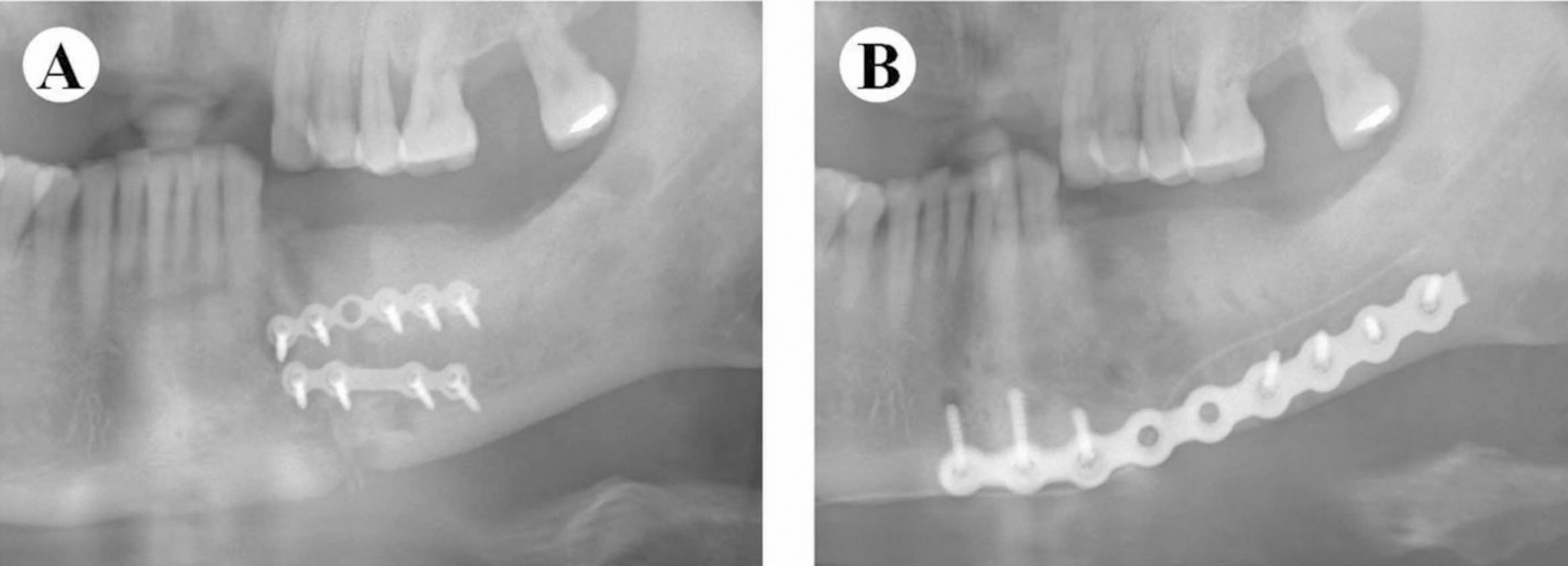
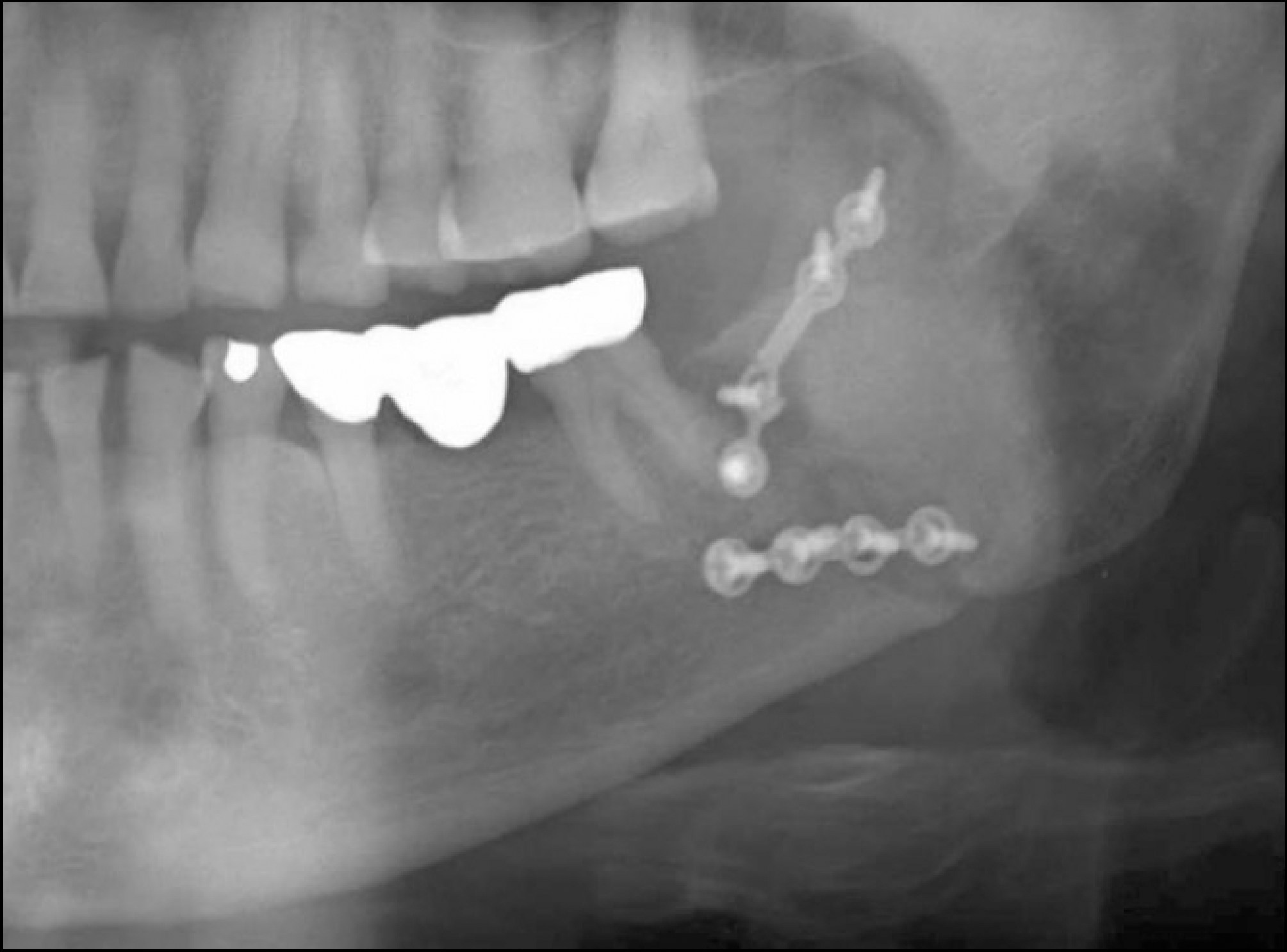
  1. The complications were classified into major and minor according to the seriousness, and the major complication rate was as low as 2.02%. Only 2 cases of re-operations (1.01%) were encountered. In the classification according to the fracture line, plate fracture was observed in both cases of mandibular symphysis fracture, and angle fractures and loosening of two screws were noted in the case of mandibular angle fracture.

  2. The complication rate was similar regardless of gender and age.

  3. The degree of bony union was satisfactory, and the complication rate was reduced as the bony union improved.

  4. More patients complained of pain as the operation time was increased.

Conclusion

The use of MMF is not always necessary if a skilled assistant is provided to help manually reduce the fracture site. Compared to otherstudies of mandibular fracture surgery using MMF, the complication rate was similar using only manual reduction and the patients' discomfort was reduced without MMF.

REFERENCES

1. Turvey TA. Midfacial fractures: a retrospective analysis of 593 cases. J Oral Surg. 1977; 35:887–91.
2. Ugboko VI, Oginni FO, Owotade FJ. An investigation into the relationship between mandibular third molars and angle fractures in Nigerians. Br J Oral Maxillofac Surg. 2000; 38:427–9.
crossref
3. Fridrich KL, Pena-Velasco G, Olson RA. Changing trends with mandibular fractures: a review of 1,067 cases. J Oral Maxillofac Surg. 1992; 50:586–9.
crossref
4. Kawai T, Murakami S, Hiranuma H, Sakuda M. Radiographic changes during bone healing after mandibular fractures. Br J Oral Maxillofac Surg. 1997; 35:312–8.
crossref
5. Horan TC, Gaynes RP, Martone WJ, Jarvis WR, Emori TG. CDC definitions of nosocomial surgical site infections, 1992: a modification of CDC definitions of surgical wound infections. Infect Control Hosp Epidemiol. 1992; 13:606–8.
crossref
6. Fordyce AM, Lalani Z, Songra AK, Hildreth AJ, Carton AT, Hawkesford JE. Intermaxillary fixation is not usually necessary to reduce mandibular fractures. Br J Oral Maxillofac Surg. 1999; 37:52–7.
crossref
7. Bell RB, Wilson DM. Is the use of arch bars or interdental wire fixation necessary for successful outcomes in the open reduction and internal fixation of mandibular angle fractures? J Oral Maxillofac Surg. 2008; 66:2116–22.
crossref
8. Coletti DP, Salama A, Caccamese JF Jr. Application of intermaxillary fixation screws in maxillofacial trauma. J Oral Maxillofac Surg. 2007; 65:1746–50.
crossref
9. Gaujac C, Ceccheti MM, Yonezaki F, Garcia IR Jr, Peres MP. Comparative analysis of 2 techniques of double-gloving protection during arch bar placement for intermaxillary fixation. J Oral Maxillofac Surg. 2007; 65:1922–5.
crossref
10. Williams JG, Cawood JI. Effect of intermaxillary fixation on pulmonary function. Int J Oral Maxillofac Surg. 1990; 19:76–8.
crossref
11. Roccia F, Tavolaccini A, Dell'Acqua A, Fasolis M. An audit of mandibular fractures treated by intermaxillary fixation using intraoral cortical bone screws. J Craniomaxillofac Surg. 2005; 33:251–4.
crossref
12. Gordon KF, Reed JM, Anand VK. Results of intraoral cortical bone screw fixation technique for mandibular fractures. Otolaryngol Head Neck Surg. 1995; 113:248–52.
crossref
13. Coburn DG, Kennedy DW, Hodder SC. Complications with intermaxillary fixation screws in the management of fractured mandibles. Br J Oral Maxillofac Surg. 2002; 40:241–3.
crossref
14. Ellis E 3rd, Walker LR. Treatment of mandibular angle fractures using one noncompression miniplate. J Oral Maxillofac Surg. 1996; 54:864–71.
crossref
15. Dimitroulis G. Management of fractured mandibles without the use of intermaxillary wire fixation. J Oral Maxillofac Surg. 2002; 60:1435–8.
crossref
16. Laurentjoye M, Majoufre-Lefebvre C, Caix P, Siberchicot F, Ricard AS. Treatment of mandibular fractures with Michelet technique: manual fracture reduction without arch bars. J Oral Maxillofac Surg. 2009; 67:2374–9.
crossref
17. Jeong CH, Jeong JW, Kwon ST. The siginificance of radiographic followup of mandibular fractures. J Korean Soc Plast Reconstr Surg. 1998; 25(5):860–865.
18. Durham JA, Paterson AW, Pierse P, Adams JR, Clark M, Hierons R, et al. Postoperative radiographs after open reduction and internal fixation of the mandible: are they useful? Br J Oral Maxillofac Surg. 2006; 44:279–82.
crossref
19. Sim SY, Park HJ, Lee JM, Lee HS. An overview of pain measurements. Korean J. Meridian Acu. 2007; 24:77–97.
20. White A. Measuring pain. Acupunct Med. 1998; 16:83–7.
crossref
21. Maloney PL, Lincoln RE, Coyne CP. A protocol for the management of compound mandibular fractures based on the time from injury to treatment. J Oral Maxillofac Surg. 2001; 59:879–84.
crossref
22. Anderson T, Alpert B. Experience with rigid fixation of mandibular fractures and immediate function. J Oral Maxillofac Surg. 1992; 50:555–60.
crossref
23. The Korean Association of Oral and Maxillofacial Surgeons. Textbook of oral and maxillofacial surgery. 2nd ed.Seoul: Dental & Medical Publishing Co.;2005.

Fig. 1.
Open reduction of mandibular symphysis fracture using manual reduction without arch bar. A. Preoperative occlusion B. Preoperative computed tomography, C. Intraoperative photograph showing repair of fracture, D. Postoperative radiograp after fixation.
jkaoms-37-255f1.tif
Fig. 2.
Plate fracture in panorama radiograph.
jkaoms-37-255f2.tif
Fig. 3.
A case of delayed union after operation. A. Delayed union in panorama radiograph after 4 months, B. Partially bony union after 1 year without re-operation.
jkaoms-37-255f3.tif
Fig. 4.
Re-operation in a case of osteomyelitis. A. Panorama radiograph after 3 months, B. Re-operation by using reconstruction plate.
jkaoms-37-255f4.tif
Table 1.
Patient characteristics included in this study
Variables No. of patients (%)
Male/female 146/52
Mean age (range) 32.3 (15–69)
Mechanism of fracture  
 Assault 44 (22.2)
 Motor vehicle accident 34 (17.2)
 Fall 76 (38.4)
 Sports 17 (8.6)
 Work 16 (8.1)
 Miscellaneous 11 (5.6)
Outcome of teeth in fracture line  
 Teeth along fracture line 49 (24.7)
 Impacted teeth along fracture line 66 (33.3)
 Delay to surgery (after 3 days) 60 (30.3)
Table 2.
Patient distribution according to fracture types
Fracture type No. of patients (%)
Unilateral subcondyle 16 (8.1)
Subcondyle and symphysis 28 (14.1)
Subcondyle and angle 10 (5.1)
Unilateral body 8 (4.0)
Body and angle 7 (3.5)
Body and symphysis 8 (4.0)
Bilateral body 4 (2.0)
Unilateral angle 43 (21.7)
Angle and symphysis 35 (17.7)
Bilateral angle 8 (4.0)
Symphysis 31 (15.7)
Total 198 (100.0)
Table 3.
Fracture line distribution according to fracture sites
Fracture site No. of fracture Line (%)
Symphysis 102 (34.0)
Body 31 (10.3)
Angle 113 (37.7)
Subcondyle 54 (18.0)
Total 300 (100.0)
Table 4.
Kawai classification according to bone healing pattern
Kawai Classification
Grade 1: No radiograhic changes
Grade 2: Bone resorption was observed at the fracture site
Grade 3: External or internal callus formation (Osteogenesis)
Grade 4: Bony union was observed
Table 5.
Definition of superficial surgical site infection in CDC
CDC definition of superficial surgical site infection
Infection occurs within 30 days of procedure
Involves only skin or subcutaneous tissue around the incision and at least one of the following:
  • Purulent drainage from the superficial incision

  • Organisms isolated from an aseptically obtained culture of fluid or tissue from the superficial incision

  • At least one of the following signs or symptoms of infection: pain or tenderness, localised swelling, redness, or heat and superficial incision is deliberately opened by surgeon, unless culture of incision is negative

CDC: Centers for Disease Control)

Table 6.
Patient distribution according to complications
Complication (n=22) No. of patients Re-operation case
Normal result 176
Wound dehiscence 4
Infection 5
Trismus 3
Malocclusion 3
Screw loosening 1
Plate fracture 2
Osteomyelitis 2 1
Delayed union 1
Nonunion 1 1
Total 198 2
Table 7.
Complication rate according to gender
Gender No. of patients (%) Complication (%) P value
Male 146 (73.7) 17 (11.6) 0.689
Female 52 (26.3) 5 (9.6)  
Table 8.
Complication rate according to age
Age (yr) No. of patients (%) Complication (%) P value
11–20 46 (23.2) 2 (4.3)  
21–30 66 (33.3) 6 (9.1)  
31–40 34 (17.2) 4 (11.8)  
41–50 24 (12.1) 4 (16.7) 0.271
51–60 17 (8.6) 4 (23.5)  
61–70 11 (5.6) 2 (18.2)  
Total 198 (100.0) 22 (11.1)  
Table 9.
Plate & screw distribution and grade∗ based on radiographic examination according to fracture site
Fracture site Total plates Total screws No. of plate fracture No. of screw loosening Grade Complication (%)
Symphysis 202 949 1 0 3.84±0.39 5 (4.9)
Body 59 284 0 0 3.68±0.79 4 (12.9)
Angle 214 884 1 2 3.87±0.41 9 (8)
Subcondyle 101 391 0 0 3.80±0.45 4 (7.4)
Total 576 2508 2 2 3.83±0.47 22 (7.3)

grade∗: Kawai classification according to bone healing pattern. Data is presented by means±SD.

Table 10.
Correlations of grade∗, time to surgery, OP time, NRS with complication
Spearman test Grade∗ Time to surgery OP time NRS
Immediate post-OP Discharge
Complication Correlation efficient (r) −0.249 −0.114 0.033 0.035 0.048
P value 0 0.094 0.623 0.603 0.481

(OP: operation, NRS: numeric rating scale)

grade∗: Kawai classification according to bone healing pattern.

Table 11.
Complication rate according to time to repair, OP time, NRS
Fracture type Time to surgery (day) OP time (min) NRS Complication (%)
Immediate post-OP Discharge
Unilateral subcondyle 2.63±1.96 79.06±23.96 6.81±0.98 1.63±1.26 1 (6.3)
Subcondyle and symphysis 3.18±1.72 104.11±34.45 6.57±1.32 2.43±1.20 4 (14.3)
Subcondyle and angle 3.50±2.17 137.70±16.15 6.40±1.90 2.10±0.74 1 (10.0)
Unilateral body 3.00±2.73 60.63±14.50 6.38±1.19 2.25±1.04 1 (12.5)
Body and angle 2.86±2.12 78.57±16.00 6.29±1.98 2.14±0.69 2 (28.6)
Body and symphysis 2.38±0.74 60.25±21.19 6.38±0.52 2.38±0.52 0 (0.0)
Bilateral body 1.75±0.96 75.00±15.81 6.75±0.96 1.75±0.50 1 (25.0)
Unilateral angle 2.51±1.42 44.02±17.10 5.42±1.26 1.33±0.57 5 (11.6)
Angle and symphysis 2.31±1.66 65.34±10.60 5.63±0.84 1.69±1.05 5 (14.3)
Bilateral angle 2.88±2.36 131.88±13.35 5.38±1.60 1.50±0.53 1 (12.5)
Symphysis 2.90±1.68 36.94±14.53 5.23±1.15 1.58±0.96 1 (3.2)
Total 2.72±1.74 69.46±35.60 5.88±1.32 1.77±0.98 22 (11.1)

(OP: operation, NRS: numeric rating scale)

Data is presented by means±SD.

Table 12.
Correlations of grade∗, time to surgery, NRS with OP time
Spearman test Grade∗ Time to surgery NRS
Immediate post-OP Discharge
OP time Correlation efficient (r) −0.093 0.066 0.283 0.199
P value 0.172 0.335 0.000 0.003

(NRS: numeric rating scale, OP: operation)

grade∗: Kawai classification according to bone healing pattern.

TOOLS
Similar articles