

 PDF
PDF ePub
ePub Citation
 Print
Print


Abstract
Plasmacytoma is a rare malignant neoplasm in the head and neck region and comprises approximately 3% of all plasma cell tumors. This lesion is a unifocal, monoclonal, neoplastic proliferation of plasma cells that usually arises within the bone. Infrequently, it is observed in soft tissue, in which case, the term extramedullary plasmacytoma is used. Approximately 80-90% of extramedullary plasmacytomas involve the mucos-Associated-Lymphoid Tissue of the upper airways with 75% of these involving the nasal and paranasal regions. The plasmacytoma is usually detected in adult males, with an average age at diagnosis of 55 years. The male-to-female ratio is 3:1. Radiographically, the lesion may be seen as a well-defined, unilocularradioluceny with no evidence of a sclerotic border. Some investigators believe that this lesion represents the least aggressive part of the spectrum of plasma cell neoplasms that extend to multiple myeloma. Therefore, plasma cytoma is believed to have clinical importance. We report a case of extramedullary plasmacytoma in the right maxillary sinus of a 59-year-old male with review of the relevant literature.
REFERENCES
1. Attanasio G, Viccaro M, Barbaro M, de Seta E, Filipo R. Extramedullary plasmacytoma of paranasal sinuses. A combined therapeutic strategy. Acta Otorhinolaryngol Ital. 2006; 26:118–20. discussion 120.
2. Cumbo V, Gallina G, Messina P. Extramedullary plasmacytoma of the maxillary sinus: report of a case. J Oral Maxillofac Surg. 1988; 46:406–9.

3. Majumdar S, Raghavan U, Jones NS. Solitary plasmacytoma and extramedullary plasmacytoma of the paranasal sinuses and soft palate. J Laryngol Otol. 2002; 116:962–5.
4. Bruns F, Janssen S, Laenger F, Dobbelstein C, Meyer A. Extramedullary plasmocytoma: a rare case with bifocal manifestation at uncommon sites. Anticancer Res. 2010; 30:1779–81.
5. Gross M, Eliashar R, Maly B, Sichel JY. Maxillary sinus plasmacytoma. Isr Med Assoc J. 2004; 6:119–20.
6. Tamada A, Araki T, Makimoto K, Takatsuki K. Plasmacytoma in nasal cavity and maxillary sinus. Arch Otorhinolaryngol. 1982; 237:83–91.
7. Megat Shiraz MA, Jong YH, Primuharsa Putra SH. Extramedullary plasmacytoma in the maxillary sinus. Singapore Med J. 2008; 49:e310–1.
8. Greenberg P, Parker RG, Fu YS, Abemayor E. The treatment of solitary plasmacytoma of bone and extramedullary plasmacytoma. Am J Clin Oncol. 1987; 10:199–204.
9. Jaffe ES, Harris NL, Stein H, Vardiman JW. World Health Organization Classification of tumours: pathology and genetics of tumours of haematopoietic and lymphoid tissues. Lyon: International Agency for Research on Cancer (IARC) Press;2001.
10. Barnes L. Surgical pathology of the head and neck. 2nd ed.New York: Marcel Dekker;2000.
11. Michalaki VJ, Hall J, Henk JM, Nutting CM, Harrington KJ. Definitive radiotherapy for extramedullary plasmacytomas of the head and neck. Br J Radiol. 2003; 76:738–41.
12. Lomeo PE, McDonald JE, Finneman J, Shoreline . Extramedullary plasmacytoma of the nasal sinus cavities. Am J Otolaryngol. 2007; 28:50–1.
13. Soutar R, Lucraft H, Jackson G, Reece A, Bird J, Low E, Samson D. Guidelines on the diagnosis and management of solitary plasmacytoma of bone and solitary extramedullary plasmacytoma. Br J Haematol. 2004; 124:717–726.
14. Katodritou E, Speletas M, Pouli A, Tsitouridis J, Zervas K, Terpos E. Successful treatment of extramedullary plasmacytoma of the cavernous sinus using a combination of intermediate dose of thalidomide and dexamethasone. Acta Haematol. 2007; 117:20–3.
15. Tsang RW, Gospodarowicz MK, Pintilie M, Bezjak A, Wells W, Hodgson DC, et al. Solitary plasmacytoma treated with radiotherapy: impact of tumor size on outcome. Int J Radiat Oncol Biol Phys. 2001; 50:113–20.
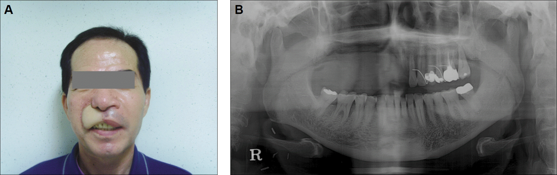
Fig. 1.
Extraoral view showing facial swelling on right buccal cheek. A: Frontal view, B: Lateral view.
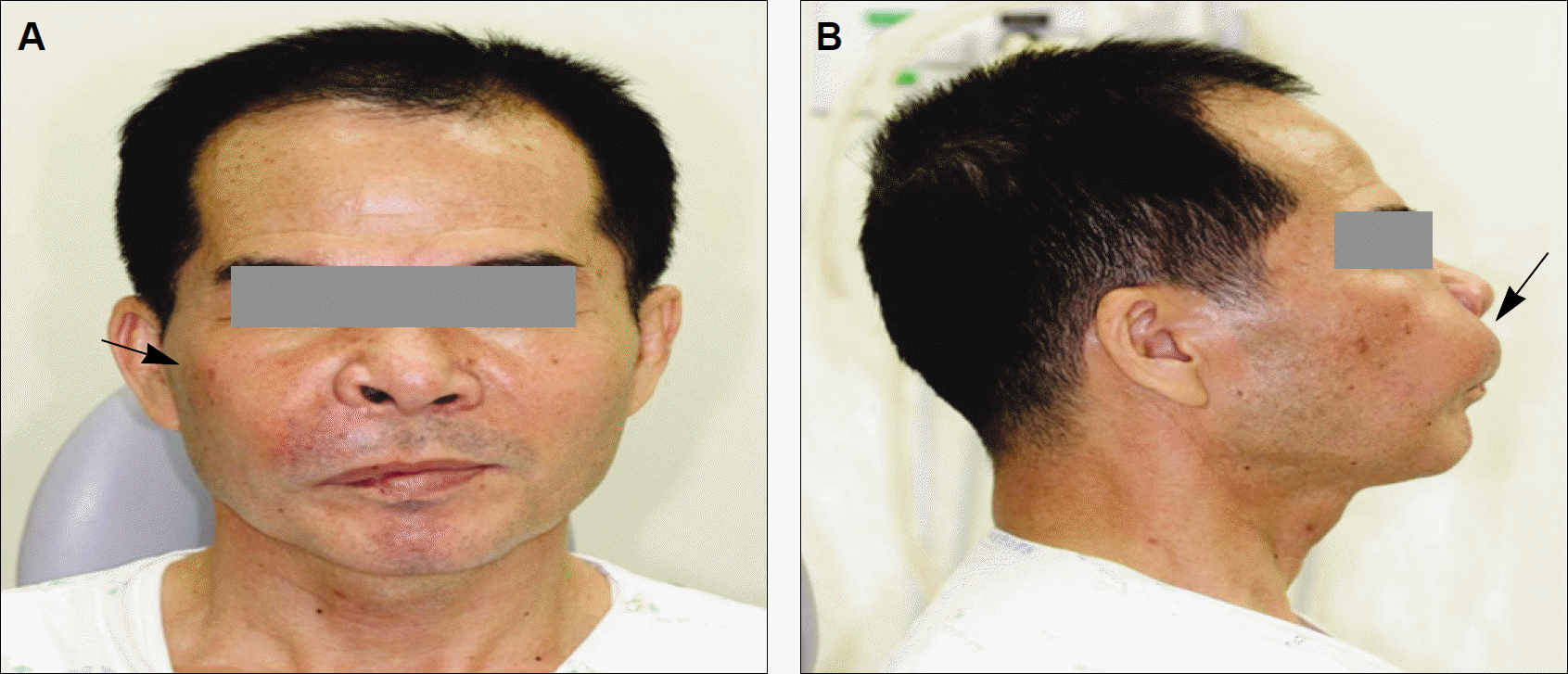
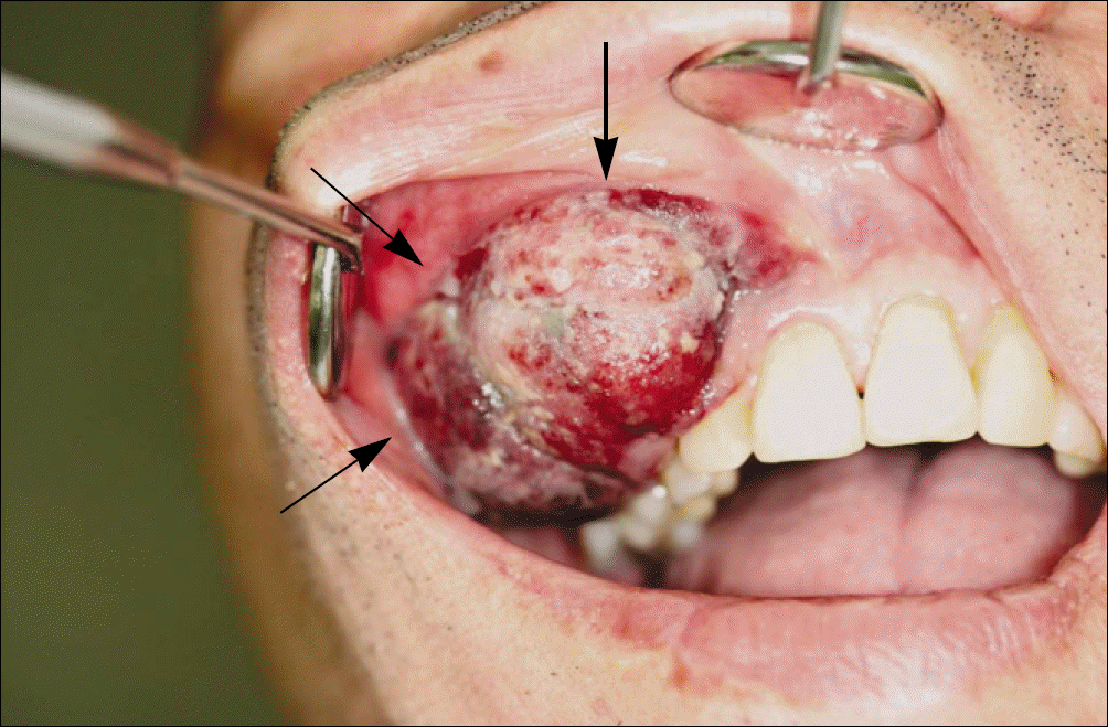
Fig. 4.
Axial and coronal computed tomography images showing resorpted buccal plate of right maxillary sinus and destructive bony change. A: Axial view, B: Coronal view.
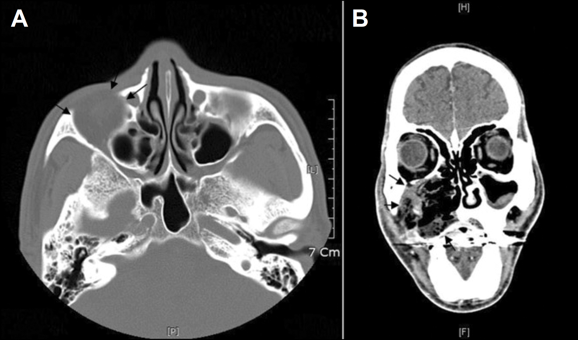
Fig. 5.
Axial and coronal MRI showing tissue mass on right buccal cheek and maxillary sinus. A: Axial view, B: Coronal view. (MRI: magnetic resonance imaging)
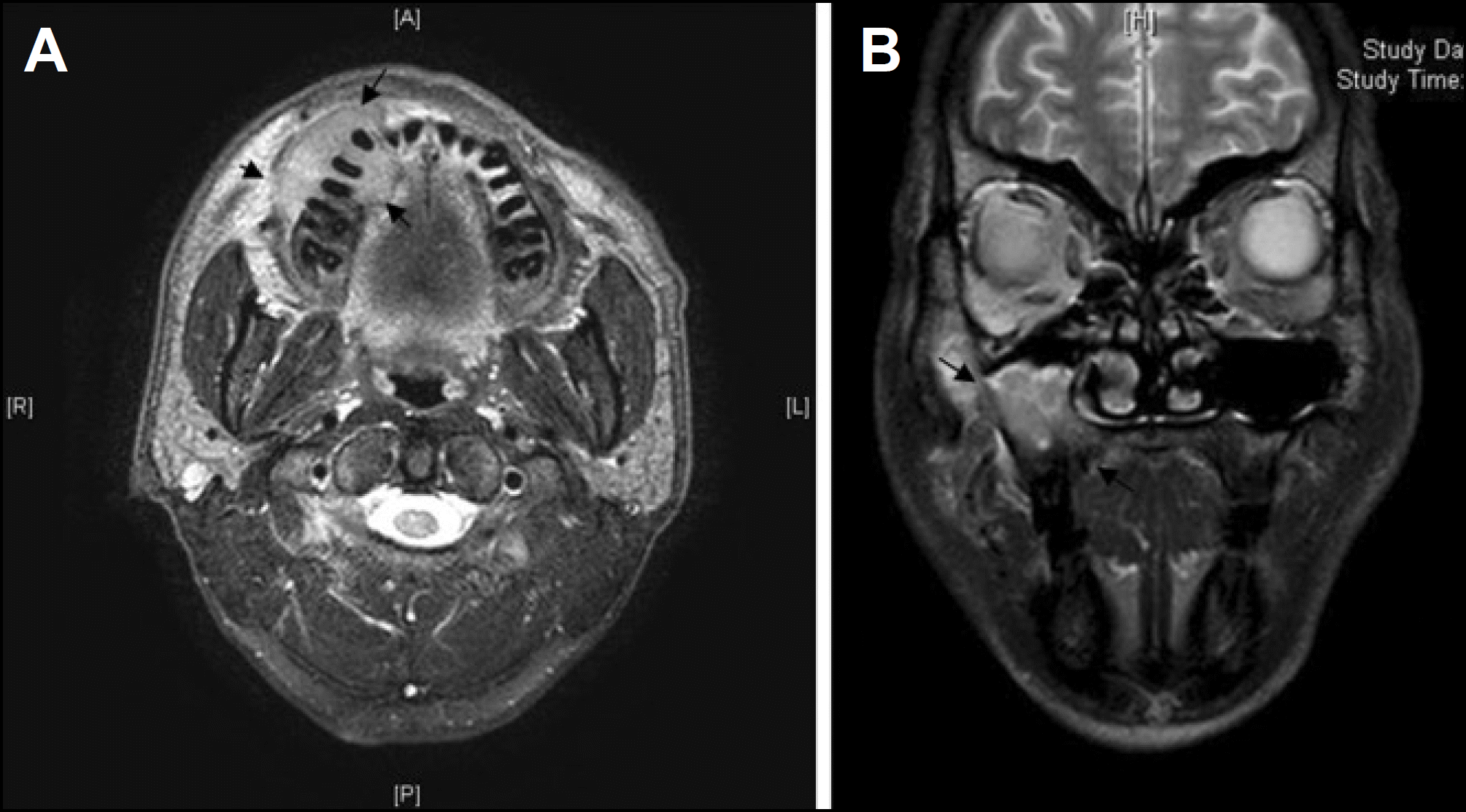
Fig. 6.
Enlarged mass (arrow) compared with previous MRI. A: Axial MRI on first visit, B: Coronal view on second visit. (11 days after) (MRI: magnetic resonance imaging)

Fig. 7.
F18-FDG PET showing uptake of FDG on right buccal cheek and maxillary sinus.(F18-FDG PET: F18-fluorode-oxyglucose positron emission tomography)
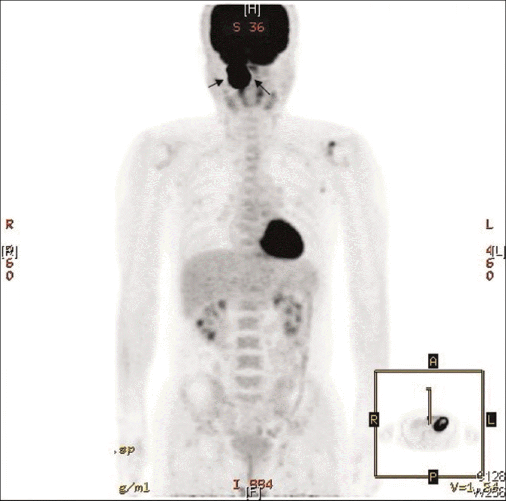
Fig. 8.
Perioperative images. A: Preoperative margin design, B: After mass removal, C: Before reconstruction, D: After reconstruction on extraoral soft tissue defect.
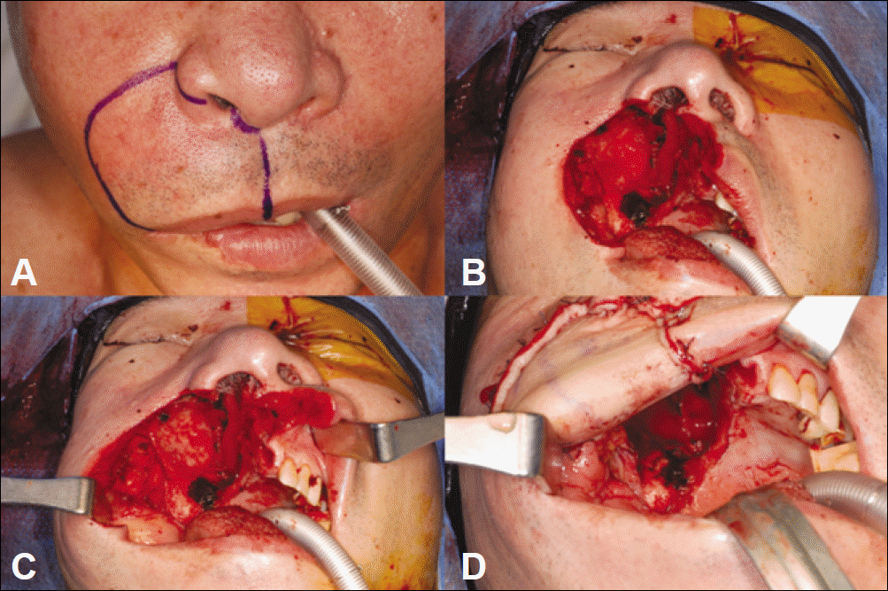
Fig. 9.
Mass removal and reconstruction using RFFF. A: Removed mass by partial maxillectomy, B: Flap design on right forearm, C: RFFF, D: After reconstruction on intraoral and extraoral soft tissue defect using one RFFF. (RFFF: radial free forearm flap)
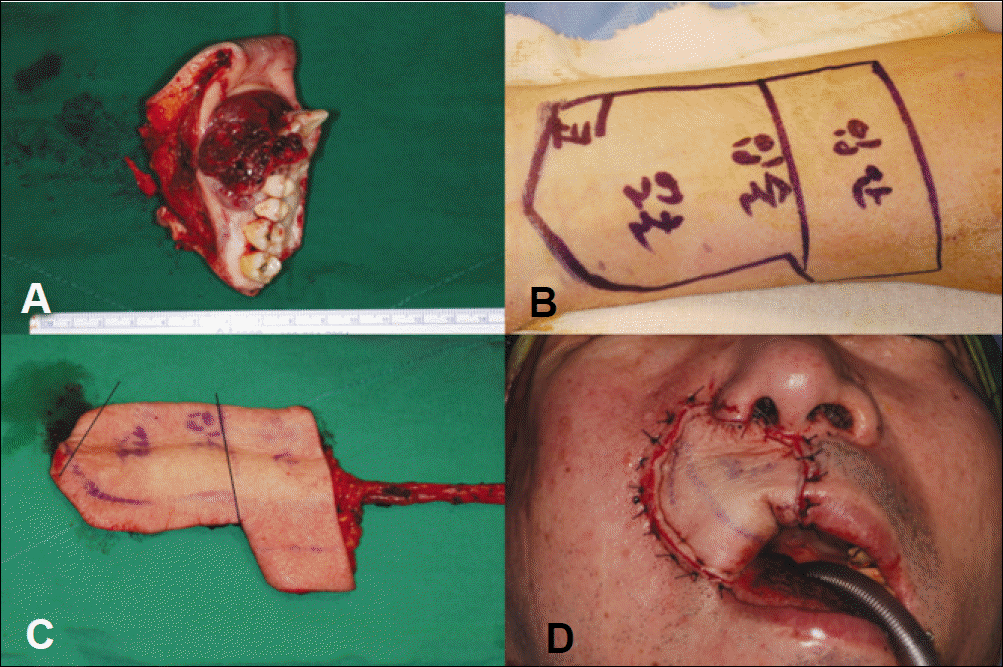
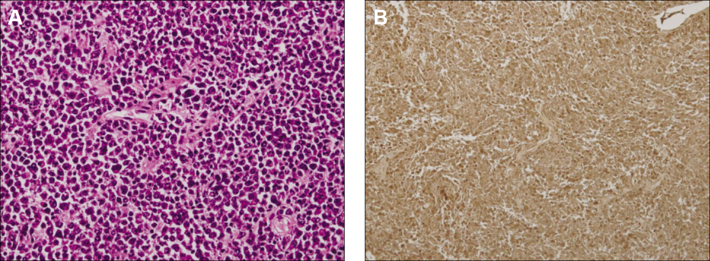
Fig. 10.
Histologic findins. A: Dense, homogenous infiltration of plasma cells showing varying degrees of dysplasia, including prominent nucleoli, dispersed chromatin, irregular nuclei and multinu-cleation.(H&E staining, original magnification ×200) B: IgG(+), Kappa(-), Lambda(-).(Immunohistochemical staining ×100)
 XML Download