

 PDF
PDF ePub
ePub Citation
 Print
Print


Introduction
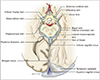
The basal vein of Rosenthal, or simply the basal vein, is a paired deep cerebral vessel that originates in the premesencephalic cistern, courses through the ambient cistern lateral to the midbrain, and terminates in the quadrigeminal cistern (Fig. 1) [1]. In the quadrigeminal cistern, the basal vein unites with the deep cerebral veins to form the great vein of Galen, which then drains to the straight sinus and, subsequently, the confluence of sinuses [1]. The basal veins receive tributaries arising from the midbrain, hippocampal gyrus, interpeduncular fossa, and the inferior horn of the lateral ventricle [1]. In this report, we discuss an anatomical variation of the left basal vein in which it drains directly into the confluence of sinuses.
Case Report
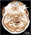
An adult female presented with headaches and was evaluated for intracranial pathology. The initial computed tomography screening was suspicious for a subarachnoid hemorrhage. Therefore, computed tomography angiography was obtained, which revealed a left basal vein that traveled posteriorly and emptied into the confluence of sinuses (Fig. 2). Aneurysm screening was negative. Intracranial pathologies could not be found nor were other anatomical variations noted in this patient.
Discussion
Variations of the basal vein of Rosenthal are thought to have developmental significance and thus several potential embryonic outcomes that may explain the anatomical variation observed in this case. Padget [2] investigated the embryological origins of the basal veins and determined that segments arise from anastomoses between differentiated parts of the telencephalic, diencephalic, and mesencephalic veins [3]. As the embryonic stage progresses, these structures further develop into the deep telencephalic, ventral diencephalic, dorsal diencephalic, and mesencephalic veins [3]. Additionally, the posterior aspect of the basal vein drains into a junction formed by the dorsal diencephalic and internal cerebral veins or the great vein of Galen. Anterior and middle cerebral veins arise from the telencephalic segment while the ventral diencephalic vein drains from the primitive tentorial sinus. Such changes throughout the embryonic stage form tributaries that can either regress later in adulthood or contribute to basal vein variants such as seen in our case [3].
Posterior drainage of the basal veins directly into the confluence of sinuses is a unique occurrence as its development would occur during the embryonic period [4]. Most likely, this variation resulted from the early basal vein draining into a persistent embryonic tentorial sinus. This sinus directs outflow into the confluence of sinuses, straight sinus, or transverse sinus [4].
The relationship between basal vein variants and complications such as perimesencephalic nonaneurysmal subarachnoid hemorrhage (PN-SAH) are important to note as they may carry clinical significance. While the etiology of PN-SAH has yet to be determined, the majority of complications are associated with anatomical variations of the basal vein [5]. Therefore, development of the basal vein of Rosenthal and subsequent variations are clinically, embryologically, and anatomically important. Understanding potential variations may help clinicians better navigate the deep cerebral system and account for pathologies that might arise from such embryological derailments [67].
 XML Download