

 PDF
PDF ePub
ePub Citation
 Print
Print


Introduction
The tensor fasciae suralis is an anomalous muscle that originates from semitendinosus, semimembranosus or short head of biceps femoris [1]. It varies in length and gets inserted to the crural fascia or the superficial part of tendocalcaneus [2]. The muscle takes its innervation from tibial component of sciatic nerve. As the fibers are from one of the hamstrings, it is also called ischioaponeuroticus. Reported clinical presentation of the tensor fasciae suralis include symptoms of neuromuscular compression in the popliteal region and palpable mass in the popliteal fossa [3]. Contraction of the muscle may lead to sciatic, tibial or sural nerve neuropathy. The anatomical relations of the muscle explain the clinical significance. This report presents a rare variation in the popliteal anatomy with an uncommon origin and innervation of tensor fascia suralis muscle.
Case Report
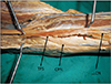
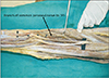
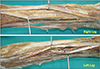
During routine dissection classes for undergraduate medical students, we found an anomalous muscle in the roof of popliteal fossa just beneath the popliteal fascia in the left lower limb of a male cadaver aged about 58 years. A thorough examination after careful dissection revealed that the muscle was taking its origin from the lower part of linea aspera of the left femur just next to the lowermost fibers of origin of short head of biceps femoris. The muscle was of 16 cm in length and 1 cm in breadth in its widest part. The ribbon like muscle was found to be inserted into the crural fascia just superficial to the lower part of lateral head of gastrocnemius. The crural fascia was found to be spitted to enclose the lower end of tensor fascia suralis. A long muscular branch of common peroneal nerve was found innervating the tensor fascia suralis. The muscle was found crossing the common peroneal nerve and lateral cutaneous nerve of the calf superficially. The variations have been shown in Figs. 1, 2, 3.
Discussion
The anomalous tensor fasciae suralis is a clinically significant accessory muscle. It may be discovered as an incidental finding during imaging or as a swelling simulating Baker's cyst or soft tissue tumors in popliteal fossa during clinical examinations. Due to the muscle's location in relation to the neuromuscular elements in the popliteal fossa the tensor fasciae suralis has been speculated to cause compression to the same [4].
Some researchers postulated that tensor fasciae suralis arises from muscle primordia that failed to disappear in lower limb [1]. The supernumerary muscles in the region of popliteal fossa were named as tensor fasciae suralis from the year 1813, though the origin and insertion varied. But in general all those muscles were taking its origin from one among the hamstrings muscles and was innervated by tibial component of sciatic nerve [5].
Our finding is different from those reported cases as this is the first one taking its origin from the linea aspera along with short head of bicpes femoris and innervated by common peroneal nerve. As the muscle was found in the extreme lateral margin of popliteal fossa and covering common peroneal and lateral cutaneous nerve of the thigh, the clinical presentation may vary. There can be pain or altered sensation along the distribution of above said nerves. Though the muscle might not give significant contribution to the knee movement, it can be considered as a weak flexor of the knee as it crossed the knee from the posterior aspect. The muscle could be used in muscle graft surgeries.
In conclusion, origin of tensor fascia suralis from linea aspera has not been reported before. Hence we postulate that this muscle cannot be named as ischiaponeuroticus, but femeroaponeuroticus. The innervation of this muscle from common peroneal nerve is emphasizing the fact that it is not ischioapneuroticus as reported earlier. But this muscle can be considered as a variant of tensor fascia suralis because of its nature of insertion and probable functional anatomy. This muscle may cause compression neuropathy to the common peroneal and lateral cutaneous nerve of the calf. Knowledge of this variation could be important to orthopedic surgeons, radiologists, plastic surgeons, and physiatrists.
 XML Download