

 PDF
PDF ePub
ePub Citation
 Print
Print


Introduction
The aortic arch (AA) is one of the three thoracic portions of the aorta, the only one found in the superior mediastinum (SM). It begins when the aorta rises from the pericardial sac and courses upward, backward and to the left passing through the SM. It ends on the left side at the level of the T4–T5 vertebrae. Three branches arise from the superior border of the AA, the brachiocephalic trunk (BCT), the left common carotid artery (LCCA), and the left subclavian artery (LSA), all being crossed anteriorly by the left brachiocephalic vein. These branches may arise from the beginning of the arch or the upper part of the ascending aorta with varying distances between them. The BCT later divides into the right common carotid artery (RCCA) and the right subclavian artery (RSA) [1]. This branching type was firstly classified as the normal pattern type A by the anatomist Buntaro Adachi (1865–1945) in 1928 [2].
AA, as a continuation of the ascending aorta, constitutes a crucial vascular structure for intrathoracic and vascular surgery, as well as for the conventional and interventional radiology. Altered congenital variations in the human vascular pattern may occur not only in the peripheral branches, but in the large vessels as well. AA is not an exception, presenting a plethora of possible vascular combinations with various frequencies, while its normal branching occurs in only 70% of individuals [1].
In this report we present a rare case of common origin of the carotid arteries (COCA), involving the origin of the LCCA from the initial portion of the BCT.
Case Report
An embalmed (formalin-fixed) male human cadaver of Caucasian (Hellenic) origin was examined during routine educational dissection in cadavers donated at the Anatomy Department of the Medical School of the University of Athens. The donated cadaver belonged to 77 years of age male, presenting no known heart disease. The cadaver derived from body donation with informed consent (with signature authentication) by the donator himself.
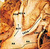
After removal of the anterior thoracic wall, fat tissue and the pericardium covering the ascending aorta and the great vessels, the AA was exposed. Then the BCT, the left and the right subclavian arteries (SCA) and the left and right carotid arteries were dissected. The BCT originated as expected from the AA and gave rise to the RCCA and RSA. After a small course the BCT gave also rise to the LCCA from its initial portion, forming a small common trunk (Fig. 1). The LCCA turned to the left side of the neck and followed the expected course and branching. The LSA rose from the left edge AA independently, before AA continued as descending aorta (Fig. 1). The cadaver presented no other vascular variations.
Discussion
Although the BCT as a nomination describes the very essence of the artery, that is the blood supply to the right arm, the head and the neck in human body, we also mention the Hellenic term “anonymous artery” as officially introduced since the rebirth of the modern Greek Anatomic School in 1843 [3]. Anonymous artery as a definition still is used among the modern anatomists, especially among many Greek and Italian anatomists [referring to the BCT or the innominate artery (Latin term meaning anonymous, nameless)] [456].
A series of anatomical variations of AA are mentioned in the literature concerning distances of the arising points or the course of the vessels, alternation in branching, even vascular agenesis. Primary branches of the AA may be reduced more commonly to two, with the LCCA arising from the BCT (7%–27%), or even more rarely to only one branch [7]. Seldom, the LCCA and SCA might all arise from a left (aberrant) brachiocephalic artery [89]. In other cases, the RCCA and SCA arise separately, in which case the branches from the left end of the AA passes behind the oesophagus. Other variations in vascular patterns concern symmetrical right and left BCTs, with an incidence of 1.2%, atresia or hypoplasia with an incidence of 1.8% to 3.1%, while sometimes a “right aorta” pattern result to reversed arrangements of the three normal branches of the AA [7891011].
The most common AA branching variation observed is the occurrence of a common trunk from which both BCT and LCCA originate. This pattern is referred as “bovine type arch”. Two subtypes of this variant are proposed, the first one with a long bovine trunk with a 4 to 6 cm length described as LCCA arising from BCT (type A) and the second with a short bovine trunk described as the common origin of LCCA and BCT [712]. These types of COCA depict an AA variant which may be found in approximately 11% among the Caucasian white population, with an even higher incidence reaching up to 25% reported for the African-American black population [9101314]. However, the presence of a LCCA arising from the initial portion of the BCT, with the same point of origin from the AA, is a variation with an incidence of 0.2% only [7]. Such an anomaly in the AA was recognised by Paraskevas et al. (2008) [7], Shiva Kumar et al. (2010) [13], and Patil et al. (2012) [14]. The first team presented the variation as a common trunk arising from the initial portion of the BCT giving though no specific name. The second named the common trunk as brachiocephalic and left carotid trunk, while the third named only the common point of origin as a great trunk [71314]. In the present case we report also such a variation, including a common origin of both carotid arteries, that is a COCA variant, combined with an egress point of the LCCA from the initial portion of the BCT.
Variations in the AA branching pattern arise from aberrantly developing patterns of embryonic aortic and pharyngeal arches, some of which persist although they normally disappear or disappear instead of persisting [89]. An anomalous branching pattern of the AA may alter the cerebral haemodynamics, leading to possible relevant symptoms, as for example the “subclavian steal syndrome” [15]. Such a complication does not appear to constitute a probable eventuality in the case of a variation as this presented in this report.
Anatomical branching variants of the AA, thus, vascular deviations from the normal anatomy, are due to the abnormal involving in the transformation of the embryonic AA system into the adult arterial pattern. Surgical and radiological maneuvers demand an excellent knowledge of the AA topographical anatomy and its variations. Ignorance may cause serious surgical complications during procedures performed in the SM and at the base of the neck, while it may also cause brain damage and severe to fatal haemorrhagic incidents.
 XML Download