

 PDF
PDF ePub
ePub Citation
 Print
Print


Introduction
The lateral antebrachial cutaneous nerve (LABCN) is the direct continuation of the musculocutaneous nerve which is derived from the lateral cord of brachial plexus (C5–7) [1]. LABCN lies lateral to the tendon of biceps brachii in the cubital fossa at the level of the interepicondylar line of the humerus. LABCN gives cutaneous supply to anterolateral surface of forearm upto the base of the thenar eminence and does not supply beyond that [2].
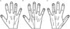
The dorsal cutaneous surface of hand is most commonly Biology supplied by the superficial branch of radial nerve (SBRN) and the dorsal cutaneous branch of ulnar nerve (DBUN). The DBUN usually innervates the dorsal medial surface of hand and medial one and half digits while SBRN supplies the dorsal lateral surface of hand and lateral three and a half digits (Fig. 1A) [1].
Due to rapid increase in use of local anaesthesia and surgeries involving the hand, wrist and digits, it is essential to know the sensory innervation patterns in the dorsum of the hand. In most of surgeries of hand and wrist dorsal approach is preferred. So after getting information about this type of variation in sensory innervations we can demarcate a safe zone for incision over dorsum of hand and wrist [3].
The aim of the present study is to report the bilateral variation of sensory innervations of the dorsum of the hand.
Case Report
During routine upper limb dissection for undergraduate students in the department of Anatomy at North DMC Medical College, Delhi, we found an additional unusual supply to the dorsum of both hands by the LABCN in a 68-year-old male cadaver.
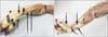
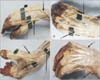
On dorsum of the right hand, the first digit and lateral half of second digit were supplied by the LABCN, medial side of second digit and lateral side of third digit were supplied by SBRN and medial side of third digit, fourth and fifth digits were supplied by DBUN (Figs. 1B, 2A, 3A, B).
However, on dorsum of the left hand, only the lateral side of the first digit was supplied by LABCN. The medial side of first digit, the second and third digits as well as the lateral side of fourth digit received branches from SBRN, medial side of fourth digit and the fifth digit were supplied by DBUN (Figs. 1C, 2B, 3C, D).
Discussion
In this case report, LABCN does not end in the forearm but reaches forward to give contribution in innervation of dorsum of hand along with SBRN and DBUN. This type of variation seen in this study may be due to some abnormality in limb muscles and the peripheral nerves development during the embryonic life. The upper limb muscles develop from the mesenchyme of paraxial mesoderm and the brachial plexus is formed due to addition of growth cones motor axon at the base of the limb bud [4]. At fifth week of embryonic life, the developing brachial plexuses give rise to peripheral nerves into the mesenchyme of the limb buds which grow distally to reach the muscles and skin of upper limb bud [56]. The development of these axons in limb buds is highly coordinated and regulated by some factors such as brain-derived neurotropic growth factor, c-kit ligand, neutrin-1, neutrin-2, etc. [7]. These variations may be due to altered neuronal signalling between the limb bud and the neuronal growth cones or factors at the time of fission of brachial plexus cords [78].
The LABCN variation was first described in 1911, where the radial nerve below the elbow gave only the posterior interosseous branch and the SBRN was absent. So, cutaneous supply of the dorsum of hand was given by LABCN and ulnar nerve [9]. Then in 1918, Stopford [10] studied more than 1,000 cases and concluded that there are very considerable variations in the radial nerve distribution and described three most common pattern of sensory distribution of the dorsum of hand. Since then many cases of partial or total communication between the LABCN and the SBRN on the dorsum of the hand have been reported. In 1985, Mackinnon and Dellon [11] studied the anatomical relationship between the LABCN and the SBRN in 53 cadavers. They found that there was significant anatomical overlap between the SBRN and the LABCN at the level of the wrist, with partial or complete overlap of the LABCN and the SBRN occurred in 75% of the cases and an interconnected plexus in 32% of the cases [11]. In 1987, Bourne et al. [12] were studied the LABCN course and relation on 20 specimens and observed that the nerve emerges from the lateral aspect of the biceps tendon at the level of the interepicondylar line. Bergman et al. [13], 1988 studied variations in the formation, distribution and termination of the musculocutaneous nerve. In 2007, Huanmanop et al. [14] concluded that connections between the SBRN and the LABCN are common but replacements are rare and these variations are important to know in cases of peripheral nerve injuries. Several researchers studied branching patterns of the musculocutaneous nerve and radial nerves [1516]. Tryfonidis et al. [17] reported the variation in the course of the SBRN. In 2005, Beldner et al. [18] reported the relation of the LABCN with the cephalic vein. They observed that the LABCN lied either superficial (31 specimens) or deep to the cephalic veins (5 specimens), and divided into two parts: one branch dorsal and one branch volar to the cephalic vein (1 specimen). Due to close approximation to the cephalic vein, the LABCN is susceptible to injury during venepuncture. Hence, study of the variations in the LABCN will help in understanding peripheral neuropathy caused by venepuncture. Sontakke et al. [19] described unilateral variant origin of musculocutaneous nerve.
In summary, the present study shows unusual bilateral sensory innervation of the dorsum of hand by LABCN. The knowledge of this variation assumes vital importance for reconstructive surgery of hand to avoid damage to these nerves and in understanding the peripheral neuropathy of unusual clinical symptoms.
 XML Download