

 PDF
PDF ePub
ePub Citation
 Print
Print


Introduction
The word styloid process has been originated from the word 'stylos,' which means, the pillar, in Greek language [1]. This process belongs to the temporal bone of the skull and it lies anterior to the stylomastoid foramen. Being cylindrical in shape, the styloid process gradually tapers towards the apex just like a pinnacle. Its apex is located next to the tonsillar area in the lateral wall of pharynx, between external and internal carotid arteries. Its tip provides attachment to the stylohyoid ligament. There are few structures blended to the stylos process, which are in relation to the nerves and vessels. The stylopharyngeus, stylohyoid and styloglossus are the muscles which attach to the base, middle part and tip of the styloid process respectively. These muscles get the innervations from the 9th, 7th, and 12th cranial nerves [2]. Spinal accessory and vagus nerves run medial to the styloid process. The facial nerve runs anteromedial to this process before piercing the substance of the parotid gland. Glossopharyngeal nerve curves in close proximity to the stylos process. The styloid process and the hyoid bone are connected by the stylohyoid ligament, which forms the anatomical basis for the glossopharyngeal neurological symptoms which are seen in styloid process syndrome [3]. The clinical features and the patient complaints associated with the long styloid process are referred as Eagle's syndrome. The objective of the present investigation was to study the prevalence of elongated styloid process in the Indian population and to study its morphology. The morphometric data of the styloid process were collected at various points and variations of the dimensions of styloid process are studied. The embryological and clinical implications of the elongated styloid process are discussed.
Materials and Methods
The present study included 110 human dried skulls of Indian origin, which were available in the department of anatomy. The dried skulls which had damaged styloid process were excluded from the present study. The styloid processes were measured for their length, thickness at the base, mid-point and the tip. Interstyloid distance of the skull was also measured at their base, mid-point and the tip. The skulls were macroscopically observed on both the sides for the elongation of the temporal bone, styloid process. Lengths of the styloid processes were measured by using the digital vernier caliper. The measurements were taken from the point of emergence of the process (base) until to the tip. The data were recorded and tabulated. The thickness of the styloid process was measured at its base, midpoint and the tip by using the digital Vernier caliper. The interstyloid distance of skull between the right and left styloid process was also measured at the same 3 points. The values were statistically analyzed among the right and left sides by using the student t test (paired samples t test). The significance is given as the P-value less than 0.05. The SPSS version 15 (SPSS Inc., Chicago, IL, USA) was utilized for performing the student t test. The data were given as mean±standard deviation. The ossified stylohyoid ligament was also considered as the continuation of the styloid process. The process was considered elongated if its length is more than 30 mm.
Results
The morphometric data of the styloid process obtained in the present study is represented in Table 1. The data was statistically compared among the right and left sides (Table 1). The analysis was statistically not significant (P>0.05). The mean interstyloid distance of the skull at the base, mid-point and tip of the styloid processes is given in Table 2.
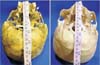
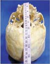
Among our specimens, only 5 skulls (4.5%) exhibited the elongated styloid process. Three skulls (2.7%) had unilateral elongation (Fig. 1) and 2 skulls (1.8%) showed bilateral elongation (Fig. 2). All the 3 unilateral elongations were observed on the left side. The variant styloid processes were unusually long (more than 30 mm), stout and strong. The elongated styloid processes were rounded up to half of their length and gradually tapered with a pointed tip (Figs. 1, 2). Table 3 show the morphometric data of the unilateral elongated styloids which were observed in the present study. The data of the bilaterally elongated styloid process is given in Table 4.
Discussion
The elongation of styloid process with or without ossified stylohyoid ligament is considered as Eagle's syndrome. This syndrome is also called as styloid neuralgia, elongated styloid syndrome, styloid-carotid syndrome and styloid-stylohyoid syndrome [4]. The stylohyoid chain extends between the temporal and hyoid bones and is generally divided into four sections as tympanohyal, stylohyal, ceratohyal, and hypohyal [1]. Eagle, who described this syndrome complex, divided it into two categories. The classical type is presented as foreign body sensation in the throat, pain in the throat and the ear ache. The other type is the styloid process compressing the carotid arterial system and presenting as dizziness and headache [5]. A variety of head and neck signs and symptoms are related to the elongated styloid process and its stylohyoid chain component. The dimension of styloid process usually varies, ranging up to 25 mm. The elongated styloid process can be clinically detected by palpating the tonsillar fossa and is diagnosed by taking the X-ray lateral view of the neck, orthopantomogram (OPG) or a computerized axial tomogram (CT). Although there is no gender predilection for the elongated styloid process, the symptoms tend to be more common in the middle aged females [67]. Eagle's syndrome is a congenital anomaly which is sometimes misdiagnosed and is found incidentally during the radiological imaging. The clinical symptoms in patients with Eagle's syndrome are because of compression of the surrounding trigeminal, facial and glossopharyngeal nerves. During the development, the hyoid smaller horn, styloid process, and the stylohyoid ligament are developed from the cartilage of second pharyngeal arch (Riechert's cartilage). On some occasions, the stylohyoid ligament goes for ossification and will lead to the formation of elongated styloid process [2]. Elongated styloid process results in a variety of symptoms which range from chronic facial and neck pain, dysphagia, tinnitus, referred pain in the ear, glossopharyngeal neuralgia, orbital pain and radiating pain into the maxillary regions [8910]. It may also cause stroke when it compresses the carotid arterial system [1112]. The inflammation and degeneration changes of the stylohyoid ligament which occurs in its tendon and the rheumatic styloiditis may also contribute to this syndrome [13].
The length of styloid process of temporal bone varies from population to population. Eagle [3] reported that a normal styloid process measures between 25 mm to 30 mm and any length more than the above mentioned values, is considered as the pathogenic factor for Eagle syndrome. Keur et al. [2] suggested that the styloid process length and its mineralized stylohyoid ligament, if appears more than 30 mm in a radiograph film, is considered as a significant predisposing factor. However, Jung et al. [14] suggested that, a styloid process of only more than 45 mm length should be considered to be elongated. In the present study, the length of styloid processes ranged from 18 mm until 50 mm. The present study observed that the mean length of the styloid process was 17.8±9.3 mm and 18.2±5.6 mm for the right and left sides, respectively. This data is smaller in comparison to the data from North Indian population reported by Rathva et al. [15]. Rathva et al. [15] reported that the length of styloid process was 43.8±11.1 mm and 43.5±10.4 mm for the right and left sides in their specimens. This variation in the data from Indian samples may be because the difference in the method which was used to measure the parameter as they performed the measurements by using digital image analysis using the adobe photoshop. In the present study, digital Vernier caliper was used to perform the measurements. However, in an another study by Patil et al. [16], which used the Adobe photoshop for the measurements, the data were 13.9±8.1 mm and 12.9±8.7 mm for the right and left sides, respectively. The findings of the present study are similar to Patil et al.'s data [16]. Among all the skulls, seven of the styloid processes measured more than 30 mm and were considered as elongated. This was observed in 5 among 110 skulls with a prevalence rate of 4.5%. The prevalence of elongated styloid process in the earlier studies were 1% [4], 4% [17], 8.2% [18], and 28% [19]. The prevalence rate of the present study (4.5%) is almost similar to the rate observed by Eagle (4%) [19]. Other Indian studies by Rathva et al. [15] reported the prevalence of elongated styloid process as up to 2%. The present study observed that the styloid process elongation was observed more commonly on the left (5:2).
The present study performed the thickness of the styloid process at the base, mid-point, and the tip. This much detailed measurements on the thickness was not reported earlier in the literature. The thickness data was compared statistically and it was found that the data was not significant statistically with respect to the right and left sides. The mean thickness at the styloid process base was 4.4±1.2 mm and 4.4±0.9 mm on the right and left sides. The same parameters were 3.2±0.4 mm and 3.8±0.7 mm; 1.5±0.6 mm and 1.4±0.5 mm at the mid-point and tip of the styloid processes, respectively. There were few reports [20] which observed 6-12 mm thickness at the base of styloid process. The present study observed 4.5 mms as the maximum thickness. In the present study, the interstyloid distance of the skull at the base, mid-point, and the tip of styloid process were 68.9±4.3 mm, 62.7±1.1 mm, and 60.7±2.4 mm, respectively. These data are different in comparison to the data by Rathva et al. [15]. Rathva et al. [15] reported the same parameters as 75 mm, 55 mm, and 30 mm, respectively. This difference is may be because of the methods used as the present study was performed by using the digital Vernier caliper and Rathva et al. [15] used the scion image analyzer. Our findings are almost similar to the data from Patil et al. [16]. According to Patil et al. [16], the mean of distance between the bases of two styloid process was 69±5.2 mm and the same parameter between the tips of two styloid processes was 64±6.1 mm.
The impression of the styloid process elongation can be made by analyzing the clinical history given by the patient and by digitally palpating the tonsillar fossa. Touching the tonsillar area exacerbates the symptoms and is relieved by the local infusion of a local anesthetic agent into the tonsillar area, which is indicative of Eagle's syndrome [6721]. The diagnosis is confirmed taking the X-ray, OPG and the skull base CT scan. The treatment of elongated styloid process is performed by the surgery, the elongation will be made short by cutting it with the trans-tonsillar and external approaches, considering avoiding injury to the surrounding neurovascular structures [22]. It has been reported that, inspite of the precautions taken during the surgery, the structures attached to this process get injured at their attachments, which may compromise the stability of hyoid bone.
The etiology and pathogenesis of Eagle's syndrome is poorly understood and is same with the calcification of the stylohyoid ligament. However, there are many theories which are described. The well accepted one is the ossification of the stylohyoid ligament or mineralization of the cartilage which is present embryologically during the development of the styloid process. The solidification of these structures may result in a variety of symptoms referred as Eagle's syndrome. Steinmann [23] proposed various theories to explain the ossification of stylohyoid ligament which include the theory of reactive-hyperplasia-metaplasia-anatomical variation. The most accepted theory is the congenital theory. According to the congenital theory, mechanical stresses during the intrauterine life may sometimes lead to stretching of the second branchial arch and the styloid process elongation.
The symptoms of elongated styloid process include dysphagia, pain in the throat, foreign body sensation and pain in the face. It may lead to transient ischemic attacks and cerebrovascular accidents due to the compression of the internal carotid artery and lack of arterial supply to the brain. It is also responsible for the difficult endotracheal intubations which are experienced by the anesthesiologists [24]. The clinical implication also includes the course of the vertebral artery which may be distorted in such situations. The Eagle's syndrome should not be misdiagnosed as trigeminal neuralgia [24]. This can be easily differentiated by taking the pain history as the Eagle's syndrome pain is persistent and the trigeminal neuralgia pain is intermittent in nature. Eagle's syndrome may mimick symptoms of phantom limb (globus hystericus) and may cause psychosomatic stress to the patient [25]. Differential diagnoses of Eagle's syndrome include unerupted molar tooth, dental prosthesis implantation, diseases of the temporo mandibular joint, tumors in the oropharynx and laryngopharynx [26]. It has been reported that there is a minimal quantity of elongation in the temporal bone styloid process as the age advances. This is especially noticed in the postmenopausal ladies and is due to the hormonal changes which occur after the menopause [27].
The morphology and the variations of the craniofacial bones have been studied since the beginning of the century [28]. The morphometric data of the styloid process is important to the neurologists, neurosurgeons, radiologists and otorhinolaryngologists. The data on the interstyloid distance is clinically important as this space accommodates important structures of neck like cranial nerves, larynx, esophagus, arteries, and veins. The surgical anatomy of the elongated styloid process is important to the neurosurgeons and radiologists while interpreting the computed tomogram scans and magnetic resonance images. The morphological knowledge of Eagle's syndrome is enlightening to all the health care professionals who are involved in the diagnosis and management of chronic head and neck pain. The knowledge is important to the physicians, dentists, neurologists, neurosurgeons, and otorhinolaryngologists. In this context, the present study has provided important data about the styloid process of the temporal bone. The dimensions of the styloid process and its stylohyoid chain are essential to the anatomists, anthropologists, forensic experts and clinicians. We believe that the present study has provided additional information on the frequency of elongated styloid process in the Indian population. In the present study, the styloid process has been studied morphologically with emphasize on their embryological and clinical implications. We opine that the present study can be more accurate with a large number of samples. The limitation of the present study includes the gender related variation which was not taken into consideration.


 XML Download