

 PDF
PDF ePub
ePub Citation
 Print
Print


Introduction
Vitamin C is an essential micronutrient synthesized from glucose. In the cell, vitamin C serves as a co-factor for enzymes involved in the biosynthesis of several molecules such as collagen fibers, carnitine, catecholamine, and some other neurotransmitters (De Tullio, 2002; Patak et al., 2004), and also acts as a physiological antioxidant (Long & Santos, 1999). These activities of vitamin C affect a wide range of biological processes (Mandl et al., 2009).
Vitamin C also affects immune responses in several aspects. There are some reports indicating that vitamin C plays a role in modulating the antibody isotype switching. Administration of vitamin C lowered IgE levels in patients with chronic granulomatous disease (Anderson & Dittrich, 1979) and in patients with bronchial asthma (Anderson et al., 1980). Vitamin C was also reported to lower IgG levels in calves but to increase in adult cows (Cummins & Brunner, 1989). Previously, we demonstrated that administration of a mega-dose vitamin C to mice, at the time of immunization, remarkably lowered serum titers of two antigen-specific Th2 isotypes, IgG1 and IgE (Noh et al., 2005). As is well known, antibody production results from concerted interactions among several immune cells including B cells, T cells, and dendritic cells. If vitamin C does modulate antibody production in B cells, either via its chemical or its antioxidant activities, it will be important to determine which immune cells vitamin C directly affects to influence this process. Because in vivo administered vitamin C modulates T cell behaviors such as proliferation and cytokine secretion (Noh et al., 2005), it could affect T cells primarily. Or, it is equally possible that vitamin C acts directly on dendritic cells to modulate the B cell functions by way of T cells (Pearce et al., 2006).
Alternatively, vitamin C may influence antibody production as a direct effect on B cells by modulating the level of intracellular reactive oxygen species (ROS) in these cells. Some reports have suggested that antioxidants might regulate B cell activation and functions including isotype switching. N-acetyl-L-cysteine (NAC), an antioxidant, was shown to inhibit both the up-regulation of CD40 and the isotype switching to IgE by IL-4 (Yanagihara et al., 1997), and also to affect in vitro B cell proliferation induced by CD40 ligation (Lee, 2003). NAC treatment of B cells from Atm-/- mice was shown to lower the increased levels of intracellular ROS and thus to rescue the impaired immunoglobulin isotype switching in these mice (Ito et al., 2007). The tabacco polyphenols, quercetin and rutin, which are antioxidants, were shown to inhibit antigen-presenting functions of B lymphoma cells (Gong & Chen, 2003). Based on these findings, the role of ROS in B cell functions has been suggested, and thus vitamin C, an important physiological antioxidant that reduces intracellular ROS levels (Blumenthal et al., 2000; Guaiquil et al., 2001), would directly modify their behaviors and functions.
It has never been studied whether vitamin C directly affects B cells with respect to isotype switching. Furthermore, even the effects of vitamin C on B cell activation such as proliferation and surface molecule expression have not been reported. In the present study, we isolated mouse splenic B cells and evaluated the direct effects of vitamin C on these cells following in vitro activation.
Materials and Methods
Experimental animals
Eight to ten-week-old male Balb/c mice were purchased from BioLink (Seoul, Korea) and were kept in animal facilities of Seoul National University, College of Medicine (Seoul, Korea). They were reared in a 12 hour light-dark cycle and were given usual mice chow and tap water ad libitum. This animal study was approved by our institutional IACUC (approval number; SNU-060926-3) and all the procedures were performed following the SOP of our Institute.
Immunization with keyhole limpet hemocyanin (KLH) and quantification of specific antibody titers in vivo
Mice were immunized twice, on days 1 and 21, with an intraperitoneal injection of 100 µg of KLH (Sigma Chemical Co., St Louis, MO) in 200 µl PBS. Vitamin C (as L-ascorbic acid, Sigma), either at a dose of 0.625 mg/mouse or 5 mg/mouse was given daily by intraperitoneal injection for 3 days before primary immunization and also every day after primary immunization for 10 more days. This protocol is based on our previous experiment (Noh et al., 2003). No vitamin C was administered around secondary immunization. Seven days after the secondary immunization, blood samples were drawn from the orbital plexus under anesthesia. Sera were obtained and then stored at 4℃ until used.
Titrations of KLH-specific antibodies in sera were determined by ELISA as follows. Ninety six well ELISA plates (Nunc, Rochester, NY) were coated with 100 µl of 2 µg/ml KLH/well. Plates were incubated for 5 hours at 37℃, and then overnight at 4℃. The subsequent steps were identical to those used below for the analysis of antibody production in cultures, but there were two differences: the serum samples were diluted 1 : 20 (in 1% skim milk/PBS containing 0.05% Tween 20), and KLH-specific anti-serum was used as a standard in these experiments. Titers were expressed as relative values to standard serum. The relative value of the standard serum was regarded as 2.5.
Isolation and activation of resting B cells in culture
The mice were sacrificed by cervical dislocation. Their spleens were obtained, chopped, and minced so that the cells could be harvested. The RBCs were lysed using lysis buffer (0.83% NH4Cl in 0.01 M Tris-HCl buffer, pH 7.5). The adhering cells were removed by incubating in a 100 mm culture dish for 1 hour in a 37℃ humidified CO2 incubator. After then, B cells were isolated by negative selection using MACS CD43 (Ly-48) MicroBead (Miltany Biotech, Germany) following the manufacturer's manual. The purity was over 95% as was assessed by FACS analysis using FITC-conjugated anti-mouse CD19 antibody (BD Pharmingen, San Diego, CA). Isolated B cells were cultured in RPMI 1640 medium containing 2 mM L-glutamine, 1 mM sodium pyruvate, 0.1 mM non-essential amino acid, 10 mM HEPES (pH 7.4), 100 µg/ml streptomycin and 100 U/ml penicillin, 5×10⁻⁵ mM 2-ME, and 10% FBS. All cultures were carried out in a humidified CO2 incubator at 37℃. B cells were activated by applying goat F(ab')2 anti-mouse IgM (1 µg/ml, Southern Biotech, Birmingham, AL) and rat anti-mouse CD40 (1 µg/ml, BD Pharmingen, San Diego, CA) antibodies to the culture. This was intended to mimic in vivo B cell activation by ligation of B-cell receptors and CD40 (Baumgarth, 2000).
Apoptosis assay
To assess whether experimental concentrations of vitamin C used in the culture media would provoke B cell apoptosis, we performed Annexin V/PI staining and flow cytometric analysis. B cells were seeded in a 24-well plate at a density of 1×10⁶ cells/well. Parallel cultures of cells were pre-treated with L-ascorbic acid (Sigma, St. Louis, MO) at a concentration of 0, 0.0625, 0.125, 0.25, 0.5 or 1 mM for 1 hour, and then activated. Twenty-four hours after activation, B cells were harvested, washed with PBS, and centrifuged. Supernatants were discarded and 400 µL of Annexin V binding buffer (BD Pharmingen, San Diego, CA) at working dilution was added and the cells were re-suspended. Each sample was added with 2 µL (per 1×10⁶ cells) of Annexin V-FITC (BD Pharmingen) and incubated for 15 minutes at room temperature with mild agitation. After then, 0.1 µg (per 1×10⁶ cells) of propidium iodide (PI, BD Pharmingen) in 2 µL volume was added to each sample. Cells were analyzed using a flow cytometer (Becton Dickinson, San Jose, CA).
Measurement of intracellular ROS levels
To evaluate antioxidant effects of vitamin C, intracellular ROS levels were measured using the fluorescence probe, 2',7'-dichlorofluorescein diacetate (DCFH-DA) (Wan et al., 1993). B cells were seeded 1×10⁶ cells/ml in a 24-well plate. The cells were incubated with 50 µM DCFH-DA (Sigma, St. Louis, MO) for 30 minutes at 37℃ and then washed with ice-cold PBS. Next, the cells were seeded in 96-well plates at a cell density of 4×10⁵ cells/well and pretreated for 1 hour with a specific concentration of L-ascorbic acid (Sigma). Each culture, in parallel, was exposed to a single concentration of vitamin C at increments within the range from 0.0625 mM to 1 mM. Next, the cells were activated as described above and the fluorescence was measured for 1 hour by using Cytofluor 2350 plate reader (Millipore, Bedford, MA). Excitation and emission frequencies were 485 nm and 538 nm, respectively. Values for each time point were obtained by subtracting the value at time 0 from each raw value.
Proliferation assay
B cells were seeded in 96-well plates at 4×10⁵ cells/well. Parallel cultures of cells were pre-treated with vitamin C at a concentration of 0, 0.0625, 0.125, or 0.25 mM for 1 hour, and activated for 48 hours as described above. One µCi/well of [3H] thymidine (Amersham Pharmacia Biotech, Oslo, Norway) was then added to the culture and incubated for additional 18 hours. The cells were harvested on glass-fiber filters using a cell harvester (INOTECH). Radioactivity was counted with a scintillation β-counter (MicroBeta Trilux, Perkin Elmer). All samples were prepared in triplicate.
Analysis of the expression of cell surface molecules using flow cytometry
Purified B cells were plated at 1×10⁶ cells/well in 24-well plates. Cells were pretreated with vitamin C for 1 hour, and then activated for 24 hours. The cells were then harvested, double-stained for B220 and for the expressions of surface molecules, CD80 or CD86. Anti-B220-PE, CD80-FITC, and anti-CD86-FITC antibodies (BD Pharmingen, San Diego, CA) were used at 2 µg/1×106 cells. After 30 minute incubation at room temperature, the cells were washed and analyzed using a flow cytometer (Becton Dickinson, San Jose, CA).
Analysis of antibody production in cultures
B cells were seeded in a 24-well plate at a density of 1×10⁶ cells/well, activated as mentioned above, and cultured for 6 days. Before activation, B cells were pre-treated with vitamin C at a specific concentration, and supplemented with the same concentration of vitamin C every 2 days until the end of culture. Supernatants were collected and then stored at 4℃ until used. The titers of total, IgG1, and IgG2a antibodies in the supernatants were determined by ELISA as follows. Ninety six well ELISA plates (Nunc, Rochester, NY) were coated with 100 µl goat anti-mouse polyvalent immunoglobulin (diluted at 1 : 1,000, Sigma, M8019). Plates were incubated for 5 hours at 37℃, and then incubated overnight at 4℃. Plates were then briefly washed and then blocked with 1% skim milk in PBS for 1 hour at room temperature. A neat concentration of supernatants was applied to the first row of the ELISA plate, and 4-fold serially diluted. All samples were prepared in duplicate. The plates were incubated for 2 hours at room temperature, followed by incubation with alkaline phosphatase-conjugated secondary antibodies for each isotype for 1 hour, and by p-nitrophenyl phosphate substrate (Sigma, St. Louis, MO) solution. OD values were measured at 405 nm. The secondary antibody used was as follows; goat anti-mouse polyvalent immunoglobulin (1 : 1,000 diluted, Sigma), goat anti-mouse IgG1, or IgG2a antibody (1 : 1,000 diluted, Southern Biotech, Birmingham, AL). A single batch of pooled sera from mice immunized with keyhole limpet hemocyanin in the previous experiments was used as a standard for the titration of total immunoglobulin, and the titers in the supernatant were expressed as relative values to this standard serum. In this case, the relative value of the standard serum was regarded as 1,000. For IgG1 and IgG2a titrations, purified IgG1 or IgG2a antibody of a known concentration was used respectively.
Results
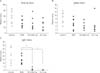
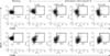
Injected vitamin C decreased the antigen-specific IgG1 titer in mice sera
Before evaluating direct effects of vitamin C on B cells in vitro, we repeated our previous in vivo experiment (Noh et al., 2005) to reproduce the in vivo effects of supplemented vitamin C on the humoral immune responses. Mice were immunized with KLH in one of four ways: 1) with no other treatment (normal control), 2) with concomitant injections of PBS (experimental control), 3) with 0.625 mg of vitamin C, or 4) with 5 mg of vitamin C, as was described in Materials and Methods. These doses are equivalent to 1.5 g and 12 g/60 kg body weight in human, respectively. Each group of mice was given a second immunization on day 21. For each group, the titers of the total KLH-specific antibodies were determined (Fig. 1A). The average titers in the three injected groups (groups 2~4) were lower than that of normal control group. However, the difference was not statistically significant (P>0.05). The titers of KLH-specific IgG2a (Fig. 1B) showed a similar pattern to that of the total antibody titers. That is, the average IgG2a titers in all three injected groups were lower than that of normal control group. In this case, the difference was statistically significant (P<0.05). However, statistically significant differences were not observed between the PBS-injected group and vitamin C-injected groups. The titers of IgG1 (Fig. 1C) showed a different pattern. For each group, the tires were 1) 20.6±9.6, 2) 13.9±5.7, 3) 2.1±1.4, and 4) 0.7±0.3. The groups of mice injected with vitamin C (3 and 4) had titers that were significantly lower than those of both control groups (1 and 2) (P<0.05). These results revealed that vitamin C modified humoral immune responses. Thus, we further evaluated whether vitamin C directly affects B cell functions.
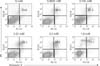
Vitamin C slightly induced apoptosis of activated B cells dose-dependently
Vitamin C at certain concentrations in culture media is known to be cytotoxic leading to apoptosis in many cell types (Duarte & Lunec, 2005). Therefore, we tested a range of vitamin C concentrations to define a range that could be safely used on B cells without affecting viability. B cells were pre-treated with increasing concentrations of vitamin C for 1 hour and activated with anti-IgM and anti-CD40 antibodies. Twenty-four hours after activation, cells were stained with Annexin V and propidium iodide. With this method, the viability of cells cultured in the absence of vitamin C was 93.6%. The viability of cells cultured with vitamin C decreased by about 3% with doubling of the vitamin C concentration, reaching 77.1% at a vitamin C concentration of 1 mM (Fig. 2).
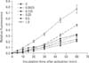
Vitamin C decreased intracellular ROS levels in activated B cells
At higher concentrations, vitamin C could exert a pro-oxidant effect instead of an anti-oxidant effect (Clément et al., 2001). Thus we next evaluated the concentration range with which vitamin C acts as an anti-oxidant in B cells by measuring intracellular ROS levels. B cells were pretreated for 1 hour with a concentration of vitamin C within the range from 0.0625 mM up to 1 mM, and then activated. In the control culture without vitamin C, intracellular ROS levels were elevated (Fig. 3) and continued to rise over time. Cultures treated with vitamin C within the range of 0.0625 to 0.5 mM showed a decrease in the levels of ROS compared to that of the 0 mM group (P<0.05). Cultures treated with 1 mM vitamin C behaved differently. These cultures did have a lower level of ROS compared to the control culture. However, the level was still higher than the other cultures that were exposed to lower concentrations of vitamin C (P<0.05).
Because the concentrations between 0.0625 mM and 0.25 mM exhibited antioxidant properties and tolerable cell viabilities, and because these concentrations cover the physiological serum concentration of vitamin C, we used these concentrations for further experiments.
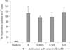
Vitamin C did not affect the proliferation of activated B cells vitro
For exploration of the effects of vitamin C on B cell functions, we first evaluated whether vitamin C exerted direct effects on the proliferation following B cell activation. Isolated B cells were pretreated with vitamin C and activated for 48 hours. Thymidine was added and the cultures were incubated for an additional 18 hours. As is shown in Fig. 4, the presence of vitamin C at any concentration did not affect B cell proliferation.
Vitamin C did not affect the expressions of CD80 and CD86 in activated B cells
As B cells are being activated, they up-regulate surface expression of co-stimulatory molecules such as CD80 and CD86. Thus we evaluated whether vitamin C given at the time of activation affected the expressions of these molecules. CD80 levels increased from 13.5% before activation to 18.4%, after activation in control cultures. CD86 levels also increased from 8.8% to 31.9% in control cultures (Fig. 5). In cultures pretreated with vitamin C, the levels of both CD80 and CD86 after activation were similar to the control group. There may have been a slight increase in CD86 levels (Fig. 5) in cultures pretreated with vitamin C. However, those results were not reproduced and so were regarded as not significant.
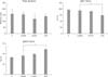
Vitamin C modified isotype switchings in B cells activated in vitro
B cells were activated and then cultured for 6 days in either the presence or absence of vitamin C in the culture media. Supernatants were collected and an ELISA was performed to quantify the titers of IgG1 and IgG2a isotypes (Fig. 6). The titer of total immunoglobulins (upper panel) was not affected by the presence of vitamin C. For IgG1 (middle panel), the titer slightly decreased as the dose of vitamin C increased up to 0.25 mM with statistical significance compared to the control group (76.6±3.0 vs 59.2±1.6, P<0.05). The titer of IgG2a (lower panel), in contrast to that of IgG1, elevated as the dose of vitamin C increased up to 0.25 mM with statistical significance compared to the control group (32.3±1.7 vs 49.3 ±1.2, P<0.05).
Discussion
This study was carried out to evaluate whether vitamin C exerts direct effects on B cells following activation. Therefore, we chose to perform our experiments using an in vitro system. This system allowed us to directly apply vitamin C to isolated B cells and monitor several properties of B cell functions upon activation. We tried concentrations of vitamin C from 0.0625 mM, which is the lower limit of physiological serum concentration in mice (Dubick et al., 1983; Dubick et al., 2002; Kuo et al., 2004) up to 1 mM, which can be achieved by intravenous infusion for the management of malignancy in human (Padayatty et al., 2006; Hoffer et al., 2008) or intraperitoneal injection of ascorbic acid in mice (Noh et al., 2003). However, because it is known that vitamin C is oxidized in culture media to produce hydrogen peroxide, which exerts oxidative stress to the cells leading to cell death (Duarte & Lunec, 2005), and because the susceptibility to this cytotoxic effects of vitamin C in culture media differ according to cell type, we determined the upper level of concentrations that does not reveal cytotoxic effects on B cells in vitro (Fig. 2, 3) as 0.5 mM.
Vitamin C at a safe range of concentrations behaved as an antioxidant in activated B cells, but did not affect cell proliferation and surface molecule expressions. However, vitamin C modified isotype switching in cultured B cells, which was somewhat different from those observed in vivo (Fig. 1). That is, the decrease of IgG1 titers with in vivo vitamin C treatment was quite remarkable (Fig. 1), in contrast to those observed in in vitro experiments (Fig. 6), which was just slight. Furthermore, we could not observe elevated IgG2a levels in in vivo experiments, which were shown in in vitro experiments. That could suggest that the modulation of in vivo isotype switching by vitamin C administration was by influencing the immune cells other than B cells, not by directly acting on B cells.
There are some reports that could indicate direct effects of vitamin C in activated B cells to influence their functions such as proliferation and/or expression of surface molecules. For example, it can be assumed that vitamin C might change the cell behaviors by modulating NF-κB activity in activated B cells, because vitamin C has been reported to inhibit NF-κB activation in a several kind of cells (Bowie & O'Neill, 2000; Han et al., 2004), and activation of NF-κB has been reported to be important in B cell activation (Basso et al., 2004; Haxhinasto & Bishop, 2004; Weil & Israël, 2004). Furthermore, When B cells from NF-κB2 (-/-) mouse are activated κ, proliferation is reduced to half of the wild type B cells (Caamaño et al., 1998). Also, the expression of CD80, following CD40 ligation, is highly dependent to NF-κB (Bishop & Hostager, 2001). The second putative way of vitamin C to directly modulate the behaviors of activated B cells could be through its antioxidant property. Reactive oxygen species (ROS) behave as a secondary signal messenger in a variety of biological responses (Dröge, 2002; Bubici et al., 2006; Pantano et al., 2006), thus antioxidants which reduce intracellular ROS levels have been shown to modify cell behaviors. For example, NAC, a representative antioxidant, was shown to inhibit TNF-α-induced CD40 expression in the immature murine DC cell line, BC1 (Iijima et al., 2003), and blocked the expression of high levels of surface molecules such as MHCs, B7-1, and B7-2 after lipopolysaccharide treatment in human DC (Nouri-Shirazi & Guinet, 2002). Similarly, oxidant generating enzyme inhibitors, such as diphenyleneiodonium and allopurinol, suppressed high expression of MHC class II, CD40, and CD80 in rat Kupffer cells after antigen challenge (Maemura et al., 2005). Thus we expected that vitamin C would modulate the behaviors of mouse activated B cells, either by way of NF-κB activation inhibition or by lowering intracellular ROS levels.
Contrary to what we expected, vitamin C applied at non-cytotoxic concentrations did not affect either the proliferation of B cells or the surface expression of activation markers, CD80 and CD86 (Fig. 4, 5). We observed the expression of both CD80 and CD86 at 72 hours and 48 hours after activation respectively, at the times when their expression normally peaks (Bhatia et al., 2006). We found that there were no differences between cells cultured either with or without vitamin C (data not shown).
This could be explained in at least two ways. The antioxidant activity of vitamin C may not be strong enough to induce changes in these particular B cell behaviors. In our experiments, vitamin C did lower ROS levels in B cells. However, regardless of the concentration of vitamin C applied, the levels of ROS decrease were similar (Fig. 3) This may indicate that the cellular redox state reached an equilibrium level that was not enough to affect cell behaviors. Another possibility is that the above-mentioned effects of several antioxidants on B cells might be by their action in chemical reactions, not by lowering ROS levels. In fact, NAC may increase the proliferation of human tonsillar B cells via the chemical activity of its thiol residues (Jeannin et al., 1995).
Little is known about how either vitamin C or ROS might affect the process of isotype switching in B cells. Recently, it was reported that high intracellular ROS levels in Atm-/- mutant mice inhibited isotype switching. This inhibition could be rescued by NAC treatment (Ito et al., 2007). Based on this result, it may be suggested that low levels of ROS, induced by antioxidants like vitamin C, could promote isotype switching. In support of this idea, we observed increased isotype switching to IgG2a and decreased isotype switching to IgG1 in B cells cultured with vitamin C (Fig. 6). These changes of isotypes elicited by vitamin C are in accordance with the previously reported Th1 shifting effects of vitamin C (Lee at al., 2008). However, the features of isotype switching observed in vitro is somewhat different from those observed in vivo. As previously reported (Noh et al., 2005), and reproduced in the present study, intraperitoneal injection of vitamin C markedly lowered the level of IgG1 isotype, without affecting the level of IgG2a. How might vitamin C influence isotype switching in vivo? It is known that the direction of switching in B cells is mainly determined by cytokines secreted by T cells (Manis et al., 2002; Pan-Hammarström et al., 2007). It could be possible that vitamin C may act directly on T cells, which then influence isotype switching in B cells. However, it does not seem to be the case, based on our recent results (Maeng et al., 2009), in which in vitro treatment of vitamin C did not induce any changes with respect to Th1/Th2 polarization. Alternately, it is possible that vitamin C directly acts on dendritic cells, which in turn affect T cells, to modulate isotype switching. ROS are involved in dendritic cell differentiation and activation. Additionally, some antioxidants have been shown to affect certain behaviors of dendritic cells, such as cytokine secretion, surface molecule expression, and T cell priming capacity (Iijima et al., 2003; Nouri-Shirazi M & Guinet E, 2002; Lee et al., 2007). Also, ROS are generated when antigens are presented (Maemura et al., 2005) and NAC added at the time of antigen presentation modulates proliferation and cytokine secretion of T cells (Verhasselt et al., 1999). Thus, dendritic cells could be primarily affected by vitamin C and in turn modulate isotype switching by way of T cells. These need further investigations.
To summarize, vitamin C did not affect proliferation, or CD80 and CD86 expression in cultures of activated B cells. Vitamin C promoted isotype switching to IgG2a in vitro. However, the in vivo effect on isotype switching was not the same. Thus, even though it is possible that vitamin C treatment could promote isotype switching by directly acting B cells in vivo, it is more likely that vitamin C influences isotype switching by acting on other immune cells, most probably dendritic cells.
 XML Download