Journal List > J Periodontal Implant Sci > v.49(1) > 1120392

Naenni, Berner, Waller, Huesler, Hämmerle, and Thoma: Influence of wound closure on volume stability with the application of different GBR materials: an in vitro cone-beam computed tomographic study

Abstract

Purpose

To assess the influence of using different combinations of guided bone regeneration (GBR) materials on volume changes after wound closure at peri-implant dehiscence defects.

Methods

In 5 pig mandibles, standardized bone defects were created and implants were centrally placed. The defects were augmented using different combinations of GBR materials: xenogeneic granulate and collagen membrane (group 1, n=10), xenogeneic granulate and alloplastic membrane (group 2, n=10), alloplastic granulates and alloplastic membrane (group 3, n=10). The horizontal thickness was assessed using cone-beam computed tomography before and after suturing. Measurements were performed at the implant shoulder (HT0) and at 1 mm (HT1) and 2 mm (HT2) below. The data were statistically analysed using the Wilcoxon signed-rank test to evaluate within-group differences. Bonferroni correction was applied when calculating statistical significance between the groups.

Results

The mean horizontal thickness before suturing was 2.55±0.53 mm (group 1), 1.94±0.56 mm (group 2), and 2.49±0.73 mm (group 3). Post-suturing, the values were 1.47±0.31 mm (group 1), 1.77±0.27 mm (group 2), and 2.00±0.48 mm (group 3). All groups demonstrated a loss of horizontal dimension. Intragroup changes exhibited significant differences in group 1 (P<0.001) and group 3 (P<0.01). Intergroup comparisons revealed statistically significant differences of the relative changes between groups 1 and 2 (P=0.033) and groups 1 and 3 (P=0.015).

Conclusions

Volume change after wound closure was minimized by using an alloplastic membrane. The stability of the augmented horizontal thickness was most ensured by using this type of membrane irrespective of the bone substitute material used for membrane support.

Graphical Abstract

jpis-49-14-abf001

INTRODUCTION

Due to ridge alterations after tooth extraction, bone dimensions can often be inadequate at the time of implant placement. If prosthetically oriented implant placement is performed, a lack of bone frequently leads to fenestration and dehiscence-type defects. For the augmentation of these localized defects, guided bone regeneration (GBR) is routinely performed [12]. At present, various GBR techniques and materials exist and are successfully used in the clinic [345].
The concept of creating a barrier by placing a membrane over a bony defect in order to prevent epithelial ingrowth is originally derived from periodontics [678], and it subsequently made its way into implant dentistry [9]. Membranes must possess properties such as biocompatibility, barrier function, space maintenance, and ease of use [310]. Although non-resorbable membranes show high clinical success rates [1112], their main clinical disadvantage lies in increased patient morbidity, since additional surgery is required to remove the membrane. Additionally, they have been reported to be more prone to bacterial infections, potentially leading to early removal of the membrane and impaired new bone formation [1314].
In contrast, resorbable membranes have been reported to be more forgiving in terms of delayed wound healing and infection and do not require additional surgery for removal [315]. Furthermore, it has been well established that collagen membranes demonstrate successful clinical outcomes [11131415]. Both resorbable collagen and alloplastic membranes show limited stiffness compared to non-resorbable expanded polytetrafluoroethylene membranes, reducing their capability for space maintenance due to partial or total collapse of the barrier membrane [11617]. As such, resorbable membranes may not enable the same amount of volume gain over time as is possible with non-resorbable membranes [15]. This trend may additionally be attributed to the fact that their barrier function is of a temporary nature [1618]. Hence, resorbable membranes require additional stabilization through the underlying bone substitute material [19]. The stability of GBR can further be increased by applying fixation pins. This additional stabilization helps to reduce the displacement of bone substitute material in response to wound closure [2021].
Only limited evidence exists regarding the volume stability of different GBR procedures. In an in vitro model, the dissimilarity of granulated versus block material was shown [21]. Researchers have yet to investigate whether similar differences exist between xenogeneic collagen membranes and alloplastic membranes, with the latter potentially possessing a higher space-maintenance capability, or between loose xenogenic particles and in situ hardening alloplastic particles
The aim of the present study was to radiographically assess the influence of wound closure after the application of 2 bone substitute materials (xenogeneic and alloplastic) and 2 membranes (xenogeneic and alloplastic) for GBR at peri-implant bone defects.

MATERIALS AND METHODS

Study design and randomization
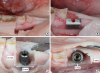
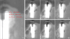
For this in vitro experiment, 5 pig mandibles were obtained from 4-month-old pigs. In each mandible, both third premolars were hemisected and the mesial roots were extracted. A standardized defect was prepared at each extraction site and an implant was placed at the centre thereof (Figure 1). According to a computer-generated randomization list, 3 different combinations of GBR materials were randomly applied to augment the peri-implant defects. Cone-beam computed tomography (CBCT) was obtained before and after suturing, and the respective horizontal thickness was measured at each site at both time-points.
Figure 1

Extraction of the mesial root of the second premolar and preparation of the bone defect. (A) Incision and hemisection of the second premolar. (B) Metallic template measuring 8 mm×6 mm×3 mm. (C) Buccal and (D) occlusal view of the standardised peri-implant bone defect showing the respective measurements.

jpis-49-14-g001
The experiment was performed by 2 surgeons. One created the bone defects, placed the implants, and performed GBR. The second researcher performed the wound closure in order to eliminate operator bias.

Preparation of the in vitro model

An intrasulcular incision was made at the buccal aspect of the second premolar. It was extended to a crestal incision at the mesial aspect. Additionally, a vertical releasing incision at the disto-buccal aspect of the second premolar was made in order to enable mobilization of the mucoperiosteal flap. A cylindrical drill was used to perform the hemi-section of the premolar and facilitate the extraction of the mesial root (Figure 1A). A metallic template measuring 8 mm×6 mm×3 mm was manufactured to standardize the bone defects (Figure 1B). The defects were created with cylindrical carbide drills. An implant with a length of 15 mm and a diameter of 4 mm (Astra Osseospeed TX®, Dentsply-Sirona Implants, Mölndal, Sweden) was inserted at the centre of each bony defect. The implant shoulder was set flush with the lingual bone crest (Figure 1C and D).

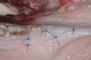
GBR/augmentation procedures and wound closure
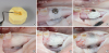
The peri-implant bone defects were augmented by randomly applying 1 of the following 3 different material combinations (Figure 2B-F):
Figure 2

Silicone mould and application of material for guided bone regeneration. (A) The silicon mould used to standardize the amount of bone substitute material. (B) Particulated demineralized bovine bone mineral used in groups 1 and 2. (C) Alloplastic bone mineral used in group 3. (D) Group 1: xenogeneic granulate+collagen membrane. (E) Group 2: xenogeneic granulate+alloplastic membrane. (F) Group 3: alloplastic granulate+alloplastic membrane.

jpis-49-14-g002
  • • Group 1: xenogeneic granulate+collagen membrane

  • Particulated demineralized bovine bone mineral (DBBM) (Bio-Oss® granules, 0.25–1 mm, Geistlich Pharma AG, Wolhusen, Switzerland)+porcine collagen membrane (Bio-Gide®, Geistlich Pharma AG) (n=10)

  • • Group 2: xenogeneic granulate+alloplastic membrane

  • Particulated DBBM (Bio-Oss® granules 0.25–1 mm, Geistlich Pharma AG)+alloplastic membrane (polylactide+acetyl tri-n-butyl citrate NF [ATBC]) (GUIDOR® bioresorbable matrix barrier, Sunstar Suisse SA, Etoy, Switzerland) (n=10)

  • • Group 3: alloplastic granulate+alloplastic membrane

  • Alloplastic in situ hardening biphasic calcium phosphate (HA, β-TCP) coated by PLGA and activated with a BioLinker (N-methyl-2-pyrrolidone solution) (GUIDOR® easy-graft® CRYSTAL, Sunstar Suisse SA)+alloplastic membrane (polylactide+ATBC) (GUIDOR® bioresorbable matrix barrier, Sunstar Suisse SA) (n=10)

In order to ensure a standardized amount of bone substitute material in all groups, the respective quantity was determined using a prefabricated silicone mould (Optosil® Comfort® Putty, Heraeus Kulzer GmbH, Hanau, Germany) (Figure 2A). In each group, the bone substitute material was adapted to the standardized bone defect measuring 8 mm×6 mm×3 mm. Substitute material was applied with a horizontal over-contour of 1 mm (Figure 2B and C). The alloplastic granulate (group 3) was activated (BioLinker® based on N-methyl-2-pyrrolidone), resulting in a sticky, but mouldable compound. Saline was applied to extract the BioLinker from the material, which subsequently led to hardening of the graft material in situ. Subsequently, the respective membrane was trimmed with scissors, with final dimensions of 12 mm×15 mm. Accordingly, it covered the respective bone substitute and the bone defect with an overlap of 2 mm at the apical, mesial, and distal borders. All membranes were fixed with 2 titanium pins (Frios Membrane Tack, DENTSPLY Sirona) at the mesial and distal apical aspects of the bone defect (Figure 2D-F).
Tension-free wound closure was performed by applying 1 horizontal mattress suture and 2 single interrupted sutures at the crestal incision (Dafilon® 5-0, B. Braun Medical AG, Sempach, Switzerland). Two additional single interrupted sutures were applied to adapt the wound margins at the vertical incision (Figure 3).
Figure 3

Wound closure. Wound closure was obtained by 1 horizontal mattress suture and 2 single interrupted sutures, each at the crestal and at the vertical releasing incision.

jpis-49-14-g003

CBCT scanning and image evaluation

The volume of the augmented area was examined before and after suturing using CBCT (I-Dixel, J. Morita MFG.CORP., Kyoto Japan), applying the following technical parameters: acceleration voltage, 90 kV; beam current, 5 mA; field of view (FOV), 10 cm×4 cm; rotation, 360°; voxel size, 0.250 mm; scan time, 17.5 seconds. The respective bone defect was positioned at the centre of the FOV by using orientational laser beams. To analyse the horizontal thickness, CBCT sections of the area perpendicular to the implant axis were enlarged using open-source image processing software (imageJ, National Institutes of Health, Bethesda, MD, USA). A transparent acetate foil displaying a printed implant and the respective levels for the implant shoulder (HT0), as well as 1 mm (HT1) and 2 mm (HT2) below the implant shoulder, was placed on each image on the computer display to facilitate the reproducibility of the measurements. The horizontal thickness of the augmented material was then measured at HT0, HT1, and HT2 on the cross-sectional images obtained from the CBCT scans (Figure 4). One examiner, blinded to the procedures, performed all measurements.
Figure 4

CBCT scan perpendicular to the implant axis before and after suturing. (A) Pre-suturing CBCT of groups 1, 2, and 3. (B) Post-suturing CBCT of groups 1, 2, and 3.

CBCT: cone-beam computed tomography, HT0: at the implant shoulder, HT1, 1 mm below the implant shoulder, HT2: 2 mm below the implant shoulder.
jpis-49-14-g004

Statistical analysis

Sample size calculation was performed using the Simple Interactive Statistical Analysis calculator (http://www.quantitativeskills.com/sisa/calculations/samsize.htm), based on a previous publication by Mir-Mari et al. [20]. The mean values and standard deviations of horizontal changes at H0 of granules alone and granules and pins were used. The standard deviation was 0.5 mm, and a difference of 0.6 (1.1–0.5) was defined as relevant. The significance level (alpha) was set to 5% and the power to 80%. Continuity correction was applied due to the expected small sample size. The study hypothesis was superiority of the test groups (groups 2 and 3) compared to the control group (group 1). The calculation revealed that a total of 27 sites (9 per group) were required. A security margin of 10% was added, resulting in a sample size of 10 per group.
The variables were described as mean, median, standard deviation, and quartiles. Mixed linear models were used for the comparison of the 3 groups due to the correlated data. Separate measurements were made for baseline values and post-treatment values, from which the changes were calculated. Post hoc tests were used for paired comparisons, applying the Bonferroni correction. The significance level was set at 5%. No correction for the multiple testing of several variables was applied for these tests of intragroup changes.

RESULTS

The descriptive data for the horizontal thickness of the augmented area before and after suturing are presented in Table 1.
Table 1

Horizontal thickness of the augmented area before and after suturing in groups 1, 2, and 3 at the different levels measured (at the implant shoulder [HT0=0 mm] and at 1 mm and 2 mm below [HT1 and HT2, respectively])
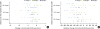
jpis-49-14-i001
Levels Suturing (mm) P values for the differences of the group means
Group 1 (n=10) Group 2 (n=10) Group 3 (n=10)
Before After P value Before After P value Before After P value Before After Change
HT0 2.55±0.53 (2.37, 2.59, 2.88) 1.47±0.31 (1.28, 1.43, 1.65) <0.001 1.94±0.56 (1.55, 1.76, 2.42) 1.77±0.27 (1.50, 1.75, 1.97) 0.723 2.49±0.73 (2.09, 2.41, 2.91) 2.00±0.48 (1.82, 1.96, 2.28) 0.002 0.055 0.011 0.020
HT1 2.44±0.56 (2.06, 2.44, 2.94 1.41±0.32 (1.28, 1.41, 1.55) <0.001 1.90±0.34 (1.76, 1.98, 2.12) 1.53±0.30 (1.44, 1.52, 1.76) 0.002 2.41±0.42 (2.06, 2.48, 2.75) 2.07±0.39 (1.85, 2.00, 2.46) 0.008 0.055 0.004 0.003
HT2 2.62±0.70 (2.34, 2.66, 2.94) 1.89±0.40 (1.52, 1.86, 2.14) <0.001 1.91±0.28 (1.60, 1.94, 2.07) 1.87±0.26 (1.68, 1.87, 2.04) 0.849 2.70±0.36 (2.33, 2.76, 2.98) 2.38±0.36 (2.26, 2.45, 2.63) 0.008 0.002 0.002 0.019
Mean values and standard deviations are given in (mm), as are values for the first quartile (Q1), median, and third quartile (Q3). P values show the results from the t-test for the changes.
HT0: at the implant shoulder, HT1: 1 mm below the implant shoulder, HT2: 2 mm below the implant shoulder.
The mean thickness of the mucosal flap before suturing was 1.94±0.56 mm (group 2), 2.55±0.53 mm (group 1), and 2.49±0.73 mm (group 3) at the level of the implant shoulder (HT0). Group 2 showed considerably less horizontal thickness of the augmented area at all measured levels (HT0, HT1, and HT2). The intergroup differences in the mean values of each group before suturing were only significantly different at 2 mm below the implant shoulder (HT2) (P=0.002) (Table 2).
Table 2

Changes in horizontal thickness for each group and comparisons among the 3 groups
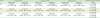
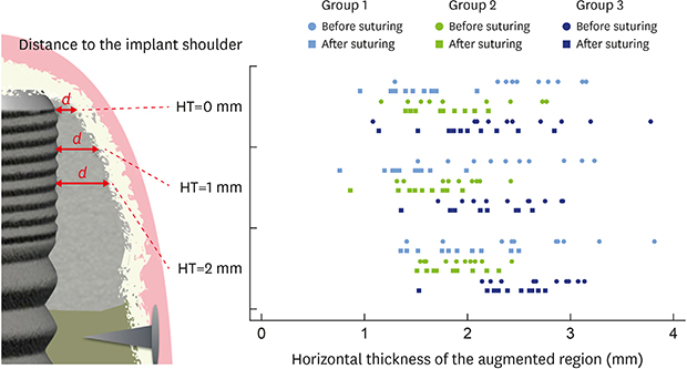
jpis-49-14-i002
Levels Group 1 (n=10) Group 2 (n=10) Group 3 (n=10) Statistical analysis: adjusted P value
Change (mm) Change (%) Change (mm) Change (%) Change (mm) Change (%)
HT0 −1.08±0.55 (−1.41, −1.07, −0.86) −39.84±17.92 (−49.03, −41.42, −33.43) −0.17±0.53 (−0.74, −0.11, 0.24) −3.03±26.19 (−28.97, −4.62, 20.73) −0.48±0.39 (−0.68, −0.43, −0.25) −17.12±12.83 (−24.65, −15.14, −12.20) 1 vs. 2: 0.033
1 vs. 3: 0.015
2 vs. 3: 0.410
HT1 −1.03±0.46 (−1.30, −0.92, −0.72) −41.11±12.72 (−52.02, −36.12, −32.26) −0.37±0.29 (−0.49, −0.40, −0.14) −18.93±13.83 (−26.94, −19.75, −6.70) −0.34±0.28 (−0.52, −0.38, −0.26) −13.47±12.60 (−21.68, −14.74, −9.48) 1 vs. 2: 0.002
1 vs. 3: 0.006
2 vs. 3: 1.000
HT2 −0.73±0.50 (−1.05, −0.61, −0.39) −25.58±13.52 (−36.61, −21.78, −17.61) −0.04±0.29 (−0.27, −0.04, 0.23) −0.93±14.97 (−13.77, −2.34, 11.14) −0.32±0.27 (−0.57, −0.34, −0.14) −11.45±10.74 (−19.14, −10.90, −5.34) 1 vs. 2: 0.011
1 vs. 3: 0.073
2 vs. 3: 0.466
Mean values and standard deviations are given in (mm), as are values for the first quartile (Q1), median, and third quartile (Q3). The P values were obtained from analysis of variance of the post hoc between-group comparisons adjusted for multiple testing.
HT0: at the implant shoulder, HT1: 1 mm below the implant shoulder, HT2: 2 mm below the implant shoulder.
After suturing, the greatest mean horizontal thickness was measured in group 3 (2.00±0.48 mm) at the level of the implant shoulder (HT0). The corresponding value in group 2 was 1.77±0.27 mm, whereas group 1 showed the least horizontal thickness (1.47±0.31 mm) (Table 1, Figure 5). The mixed model test showed significant differences among the groups at all levels (HT0: P=0.011; HT1: P=0.004; HT2: P=0.002) (Table 2).
Figure 5

Diagram showing all values for horizontal thickness of the augmented region buccal of the implant before and after suturing at the 3 levels measured. Skyblue: group 1; green: group 2; blue: group 3.
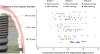
HT0: at the implant shoulder, HT1: 1 mm below the implant shoulder, HT2: 2 mm below the implant shoulder.
jpis-49-14-g005
A decrease in horizontal thickness was observed after suturing in all 3 groups. At the level of the implant shoulder (HT0), the greatest mean change (before and after suturing) was measured in group 1 (−1.08±0.55 mm), compared to −0.48±0.39 mm in group 3 and −0.17±0.53 mm in group 2 (Table 2, Figure 6). The within-group changes were highly significant in group 1 (HT0: P<0.001; HT1: P<0.001; HT2: P<0.001) and group 3 (HT0: P=0.002; HT1: P=0.008; HT2: P=0.008) at all measured levels. However, group 2 only showed statistical significance at HT1 (P=0.002) (Table 1).
Figure 6

Diagram showing the values for the absolute changes (mm) and the relative changes (%) in horizontal thickness at the different levels measured (HT0, HT1, HT2) for the 3 investigated groups. (A) Change in horizontal thickness in (mm). (B) Relative change in horizontal thickness in (%). Skyblue: group 1; green: group 2; blue: group 3.

HT0: at the implant shoulder, HT1: 1 mm below the implant shoulder, HT2: 2 mm below the implant shoulder.
jpis-49-14-g006
Intergroup differences in the changes of thickness before and after suturing were significant between group 1 and 2 at HT0 (P=0.033), HT1 (P=0.002), and HT2 (P=0.011), as well as between group 1 and group 3 at HT0 (P=0.015), HT1 (P=0.006), and HT2 (P=0.073). No statistical significance was found between groups 2 and 3 at any level (Table 2).

DISCUSSION

The results of the present study demonstrated: 1) that group 2 (xenogeneic bone substitute material and an alloplastic membrane) had less horizontal thickness before suturing despite augmentation with identical volumes of bone substitute materials in all groups; 2) that the greatest horizontal thickness after suturing occurred when a combination of an alloplastic substitute and membrane was used (group 3); and 3) that the least (non-significant) change in horizontal thickness occurred when the combination of a xenogeneic bone substitute material with an alloplastic membrane was used.
To date, the majority of pre-clinical and clinical studies have investigated the use of non-resorbable and resorbable membranes. Non-resorbable membranes have been reported to result in an increased rate of dehiscence [1314]. Even though the rate of dehiscence for resorbable membranes appears to be similar, the clinical consequences are far less dire. This is predominantly due to the fact that resorbable membranes, even in case of early exposure, do not need to be removed and may (when native collagen membranes are used) heal without further intervention [15]. Nevertheless, resorbable membranes fail to maintain space per se and can result in displacement of graft material [192022]. Thus, the use of resorbable and stiffer alloplastic membranes might combine the advantages of non-resorbable, form-stable membranes with those of resorbable collagen membranes.
Group 2 showed the greatest stability in terms of changes in horizontal thickness before and after suturing. The changes between the 2 measurements were non-significant, indicating that this group showed the most stable combination of bone substitute material and membrane. Nevertheless, group 2 did not show the highest values of overall thickness. Although a standardized amount of bone substitute material was used in all groups, the volume obtained before suturing differed. One reason for the lower values of the initial thickness in group 2 (xenogeneic bone substitute material and an alloplastic membrane) might have been compression of the loose xenogeneic granules by the stiffer alloplastic membrane. In contrast to the other 2 groups, this seems to have led to a flattening of the augmented volume prior to suturing. This effect was not observed in group 1 (xenogeneic granules and xenogeneic membrane) or group 3 (alloplastic substitute and alloplastic membrane). Thus, it seems that the alloplastic membrane did not compress the hardened alloplastic granules in a similar way. In contrast, the granules cohered by the PLGA coating after applying the BioLinker (N-methyl-2-pyrrolidone solution) appeared to have formed a more volume-stable entity even before the application of the alloplastic membrane. A limitation of the present study might thus be the fact that a fourth group consisting of alloplastic bone substitute in combination with a xenogeneic membrane was not investigated. It would also have been interesting to investigate the volumetric behaviour of the alloplastic bone substitute material without the use of a membrane. Nevertheless, due to statistical considerations and in order to avoid analysing too many parameters and groups, it was decided to investigate a total of 3 groups. Thus, the results of this study are more representative of the behaviour of the alloplastic membrane than of the substitute material. Further research, potentially including clinical studies, might explore such combinations.
A further aspect to consider regarding volume stability is the use of pins for fixation of the resorbable collagen membrane [20]. Whilst the mere covering of particulated bone substitute material with the membrane led to a significant change in volume, these changes did not occur when the membrane was held in place by 2 pins [20]. Thus, in order to maximize stability, pins were used in all 3 investigated groups to prevent displacement of the respective membrane and the underlying bone substitute. In the present study, the least change in horizontal thickness was obtained when a combination of an alloplastic substitute and an alloplastic membrane was used. These results are limited, though, by the fact that the measurements were only obtained after covering the bone substitute material with the respective membrane, and no measurements were made before the membrane was placed. Furthermore, these results are limited to an in vitro setting. Therefore, pre-clinical and clinical investigations will need to be conducted, as well as studies of the histologic outcomes and degradation of these materials.
All investigated groups showed a decrease in horizontal thickness due to suturing. The least volume loss was observed when a xenogeneic bone substitute material and an alloplastic membrane were applied. In order to maximize the overall horizontal thickness, an alloplastic substitute material in conjunction with an alloplastic membrane led to most favourable results. Regardless of the bone substitute used, the alloplastic membrane provided superior (overall volume) stability following suturing and wound closure.

ACKNOWLEDGMENTS

This study was supported by a grant from Sunstar Suisse SA, Etoy, Switzerland and the Clinic of Fixed and Removable Prosthodontics and Dental Material Science, Center for Dental Medicine, University of Zurich, Switzerland. The authors report no conflict of interest.

Notes

Funding This study was funded by Sunstar Suisse SA, Etoy, Switzerland and the Clinic of Fixed and Removable Prosthodontics and Dental Material Science, Center of Dental Medicine, University of Zurich, Zurich, Switzerland.

Author Contributions

  • Conceptualization: Nadja Naenni, Daniel Thoma, Christoph Hämmerle.

  • Data curation: Tanja Berner, Tobias Waller.

  • Formal analysis: Juerg Huesler, Tanja Berner, Nadja Naenni.

  • Funding acquisition: Nadja Naenni, Daniel Thoma.

  • Investigation: Tanja Berner, Tobias Waller.

  • Methodology: Nadja Naenni, Daniel Thoma, Christoph Hämmerle.

  • Project administration: Nadja Naenni.

  • Resources: Nadja Naenni, Daniel Thoma.

  • Software: Tanja Berner.

  • Supervision: Nadja Naenni.

  • Validation: Nadja Naenni, Juerg Huesler.

  • Visualization: Tanja Berner, Juerg Huesler.

  • Writing - original draft: Nadja Naenni, Tanja Berner, Tobias Waller, Juerg Huesler, Daniel Thoma.

  • Writing - review & editing: Nadja Naenni, Tanja Berner, Daniel Thoma, Juerg Huesler.

Conflict of Interest No potential conflict of interest relevant to this article was reported.

References

1. Hämmerle CH, Karring T. Guided bone regeneration at oral implant sites. Periodontol 2000. 1998; 17:151–175.
crossref pmid
2. Bornstein MM, Halbritter S, Harnisch H, Weber HP, Buser D. A retrospective analysis of patients referred for implant placement to a specialty clinic: indications, surgical procedures, and early failures. Int J Oral Maxillofac Implants. 2008; 23:1109–1116.
pmid
3. Benic GI, Hämmerle CH. Horizontal bone augmentation by means of guided bone regeneration. Periodontol 2000. 2014; 66:13–40.
crossref
4. Chiapasco M, Casentini P, Zaniboni M. Bone augmentation procedures in implant dentistry. Int J Oral Maxillofac Implants. 2009; 24:Suppl. 237–259.
pmid
5. Donos N, Mardas N, Chadha V. Clinical outcomes of implants following lateral bone augmentation: systematic assessment of available options (barrier membranes, bone grafts, split osteotomy). J Clin Periodontol. 2008; 35:173–202.
crossref pmid
6. Karring T, Nyman S, Gottlow J, Laurell L. Development of the biological concept of guided tissue regeneration--animal and human studies. Periodontol 2000. 1993; 1:26–35.
crossref
7. Nyman S, Lindhe J, Karring T, Rylander H. New attachment following surgical treatment of human periodontal disease. J Clin Periodontol. 1982; 9:290–296.
crossref pmid
8. Gottlow J, Nyman S, Karring T, Lindhe J. New attachment formation as the result of controlled tissue regeneration. J Clin Periodontol. 1984; 11:494–503.
crossref pmid
9. Dahlin C, Lekholm U, Becker W, Becker B, Higuchi K, Callens A, et al. Treatment of fenestration and dehiscence bone defects around oral implants using the guided tissue regeneration technique: a prospective multicenter study. Int J Oral Maxillofac Implants. 1995; 10:312–318.
pmid
10. Scantlebury TV. 1982–1992: a decade of technology development for guided tissue regeneration. J Periodontol. 1993; 64:1129–1137.
crossref
11. Jung RE, Fenner N, Hämmerle CH, Zitzmann NU. Long-term outcome of implants placed with guided bone regeneration (GBR) using resorbable and non-resorbable membranes after 12–14 years. Clin Oral Implants Res. 2013; 24:1065–1073.
crossref pmid
12. Aghaloo TL, Moy PK. Which hard tissue augmentation techniques are the most successful in furnishing bony support for implant placement? Int J Oral Maxillofac Implants. 2007; 22:Suppl. 49–70.
13. Zitzmann NU, Naef R, Schärer P. Resorbable versus nonresorbable membranes in combination with Bio-Oss for guided bone regeneration. Int J Oral Maxillofac Implants. 1997; 12:844–852.
pmid
14. Moses O, Pitaru S, Artzi Z, Nemcovsky CE. Healing of dehiscence-type defects in implants placed together with different barrier membranes: a comparative clinical study. Clin Oral Implants Res. 2005; 16:210–219.
crossref pmid
15. Naenni N, Schneider D, Jung RE, Husler J, Hammerle CH, Thoma DS. Randomized clinical study assessing two membranes for guided bone regeneration of peri-implant bone defects: clinical and histological outcomes at 6 months. Clin Oral Implants Res. 2017; 28:1309–1317.
crossref pmid
16. Rothamel D, Schwarz F, Sager M, Herten M, Sculean A, Becker J. Biodegradation of differently cross-linked collagen membranes: an experimental study in the rat. Clin Oral Implants Res. 2005; 16:369–378.
crossref pmid
17. Lundgren AK, Sennerby L, Lundgren D, Taylor A, Gottlow J, Nyman S. Bone augmentation at titanium implants using autologous bone grafts and a bioresorbable barrier. An experimental study in the rabbit tibia. Clin Oral Implants Res. 1997; 8:82–89.
crossref pmid
18. von Arx T, Broggini N, Jensen SS, Bornstein MM, Schenk RK, Buser D. Membrane durability and tissue response of different bioresorbable barrier membranes: a histologic study in the rabbit calvarium. Int J Oral Maxillofac Implants. 2005; 20:843–853.
pmid
19. Strietzel FP, Khongkhunthian P, Khattiya R, Patchanee P, Reichart PA. Healing pattern of bone defects covered by different membrane types--a histologic study in the porcine mandible. J Biomed Mater Res B Appl Biomater. 2006; 78:35–46.
crossref pmid
20. Mir-Mari J, Wui H, Jung RE, Hämmerle CH, Benic GI. Influence of blinded wound closure on the volume stability of different GBR materials: an in vitro cone-beam computed tomographic examination. Clin Oral Implants Res. 2016; 27:258–265.
crossref pmid
21. Mir-Mari J, Benic GI, Valmaseda-Castellón E, Hämmerle CH, Jung RE. Influence of wound closure on the volume stability of particulate and non-particulate GBR materials: an in vitro cone-beam computed tomographic examination. Part II. Clin Oral Implants Res. 2017; 28:631–639.
crossref
22. Friedmann A, Strietzel FP, Maretzki B, Pitaru S, Bernimoulin JP. Histological assessment of augmented jaw bone utilizing a new collagen barrier membrane compared to a standard barrier membrane to protect a granular bone substitute material. Clin Oral Implants Res. 2002; 13:587–594.
crossref pmid
TOOLS
ORCID iDs

Nadja Naenni
https://orcid.org/0000-0002-6689-2684

Tanja Berner
https://orcid.org/0000-0002-3861-1929

Tobias Waller
https://orcid.org/0000-0003-0158-1866

Juerg Huesler
https://orcid.org/0000-0002-0946-8510

Christoph Hans Franz Hämmerle
https://orcid.org/0000-0002-8280-7347

Daniel Stefan Thoma
https://orcid.org/0000-0002-1764-7447

Similar articles