

 PDF
PDF ePub
ePub Citation
 Print
Print


INTRODUCTION
There are many limiting conditions associated with implant surgery on the upper posterior area, such as maxillary sinus pneumatization and insufficient bone volume. These limitations have been addressed by the development and application of a variety of surgical techniques, including sinus augmentation. In the service of more successful clinical outcomes for these techniques, studies on growth factors have been actively carried out over the past few decades and have proven to be essential. One of these growth factors, bone morphogenetic protein (BMP), is known to play an important role in osteoblast differentiation, and its ability to accelerate new bone formation has been demonstrated in many in vivo studies.
Despite the promising effects of BMP in sinus augmentation, adverse effects such as severe swelling and ectopic or delayed bone formation have also been reported. Such adverse effects have been reported mainly to be caused by an overdose of BMP. Therefore, the current trend of research on BMP has focused on finding the appropriate dose. In particular, the sinus augmentation procedure requires a considerable amount of bone graft material compared to other dental procedures, and so there is a risk of applying more BMP than necessary, which could result in severe complications. In consideration of such problems, it is important to carry out studies to find the minimum dose of BMP for successful sinus augmentation. As a minimal dose, a 0.1 mg/mL concentration of BMP loaded onto biphasic calcium phosphate (BCP) has been used in a previous rabbit sinus augmentation study and this has proven effective for total augmentation and new bone volume [1]. The study also suggested that experiments using a dose lower than 0.1 mg/mL were necessary for determining the ideal concentration of BMP. Therefore, BMP at a concentration of 0.05 mg/mL, which is half of 0.1 mg/mL, was used for this study to determine the proper dose in a rabbit sinus model.
BCP is comprised of hydroxylapatite (HA) and β-tricalcium phosphate (TCP) at various ratios. It has been extensively investigated as a bone substitute for its space-maintaining capacity, similarity to human bone, and biocompatibility [2], especially for sinus augmentation procedures [3]. Since HA is less soluble than β-TCP, HA can act as a scaffold for maintaining grafted volume in order to prevent sinus pneumatization after augmentation, thus leading to enhanced osteoconductivity. On the other hand, the solubility of TCP allows BCP to be rapidly replaced by newly formed bone (NB) via particle biodegradation. The bioactivity of BCP—including its osteoconductivity and resorption rate—may be controlled by altering the HA/β-TCP ratio. Recently, a study has demonstrated that any BCP, regardless of the ratio between HA and TCP, is useful as a sinus graft material [3]. However, a higher proportion of TCP in BCP provided higher osteoconductivity and biocompatibility at the histological level and clinically successful outcomes for sinus augmentation procedures [4]. Thus, in the present study, BCP with an HA/β-TCP ratio of 20/80 was chosen as the graft material for sinus augmentation.
The purpose of the present study was to determine the efficacy of a minimal dose of BMP-2 with a BCP carrier in a rabbit sinus model.
MATERIALS AND METHODS
Animals
Seven male New Zealand white rabbits weighing 2.8–3.2 kg were used for this study. They were maintained in separate cages and fed a controlled diet under standard laboratory conditions. The sample size calculation was performed with G*Power software v. 3.1 (University of Dusseldorf, Dusseldorf, Germany) at an alpha level of 0.05 and a statistical power of 95%. The histometric difference in new bone area between 2 groups was set to 2.18 mm2 and the standard deviation of the outcome was assumed to be the same for both groups in accordance with the previous study [5]. The required sample size per group was 6; therefore, 7 rabbits were prepared in consideration of possible loss during histologic slide preparation. The animal selection, preparation, management, and surgical protocol were approved by the Institutional Animal Care and Use Committee of Yonsei Medical Center, Seoul, Korea.
Preparation of BMP-2 and carriers
The BCP (MBCP plus, Biomatlante, Nantes, France) used as the carrier for BMP-2 (Korea Bone Bank, Seoul, Korea) in this study had an HA/β-TCP ratio of 20/80. The BMP-2 was reconstituted and diluted in a buffer to a concentration of 0.05 mg/mL. In the BMP-2-treated group, 0.15 g of BCP was loaded with 0.1 mL of BMP-2 for 1 hour to obtain an implant volume of 5-μg BMP-2. In the control group, the BMP-2 was replaced by 0.15 g of BCP soaked in saline.
Surgical procedure and study design
All of the rabbits were anesthetized prior to the surgery by intramuscular injection with a mixture of ketamine hydrochloride (Ketalar, Yuhan, Seoul, Korea) and xylaxine (Rompun, Bayer Korea, Seoul, Korea). Local anesthesia was achieved by subcutaneous injection of 0.5 mL of 2% lidocaine (Lidocaine HCl, Huons, Seoul, Korea). After preoperative shaving and local disinfection with iodine, a straight midline incision was made on the dorsal side of the nasal bone and a full-thickness flap was elevated laterally. Standardized circular, bilateral cranial windows were prepared with the aid of a circular reamer (5.5-mm-diameter C-reamer, Neobiotech, Seoul, Korea) under cooling with saline. The windows were placed 10 mm laterally to the midline and 20 mm anteriorly to the nasofrontal suture line [6]. The circular bony discs made by the reamer were removed and the sinus membrane was carefully elevated to create a space for grafting of the materials (Figure 1).
Figure 1
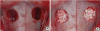
Photographs showing the surgical procedure. (A) Bilateral sinus windows were cut using a 5.5-mm circular reamer and (B) graft materials were placed into those windows: the test material (BMP-2-loaded BCP) on one side and the control material (saline-soaked BCP) on the other.
BMP, bone morphogenetic protein; BCP, biphasic calcium phosphates.
The graft materials were placed into the sinus windows, with BMP-2-loaded BCP into one (the BMP group) and saline-soaked BCP into the other (the control group), where the side was randomly assigned for each animal. When the implantation of graft materials was complete, the skin and periosteal flap were sutured layer by layer with 4-0 glyconate absorbable monofilament (Monosyn, B-Braun Aesculap, Allentown, PA, USA). The rabbits were sacrificed 8 weeks after the surgery.
Radiographic evaluations: micro-computed tomography
The specimens including the augmented area and surrounding tissues were removed and rinsed with sterile saline. All of the specimens were fixed in 10% formalin for 10 days. Radiographic images were then taken with the aid of a micro-computed tomography (micro-CT) scanner (Skyscan 1072, Skyscan, Aartselaar, Belgium) at a resolution of 35 μm (100 kV and 100 μA). The scanned images were reconstructed and the total augmented volumes of both sinuses were measured using OnDemand3D software (CyberMed, Seoul, Korea).
Histologic processing and histometric evaluations
Following the micro-CT scanning, the specimens were decalcified in 5% formic acid for 14 days and then embedded in paraffin. The embedded blocks were sectioned at a thickness of 5 μm along the most augmented area, mounted onto glass slides, and then stained with hematoxylin/eosin and Masson trichrome. All of the slides were examined with the aid of a light microscope (BX50, Olympus, Tokyo, Japan), and the fields were captured digitally.
Histometric measurements were acquired with an automated image analysis program (Image Pro Plus, Media Cybernetics, Silver Spring, MD, USA). The measured parameters included total augmented area, area of NB, and that of residual particles (RP). The fibrovascular tissues (FV) were measured in numerical values, which is the total augmented area without NB and RP. The proportion of NB, RP, and FV were calculated and compared. As a secondary variable, the length of each RP was measured in all specimens to evaluate the resorption pattern of RP between the groups. The length of RP was defined as the longest length of particle that was automatically measured by using the software.
Statistical analysis
The software program R v. 3.1.1 (University of Auckland, Auckland, New Zealand) was used for the statistical analysis. A nonparametric mixed model [7] was used to evaluate the differences between the 2 groups with respect to total augmented volume, total augmented area, area of NB, area of RP, and the proportion of NB, RP, and FV. A linear mixed model was used to compare the length of RP. The statistical significance level was set at 5%.
RESULTS
Clinical findings
During the surgery, 4 small (<1 mm) sinus membrane perforations (2 in the control group, 2 in the BMP group) occurred. Because of the small size of the perforations, additional surgical procedures for repairing the sinus membrane were not attempted. Nevertheless, the overall healing of all animals was uneventful during the 8-week recovery period. No signs of inflammation nor other pathologic symptoms were detected.
Micro-computed tomography evaluations
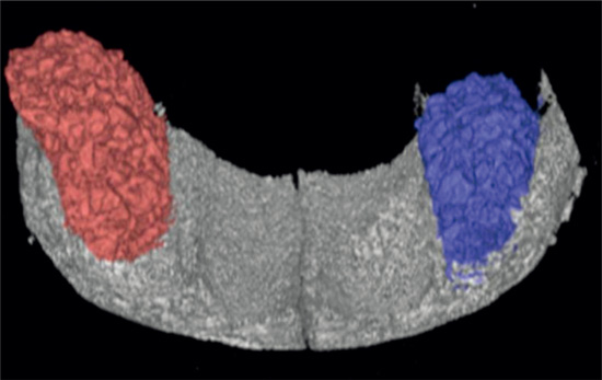
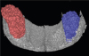
A dome-shaped augmented mass with a radiopaque matrix including RP could be observed in both groups. There was no radiolucent void within the augmented sinus cavity in both groups. Although the total augmented volume appeared to be greater in the BMP group (149.50±48.60 mm3) than in the control group (139.69±69.40 mm3) at 8 weeks after the surgery, the difference was not statistically significant (Figure 2).
Histologic and histometric evaluation
Histologically, the augmented sinus cavities had a convex form and fully filled with NB, RP, and FV. There were no signs of inflammation in both groups of sinus cavities. The sinus window regions were almost regenerated with NB in both groups. Despite the occurrence of small-sized sinus membrane perforations in 4 animals during the surgical procedure, the linings of the Schneiderian membranes were intact in all animals. The new bone formation was observed in both the central and peripheral areas of the augmented sinuses in both the BMP and control sites. Furthermore, the NB was closely attached to the residual particles, with no intervening spaces. The RP had an irregular surface, possibly due to the resorption of particles. The patterns of FV were different between the 2 groups; dense connective tissues were observed in the control group, whereas loose connective tissues were mainly observed in the BMP group (Figure 3).
Figure 3
Histological images. (A, B) Low magnification images of the BMP and control group, respectively (scale bar=1 mm); (C, D) high magnification images of the central portion of the augmented area, the BMP and control group, respectively (scale bar=100 μm); (E, F) high magnification images of the sinus membrane area, the BMP and control group, respectively (scale bar=100 μm).
NB, new bone; RP, residual particles; FV, fibrovascular tissue; SM, Schneiderian membrane, Masson trichrome staining; BMP, bone morphogenetic protein.
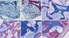
Interestingly, there was a difference in the mineralization pattern of NB between the BMP and control group. In the BMP group, the greater part of NB was mature lamellar bone with evident trabecular pattern and Harversian system. On the other hand, NB of the control group showed mostly woven bone that was partially lamellar bone (Figure 4).
Figure 4
Highly magnified histologic images of (A) the BMP group and (B) the control group. The NB of the BMP group showed more maturation than the NB of the control group (white asterisks; the Haversian canals, Masson trichrome stains; scale bar=100 μm).
BMP, bone morphogenetic protein; NB, new bone; LB, lamellar bone; RP, residual particles; WB, woven bone.
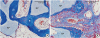
Table 1 shows the results of the histometric analysis. In the BMP group, the total augmented area appeared to be greater than in the control group, although the difference was not statistically significant. The area of NB in the BMP group (4.55±1.35 mm2) was significantly greater than that in the control group (2.99±0.86 mm2; P<0.05), while there was no statistical difference in the area of RP. Likewise, there was no difference in the length of RP between the groups.
Table 1
Histometric analysis

| Group | TAA (mm2) | Area of NB (mm2) | Area of RP (mm2) | Length of RP (mm) |
|---|---|---|---|---|
| Control group (n=7) | 10.38±2.36 | 2.99±0.86a) | 3.75±0.69 | 0.47±0.23 |
| BMP group (n=7) | 13.26±5.33 | 4.55±1.35 | 3.83±1.58 | 0.45±0.22 |
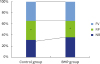
Figure 5 represents the proportion of NB, RP, and FV between the 2 groups. At 8 weeks after the grafting, notwithstanding the greater proportion of NB and FV of the BMP group, there were no significant differences. However, the proportion of RP was significantly less in the BMP group (29.40%±4.61% vs. 36.64%±4.62%; P<0.05).
Figure 5
The proportion of NB, RP, and FV. The proportion of RP in the BMP group was significantly less than the control group (P<0.05).
NB, new bone; RP, residual particles; FV, fibrovascular tissue; BMP, bone morphogenetic protein.
*Statistically significant difference between the two groups (P<0.05).
DISCUSSION
The purpose of this study was to evaluate the effectiveness of a minimal dose of BMP-2 with BCP composed of a high ratio of TCP carrier in a rabbit sinus augmentation model over 8 weeks. Although the concentration of BMP-2 used in the present study was only half of the lowest concentration of previous studies (0.05 mg/mL), enhanced osteoinductivity and accelerated bone maturation could be observed compared to the control group. It might be suggested that the use of a minimal dose of BMP-2 with BCP improves the quantity of newly formed bone and shortens the healing time following the sinus augmentation procedure.
As a principle finding of this study, more mineralization of the newly formed bone could be observed in the BMP group than in the control group. This result is in concordance with a previous in vivo study that investigated bone formation with BMP-2 in the rabbit maxillary sinus during the early healing period [1]. Furthermore, a recent clinical trial showed a significantly higher amount of mineralized bone formation in the higher expression of the BMP-4 group after maxillary sinus augmentation [8], which indicates that faster and greater bone formation would be induced by the BMPs.
There have been many other studies to determine the appropriate concentration of BMP to enhance bone formation in sinus augmentation and achieve minimal side effects at the same time. In previous studies, the new bone formation was significantly greater in the control group than in the test group, with a relatively high concentration of 1.5 mg/mL of BMP, using BCP carrier, regardless of the loading methods, over 2 weeks [59]. The researchers suggested that the extremely high concentration of BMP would inhibit bone regeneration and increase tissue swelling and inflammatory response in rabbits because of excessive initial release of the growth factor. These studies concluded that a 1.5 mg/mL concentration of BMP would be excessive in a rabbit defect model, though this concentration was approved for human use [10]. Therefore, studies with a lower dose in the range 0.10–0.15 mg/mL of BMP-2 have been conducted in rabbit sinuses with various healing periods. In another previous study with 0.1 mg/mL of BMP-2, new bone formation of the BMP group was statistically greater than that of the control group [11]. Kim et al. [1] reported similar data with the same concentration of BMP-2 in a previous study and the area of newly formed bone of the BMP group was significantly greater than that of the control group at weeks 2 and 4 of the healing period. On the other hand, there was a report that 0.15 mg/mL of BMP-2-coated BCP could not exert a significant effect despite the volumetric increase in the early phase [12]. This controversy might be due to the release kinetics of BMP according to the loading methods of BMP, such as soaking or coating. This should be further investigated in future studies.
It has been suggested that the prerequisites of optimal BMP-2 carriers are that they should be osteoconductive, noncollagenous, and biodegradable [13]. BCP particles satisfy these characteristics, and are widely accepted as an appropriate scaffold for BMP-2 carriage [14]. Synthetic substances such as BCP affect cellular and vascular ingrowth [15], and osteogenic cell attachment and proliferation [14]. Kim et al. [16] used block-type BCP as a carrier for BMP-2 in rabbit calvarium vertical augmentation models, and Jang et al. [17] used BCP as a carrier for various concentrations of BMP-2 in rats. These previous reports provide evidence for the suitability of BCP as a carrier for BMP-2.
Lim et al. [18] recently reported that BCP with low and high proportions of TCP had similar effects on bone regeneration and space-maintaining capabilities in a rabbit sinus for up to 8 weeks. Likewise, in the present study, the BCP was well maintained in its structure even at the higher ratio of TCP (80%). In a previous in vivo study, the resorption of BCP with increased inflammatory reaction has been suggested as a drawback of using high concentrations of BMP [9]. Another clinical study reported that the surface of the BCP exhibited irregular morphology in the BMP group, possibly as a result of superficial resorption of the BCP [19]. However, our finding bypassed this limitation by using a minimal concentration of BMP and producing similar area and length of RP between the groups.
There are 2 different methods (lyophilization or soaking) to load BMP onto carriers. The soaking method, which was used in this study, is the Food and Drug Administration (FDA)-approved loading method and is relatively easy and saves time considerably. However, there are concerns that this may result in uncontrolled release and uneven distribution of the BMP. A coating method using lyophilization has been shown to have a sustained release profile in in vitro models [11], but the complicated process is a shortcoming of this method. There are no studies comparing the effect of BMP according to different loading methods to the best of our knowledge. Further study is needed to evaluate the influence of these methods.
Interestingly, the density of fibrovascular tissue was different between the groups in histologic analysis. Denser connective tissue was observed in the control group than in the BMP group. Previous in vitro study has demonstrated that the pronounced adipose tissue was formed by high concentrations of BMP-2 [20]. However, in the present study, a large amount of new bone was obtained with limited adipose tissue formation at the minimal concentrations of the BMP group. Further study seems to be needed to investigate the effect of BMP-2 on the composition of soft tissue as well as its influence on the clinical success of implantation.
In the present study, there were some limitations, especially during the healing period. It is known that bone metabolism is 3–4 times faster in rabbits than in humans. Thus, a healing period of 8 weeks in rabbits could be considered equivalent to 6–8 months, or the late healing phase, in humans [21], which is enough time to regenerate the new bone. Therefore, we could only observe the end stage of bone formation after the 8-week healing period. Various periods of study are necessary to confirm the bone formation process and tissue maturation effect of BMP-2.
The results of this study showed lower values of total augmented area than previous studies that have investigated the effect of BMP-2 in the same experimental model [15911]. The smaller total augmented area could be associated with characteristics of the grafted material. In addition, the difference in the degree of sinus membrane elevation could induce the difference in total augmented volume and area. Moreover, in the present study, the hypothetical concentration of 0.05 mg/mL was selected intuitionally below the threshold concentration of 0.1 mg/mL, which had been verified in a previous study. However it should be considered that a study design that utilizes a gradient of BMP-2 concentration would be more appropriate for finding the minimal threshold concentration. Thus in continuation of the current focus of finding the minimal concentration of BMP-2 for sinus augmentation, the results of our experiment indicate the necessity of future studies using gradients of concentrations of BMP-2 lower than 0.05 mg/mL. Additional immunohistochemical analysis using antibodies such as osteocalcin would be helpful to compare the remaining osteogenic potential and mineralization of the newly formed bone during the 8-week healing period. Full clarification of this may be necessary in further studies.
In conclusion, it can be suggested that BCP soaked with a 0.05 mg/mL dose of BMP-2 significantly enhances new bone formation and accelerates bone maturation.
 XML Download