

 PDF
PDF ePub
ePub Citation
 Print
Print


INTRODUCTION
Tooth avulsion is considered a severe form of dental trauma because the involved tooth is completely displaced from the alveolar socket, thus causing damage to the periodontium of the tooth [1]. The established vascular system of the periodontium is destroyed, the immediate vacant space is filled mostly with blood clots that further disconnect the avulsed tooth from the socket, and thus the supply of blood and nutrition to it is insufficient [2]. More seriously, the periodontal ligament (PDL) may be ruptured and damaged by the traumatic pressure, and may become infected. Such damage to the PDL can cause root resorption, tooth-bone ankylosis, or tissue necrosis, and these outcomes end in transplantation failure [3,4]. The presence of healthy PDL tissues and cells is known to be a pivotal factor for both successful tooth transplantation and avulsed tooth replantation [3,5]. Many studies have therefore been performed to determine the most appropriate methods of preserving healthier PDL tissues, and to develop a socket-preparation method to promote more effective tooth replantations.
Milk, Hanks' balanced salt solution, University of Wisconsin solution, and Euro-Collins solution all have favorable preservation effects for up to 24 hours after extraction [6-10]. Meanwhile, Nethander et al. [2] introduced the two-stage surgical technique for tooth transplantation, in which they left the recipient socket to heal 14 days before implanting the tooth; this delayed tooth transplantation technique has yielded outstanding results, with lower rates of resorption and ankylosis being found in many studies [2,11-14]. However, the preservation period of the suggested storage solutions was found to be insufficient, since they were unable to maintain PDL cell viability for 2 weeks, and inflammatory or replacement (ankylosis) resorption still occurred in the delayed tooth transplantation technique [3,11,15]. Furthermore, when patients suffer from more serious injuries than dental avulsion, they may not receive the most appropriate dental treatments in some cases, even leaving the exfoliated tooth for a few days until replantation. Therefore, it seems to be essential to develop storage agents that can maintain the viability and function of PDL tissues for a much longer period, and, if it could, can control the inflammatory reactions or infections at the recipient site.
(-)-Epigallocatechin-3-gallate (EGCG), a major polyphenol of green tea, reportedly exerts various biological effects including cytostatic properties for preserving cells, antibacterial and anti-inflammatory reactions, and antioxidant and anticarcinogenic effects [16-18]. In particular, it has been demonstrated that EGCG is effective in preserving mammalian cells and tissues, such as blood vessels, corneas, nerves, islet cells, myocardium, and hematopoietic stem cells [19-22], and their excellent histological and biomechanical recovery was also demonstrated for up to 3 months after storage in EGCG [22]. It was also reported that the high cell viability of guinea pig incisor PDL cells could be maintained using EGCG [23], and we have confirmed the ability of EGCG to control the viability of human PDL (hPDL) fibroblasts [24]. In addition, EGCG was reported to improve the periodontal condition in periodontitis and periapical lesions due to its bactericidal effect against periodontal pathogens [25,26], and to inhibit the production of related cytokines and their inflammatory pathways, such as carbon tetrachloride, tumor necrosis factor-α, nuclear factor-κB, cycolooxygenase-2, and inducible nitric oxide synthase [27-30]. In addition, it was demonstrated that EGCG concentration-dependently inhibits the formation of bone-resorbing cells (i.e., osteoclasts and osteoclast-like multinucleated cells) and induces their apoptotic cell death [31,32].
Given the protective potential of EGCG treatment against destruction of the periodontium, it may be used to promote favorable tooth transplantation with lower rates of infection and less root resorption and ankylosis, which are major reasons for failure of the procedure. If the preservative and therapeutic properties of EGCG can also be applied in other dental treatments, patients may experience better rehabilitative treatment. This prompted the present study to evaluate the effect of EGCG on the preservation of teeth extracted from Beagle dogs and the maintenance of their PDL cell viability.
MATERIALS AND METHODS
Animals
Five male, 18- to 24-month-old Beagle dogs (25-30 kg) with intact dentition and healthy periodontia were used in the experiments. Animal selection, management, surgical protocol, and preparation followed routines approved by the Institutional Animal Care and Use Committee, Yonsei Medical Center, Seoul, Korea.
Tooth extraction
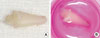
Supragingival scaling and plaque control were performed for all dogs 5 days before tooth extraction. Surgery was performed under general anesthesia induced by intravenous injection of Zoletil50 (5 mg/kg; Yuhan, Seoul, Korea) and Rompun (0.2 mg/kg; Bayer Korea, Seoul, Korea) followed by the administration of inhaled enflurane. Local infiltration anesthesia (2% lidocaine with HCl epinephrine, 1:100,000; Huons Co., Seoul, Korea) was used to reduce hemorrhage in the surgical areas. In both quadrants of the mandible, the first to third premolars (P1, P2, and P3) were the focus of this study. After an intrasulcular incision, the teeth were carefully hemisectioned with a high-speed bur and the teeth were extracted as atraumatically as possible using a forceps to protect the sound periodontal ligament tissues. Thirty teeth (P1, P2, and P3) were extracted from five Beagle dogs (Fig. 1A). Of these, 50 uncontaminated and undamaged hemisectioned-teeth were used fot the analysis (Fig. 1B).
Preservation of the tooth with EGCG
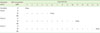
A modified version of the tooth-preparation protocol of Matsumura et al. [23] and Oh et al. [33] was followed. Of the 60 extracted hemisectioned teeth, 50 uncontaminated and undamaged hemisectioned teeth were analyzed. The teeth were divided into ten groups containing five teeth each. In brief, each tooth was lightly dipped three times in 70% alcohol and washed in phosphate-buffered solution (PBS). The coronal parts of the teeth were then soaked in povidone-iodine for 20 seconds and washed a further three times in PBS. After rinsing with α-MEM, each tooth was preserved in 3 mL of medium supplemented with 10% fetal bovine serum (FBS; Invitrogen Co., Carisbad, CA, USA), 100 U/mL penicillin, 100 mg/mL streptomycin 0% FBS (Invitrogen Co.), 0.3 µg/mL Fungizone 0% FBS (Invitrogen Co.), 22.5% Ham's nutrient mixture F-12 0% FBS (Invitrogen Co.), and 66.5% DMEM 0% FBS (Invitrogen Co.) in 24-well plates (Fig. 1B). The treatment concentrations of EGCG selected were 0, 10, and 100 µM based on our previous study [24] finding that the cell viability of cultured hPDL fibroblasts was highest in 10 µM EGCG media. Moreover, Matsumura et al. [23] demonstrated that cells with the most similar morphology and greatest viability were obtained after tooth preservation in media containing 250 µg/mL EGCG (i.e., 545.4 µM). The extracted teeth were divided into the following four groups: immediate, period 1, period 2, and period 3 (Table 1). The extracted teeth in the immediate (control) group were subjected to the 3-(4,5-dimethylthiazol-2-yl)-2,5-diphenyltetrazolium bromide (MTT) assay immediately after washing. Those in the period-1 group were subjected to the MTT assay after preservation with 0, 10, or 100 µM EGCG medium for 4 days, and then with EGCG-free medium for a further day. Those in the period-2 group were analyzed after preservation in the various concentrations of EGCG media for 8 days and in EGCG-free medium for 2 days. Finally, those in the period-3 group were analyzed after treatment for 12 days in the various EGCG media and in EGCG-free media for a further 2 days. According to Han et al. [34], EGCG conjugated to fluorescein-4-tisothiocyanate was observed at the membrane, cytoplasm, and nuclei of L929 fibroblast cells at 2 days after treatment, with the intensity increasing with increasing treatment time but beginning to reverse 2 days later. Therefore, in the present study we added EGCG-free periods after preservation in EGCG medium to analyze the recovered cell viability on the root surfaces of the teeth in each group.
Evaluation of PDL cell viability (MTT/eosin assay)
The PDL cell viability on each root surface was analyzed using a modification of the method reported by Kim et al. [35]. MTT solution (Amresco Inc., Solon, OH, USA) was added to each well of a 24-well plate at a final concentration of 0.5 mg/mL. Each treated tooth was immersed in a well. After incubating the wells for 3 hours at 37℃, the supernatant of each well was removed and replaced by 1 mL of dimethyl sulfoxide (Amresco Inc.). The plate was incubated for a further 30 minutes. After removing the teeth, the optical density (OD) of each well was measured using a microplate reader (Bio-Rad Laboratories Inc., Hercules, CA, USA) at a wavelength of 570 nm. Each OD ratio of the MTT assay was divided by the PDL tissue area of each root surface in order to eliminate the measurement errors caused by the different tissue volumes. To obtain the relative tissue volume, each tooth that had already been used for the MTT assay was stained again with eosin (Sigma-Aldrich Co., St. Louis, MO, USA) for 12 hours, and then treated with ethanol and 1% acidified alcohol for another 12 hours to extract the stained eosin. The OD of each of the extracts was then measured at 450 nm as described above. Each MTT value was divided by the relative tissue volume to obtain the final value.
RESULTS
MTT/eosin assay
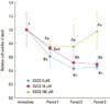
The cell viability was highest in the control group, and the number of living cells on the root surface decreased in a preservation-period-dependent manner for all treatment groups (Fig. 2). For the group treated with 0 and 10 µM EGCG, there was a statistically significant decrease in the number of living cells from immediate to period 1, although it generally decreased over the whole period. Similarly, though it did not show clear significant differences, the 100 µM EGCG group exhibited a decrease in cell viability from immediate to period 1. Interestingly, however, the cell viability of the 100 µM EGCG group increased and was recovered from period 2 to period 3. On the other hand, all the cell viabilities of the EGCG treated groups were higher than those of the untreated group, and the differences between 100 and 0 µM EGCG groups were statistically significant throughout all of the periods. A similar outcome was found between 100 and 10 µM EGCG groups (n=5, P<0.05).
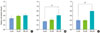
The cell viabilities related to EGCG concentration were compared for each period except for the immediate one (Fig. 3). In all periods, the lowest and highest cell viabilities were exhibited by the 0 and 100 µM EGCG groups, respectively. There was no significant difference among the three groups of immediate, and also between the 0 and 10 µM, and the 10 and 100 µM EGCG groups in all periods. More importantly, the cell viability was markedly higher in the 100 µM EGCG group than in the untreated (0 µM EGCG) group (P<0.05). Therefore, EGCG at a high concentration appeared to improve the maintenance of cell viability over the controlled conditions.
DISCUSSION
In this study, we observed improved maintenance of the viability of Beagle dog tooth cells when they were stored in medium containing EGCG, an effect that was somewhat concentration-dependent. Extension of the preservation period up to 14 days was achieved using 100 µM EGCG, compared to that of conventional (non-EGCG-containing) storage solutions, in which it lasts for 24 hours at most [6-10].
To the best of our knowledge, few studies have investigated the application of EGCG in tooth preservation. However, the efficacy of EGCG as a storage medium has been explored for other tissues and cells, such as osteoblasts, fibroblasts, endothelial cells, platelets, blood vessels, nerves, articular cartilage of joints, and hematopoietic stem cells [19-22,36,37]. These cells were successfully preserved by EGCG treatment for significantly longer periods of up to 3 months, maintaining their cell viability, biomechanical properties, structures, and functions [19-22,36,37]. Moreover, in a previous study we explored the effect of EGCG on hPDL fibroblasts and concluded that EGCG has the potential to control the cell viability of these cells. Considering these results, it is thought that the higher cell viability of the teeth observed in the present study was due to the preservative effects of EGCG, and that tooth preservation of much longer than 14 days is plausible by using EGCG as a storage agent.
The preservative potential of EGCG has been attributed to its intrinsic properties, and in particular, its molecular structure and amphipathic property [20,38]. Because of its phenolic structure, EGCG neutralizes the free radicals of reactive oxygen species by chelating with them, which cause hazard alterations of cell components, and thus protects cells and tissues from oxidative-stress-induced damage [38,39]. Furthermore, since EGCG has both hydrophilic and lipophilic properties, it combines easily with the extracellular matrix, phospholipid bilayered membranes, and any type of intracellular protein [20], as well as the cytoplasm and nucleus [34]. Thereafter, those incorporated EGCGs begin to arrest the cell cycle through blockage at specific sites on the cellular membrane or DNA, such as the cyclin genes, which are involved directly in cell-cycle regulation [40]. Then, those reactions induce cytostatic activity in the cells or tissues. This activity may be related to the mechanism underlying the maintenance of cell viability observed in the present study. Thus, the higher cell viability of EGCG-treated teeth observed herein may be the result of less oxidative damage and the cytostatic effects of EGCG.
In our study, the EGCG treatment was followed by additional EGCG-free incubations. Although EGCG molecules can be easily incorporated into cellular proteins or nuclei, they become reversibly detached with the progression of time in the absence of EGCG, thus initiating the cell cycles of stored cells [20,34,40]. In a study, cDNA microarray analysis has revealed that the cell-cycle-related genes of cells are suppressed under the influence of EGCG, but once EGCG is removed from the medium, the gene expressions are restored to their original levels [40]. Over 60% of such alterations were reversed 2 days after removal of EGCG, and restored to their initial state 8 days after eliminating EGCG [40]. However, the cell viability of the tooth surface in the present study was analyzed 1 or 2 days after removing EGCG, not up to 8 days, and thus the accomplishment of cellular recovery could not be analyzed. Therefore, the recovered cell viabilities of the EGCG-treated groups may in fact be much higher than shown by the present results with longer post-EGCG-free periods.
The promising potential of EGCG has been explored in dental parts as well as in the medical fields [16-26,36,37]. EGCG has shown to extend the lifespan of dental cells and tissues and maintain them in a stable condition [23-24]. Furthermore, it was reported to help the recovery from inflammation in periodontal and periapical lesions, acting as a bactericide and immune suppressor [25-27,30]. Thus, treatment with EGCG can be a great therapeutic strategy for tooth transplantation, since storage of avulsed teeth in a medium containing EGCG will allow sufficient time for the appropriate dental treatment. The EGCG subsequently released from the replanted tooth into its socket could reduce inflammatory reactions and bacterial infections [25-30]. These effects are not limited to the replantation of avulsed teeth; they could be applied to autogenous tooth transplantation or selected allografting of healthy teeth. However, before its clinical application, further studies are needed to determine the precise mechanisms underlying the preservative effects of EGCG on teeth and other dental tissues, and more long-term in vivo data of replants with EGCG are required.
 XML Download