

 PDF
PDF ePub
ePub Citation
 Print
Print


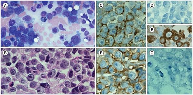
A 54-year-old woman was admitted with compression fracture of thoracic vertebral bodies. Urine protein electrophoresis revealed the presence of M protein and serum free κ/λ ratio was 0.01. Bone marrow aspiration (A) and biopsy (B) revealed diffuse infiltration of numerous large-sized blasts with basophilic cytoplasm, dispersed nuclear chromatin, high nuclear/cytoplasmic ratio and prominent nucleoli. Tumor cells showed strong expression of plasma cell (PC) antigen CD138 (C), monotypic light chain (D, κ negative; E, λ positive), negative expression of CD3, CD20, CD34, Tdt and CD30 by immunohistochemistry, mimicking a picture of plasmablastic lymphoma (PBL). PBL was excluded on the basis of CD56 positivity (F) and absence of EBV-encoded RNA (G). The diagnosis of plasmablastic plasma cell myeloma (PBPCM) was made and the patient started chemotherapy. Morphologic features usually distinguish PBL from well-differentiated plasma cell myeloma (PCM). However, highly aggressive PBPCM may show predominance of plasma blasts, which can resemble PBL cells. Although both PB myeloma and PBL express PC antigens (CD138, monotypic light chain), positivity of EBV-encoded RNA is useful in establishing the diagnosis of PBL.
 XML Download