

 PDF
PDF ePub
ePub Citation
 Print
Print


Abstract
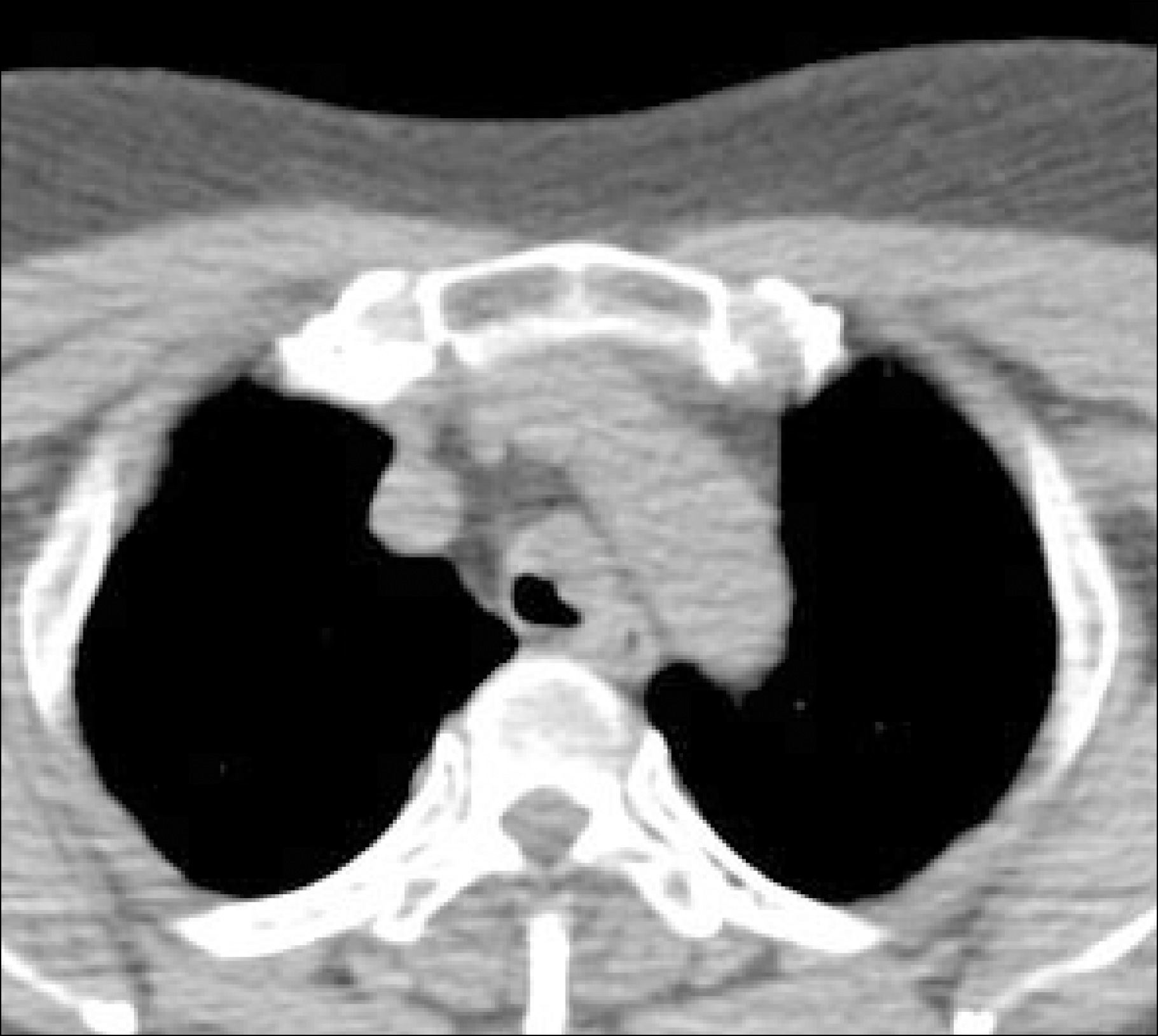
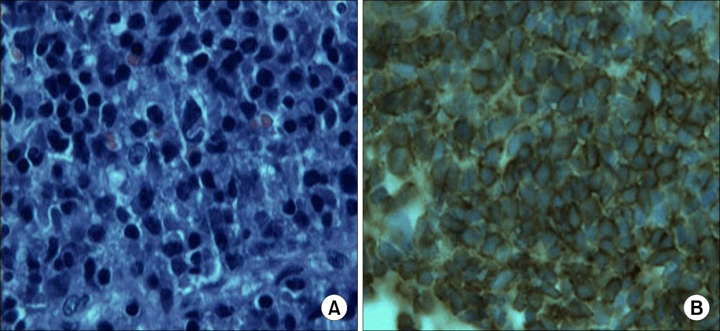
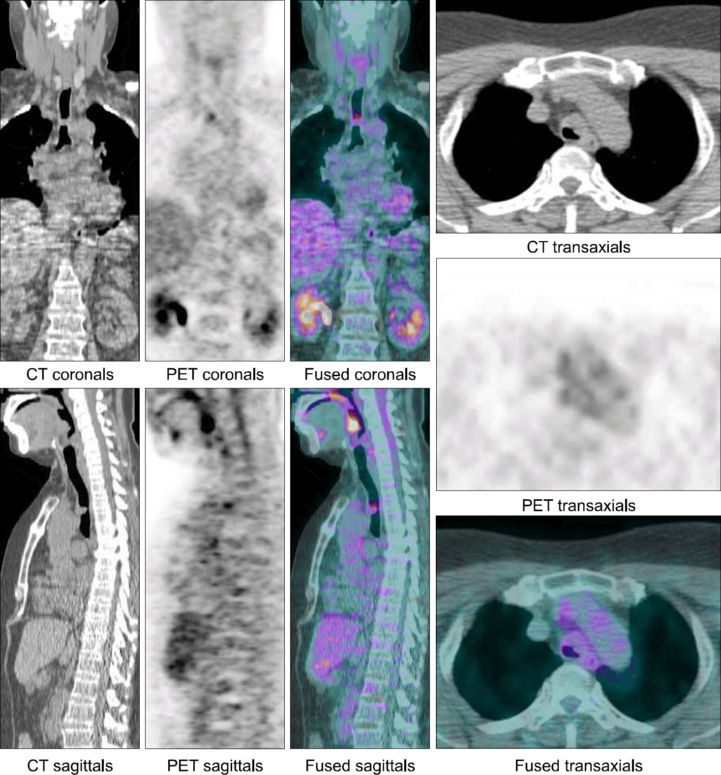
Primary extranodal non-Hodgkin's lymphomas comprise approximately 10% of all non-Hodgkin's lymphomas. However, primary tracheal non-Hodgkin's lymphoma is extremely rare, being mainly mucosa-associated lymphoid tissue lymphoma. A 65-year-old female has dry cough for one year. She was diagnosed as diffuse large B-cell lymphoma via bronchoscopic-guided biopsy. She was treated with four cycles of the R-CHOP regimen and adjuvant radiotherapy. After completion of the combined treatment, the treatment response was complete remission, and the disease free survival was 26 months.
REFERENCES
1). Macchiarini P. Primary tracheal tumours. Lancet Oncol. 2006. 7:83–91.

2). Gaissert HA., Grillo HC., Shadmehr MB, et al. Uncommon primary tracheal tumors. Ann Thorac Surg. 2006. 82:268–73.
3). lsaacson P., Wright DH. Extranodal lymphoma arising from mucosa-associated lymphoid tissue. Cancer. 1984. 53:2515–24.
4). Young GA. Lymphoma at uncommon sites. Hematol Oncol. 1999. 17:53–83.
5). Rudders RA., Ross ME., DeLellis RA. Primary extranodal lymphoma: response to treatment and factors influencing prognosis. Cancer. 1978. 42:405–16.
6). Rosenberg SA., Diamond HD., Jaslowitz B., Craver LF. Lymphosarcoma: a review of 1,269 cases. Medicine (Baltimore). 1961. 40:31–84.
7). Zhang WD., Li SY., Ouyang M., Zhong NS. Primary endotracheal non-Hodgkin's lymphoma in a Chinese woman: a case report. Chin Med J (Engl). 2005. 118:702–4.
8). Kaplan MA., Pettit CL., Zukerberg LR., Harris NL. Primary lymphoma of the trachea with morphologic and immunophenotypic characteristics of low grade B cell lymphoma of mucosa-associated lymphoid tissue. Am J Surg Pathol. 1992. 16:71–5.
9). Hahn JS., Ko YW., Min YH, et al. Statistical analysis of maliganat lymphoma in Korea. Korean J Hematol. 1995. 2:197–214.
10). Feugier P., Van Hoof A., Sebban C, et al. Long-term results of the R-CHOP study in the treatment of elderly patients with diffuse large b-cell lymphoma: a study by the Groupe d'Etude des Lymphomes de l'Adult. J Clin Oncol. 2005. 23:4117–26.
11). Jhanwar YS., Straus DJ. The role of PET in lymphoma. J Nucl Med. 2006. 47:1326–34.
12). Juweid ME., Stroobants S., Hoekstra OS, et al. Use of positron emission tomography for response assessment of lymphoma: consensus of the Imaging Subcommittee of International Harmonization Project in Lymphoma. J Clin Oncol. 2007. 25:571–8.
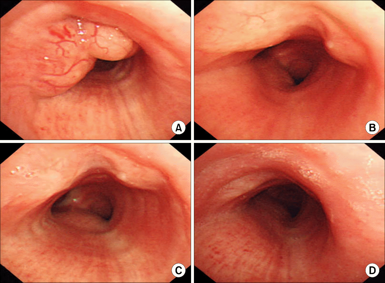
Fig. 1
Serial bronchoscopic findings. (A) At 20cm from upper incisor, multi-lobulated, multi-lobulated tumor with vascularization was seen. the tumor obstructed the tracheal lumen by about 50%. (B, C) Comparing previous bronchoscopic finding. the size of tracheal lymphoma was marked decreased at about 5cm above carina. (B) After the patient received 2 cycles of combination chemotherapy with R-CHOP regimen. (C) After the patient received 4 cycles of combination chemotherapy with R-CHOP regimen. (D) Comparing previous bronchoscopic finding. Slight flat bilobulated nodule was more decreased at about 5cm above carina after additional RT was done.
 XML Download