

 PDF
PDF ePub
ePub Citation
 Print
Print


TO THE EDITOR: Lymphomas have diverse clinical presentations, may affect multiple organ systems, and have been diagnosed across different age groups. Bone involvement is present in 16–20% of patients, and patients with bone involvement often present with pain, mass, fractures, and fever [12]. These non-specific symptoms can lead physicians to misdiagnose the patients with septic arthritis, osteomyelitis, or primary bone tumors. Histopathological examination is the gold standard for diagnosis; however, in some cases, this type of examination is difficult or impossible. Moreover, the clinical and histopathological distinction between osteomyelitis and malignancy is sometimes difficult, and misdiagnoses have been reported [3]. We report a case of lymphoma that occurred at the hip joint and mimicked osteomyelitis and septic arthritis. A 25-year-old male presented to the emergency department with a one-month history of pain in the right hip, walking difficulty, and fever. On physical examination, he had a high fever (39℃), pallor, and right hip arthralgia with decreased range of motion. The laboratory examination revealed pancytopenia (neutrophils: 500/µL, lymphocytes: 500/µL, blood hemoglobin level: 64 g/L, platelets: 36,000/µL) and elevated serum C-reactive protein (60 mg/L), erythrocyte sedimentation rate (42 mm/hr), lactate dehydrogenase (LDH, 1,928 IU/L), alkaline phosphatase (ALP, 749 IU/L), and gammaglutamyl transferase (GGT, 223 IU/L) levels. Serological tests for hepatotrophic viruses were negative. The pelvic magnetic resonance image (MRI) showed destruction of the right femoral head and edema in the right femoral metaphysis and shaft. We consulted the orthopedics department and initially diagnosed the patient with right femoral osteomyelitis and septic arthritis of the right hip joint. Three sets of blood cultures were drawn, and intravenous ampicillin/sulbactam and ciprofloxacin were started while preparing for orthopedic surgery.
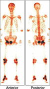
No clinical or laboratory improvement was noted after the first week of treatment, and a peripheral blood smear revealed atypical lymphocytes. A triphasic bone scan scintigraphy showed increased late-phase activities in the frontal and parietal bones, right hip joint, right femoral condyle, right proximal and distal epiphyses of the tibia, and right tarsal bones (Fig. 1). A bone marrow biopsy showed infiltration of T-cell/histiocyte-rich B-cell lymphoma (TC/HRBCL) (Fig. 2A–C), and thoracic and abdominal CT scans identified diffuse pathological lymphadenopathies. The R-CHOP regimen with radiotherapy to the right hip joint was initiated, and the patient's clinical and laboratory findings improved after the first four cycles of R-CHOP treatment. The patient achieved complete remission after eight cycles of R-CHOP treatment; however, the disease relapsed one year later, and the patient died due to septic shock after an ESHAP regimen.
TC/HRBCL is an uncommon subtype of DLBCL that predominantly occurs in younger patients and men. Bone marrow and extra-nodal involvements are more frequent in this subtype than in other types of DLBCL [4].
Lymphoma misdiagnoses that delay the appropriate treatment can be catastrophic. The lack of response to infection treatments and alternative diagnoses should promptly improve differential diagnoses. Lymphoma should be considered in patients with bone and joint pain.

 XML Download