

 PDF
PDF ePub
ePub Citation
 Print
Print


INTRODUCTION
Thrombocytosis is a common condition among children, and it is a frequent cause of referral to a pediatric hemato-oncologist or medical institution for further testing, accounting for 6–15% of hospitalizations and/or visits to an ambulatory/emergency clinic among children [123]. Thrombocytosis can be classified as primary or reactive thrombocytosis (RT), with primary thrombocytosis involving a primary bone marrow disorder, such as essential thrombocytosis, polycythemia vera, chronic myeloid leukemia, or myelodysplastic syndrome [4]. In contrast, RT is much more frequent than primary thrombocytosis and is caused by non-bone marrow related medical or surgical conditions, which are most often viral or bacterial infections [15]. There are national reports regarding the etiology and clinical characteristics of RT among children from several countries [267891011], although there are no data regarding RT among children in Korea. The present retrospective study aimed to determine the etiology and clinical characteristics of RT among children who were admitted to a single Korean center during a 10-year period, and to compare the results to those from other countries.
MATERIALS AND METHODS
Participants and ethics statement
In this retrospective study, we collected data for children <18 years old with a platelet count of >500×109/L, who were admitted to the pediatric ward in Keimyung University Dongsan Medical Center between January 2006 to December 2016. We excluded healthy newborns, premature infants who were admitted to the neonatal intensive care unit, and patients with primary thrombocytosis. The study protocol was approved by the institutional review board of Keimyung University Dongsan Medical Center (2017-11-038).
Definitions
Cases of RT were classified based on previous criteria [912] as mild (grade 1, platelet count of 501–700, ×109/L), moderate (grade 2, platelet count of 701–900, ×109/L), severe (grade 3, platelet count of 901–1,000, ×109/L), and extreme (grade 4, platelet count of >1,000, ×109/L).
Kawasaki disease (KD) is diagnosed by prolonged fever (≥5 days) and at least four of five clinical features: changes in the extremities, polymorphous exanthema, bilateral bulbar conjunctival injection without exudate, changes in the lips and oral cavity, and cervical lymphadenopathy (>1.5 cm diameter) [13]. According to the American Heart Association, atypical KD is possible in children (≥6 mo) with prolonged fever and two or three principle features or in infants with fever (≥7 days) without other explanation [13]. Refractory KD is defined as persistent fever that lasts for more than 24–36 hours after the end of initial KD treatment [14].
Statistical analysis
Patients' baseline characteristics were reported as median and range, although intragroup comparisons were performed using mean and 95% confidence interval (CI) values. Analysis of variance with Bonferroni adjustment was used to compare mean values between the groups, and intragroup comparisons of proportions were performed using the linear by linear chi-square statistic. Pearson's correlation analysis was performed to define correlations between the variables. All statistical analyses were performed using IBM SPSS version 23.0 (IBM Corp., Armonk, NY, USA), and P-values <0.05 were considered statistically significant.
RESULTS
Baseline characteristics and underlying causes of RT
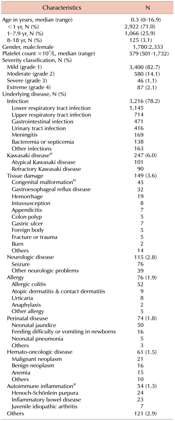
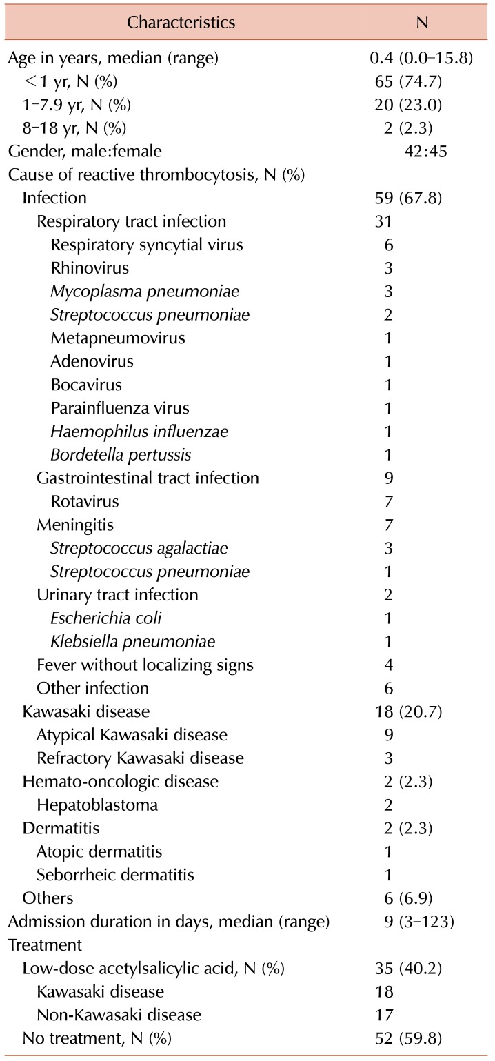
Patients' baseline characteristics and underlying causes of RT are described in Table 1. During the study period, RT accounted for 13.5% of children who were admitted to the pediatric ward (4,113/30,355). The median age of children with RT was 0.3 years, and the proportion aged <1 year was 71.0%. Among a total 4,113 children, 82.7% of cases involved mild RT, 14.1% involved moderate RT, 1.1% involved severe RT, and 2.1% of cases involved extreme RT. The most common cause of RT was infection (78.2%) and the second most common cause was KD (6.0%). The characteristics and underlying clinical conditions of the 87 children with extreme RT are described in Table 2. The median age at diagnosis of extreme RT was 0.4 years, and the proportion of children who were <1 year old was 74.7%. The most common cause of extreme RT was infection (67.8%), which most frequently involved respiratory tract infection; the second most common cause of extreme RT was KD (20.7%).
Comparisons according to RT severity
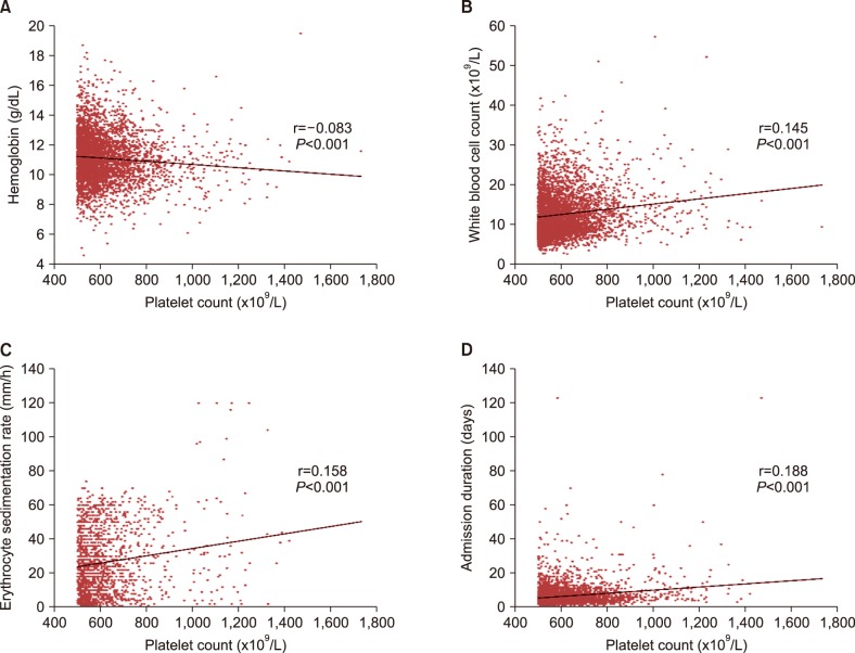
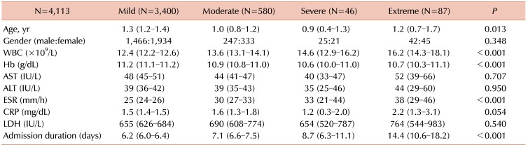
Table 3 shows the variables according to RT severity. Significant differences according to RT severity were detected for hemoglobin (Hb) levels, white blood cell (WBC) count, erythrocyte sedimentation rate (ESR), and admission duration. No significant differences were detected in terms of age, gender ratio, liver function results, C-reactive protein levels, and lactate dehydrogenase levels. Correlation analysis revealed a negative correlation between platelet count and Hb level (r=−0.083, P<0.001), as well as positive correlations between platelet count and WBC count (r=0.145, P<0.001), between platelet count and ESR (r=0.158, P<0.001), and between platelet count and admission duration (r=0.188, P<0.001) (Fig. 1). After controlling for patient age, disease category, liver function, C-reactive protein levels, and lactate dehydrogenase levels, the negative correlation between platelet count and Hb level persisted (r=−0.251, P=0.008). In addition, independent positive correlations persisted between platelet count and WBC count (r=0.305, P=0.001), between platelet count and ESR (r=0.252, P=0.007), and between platelet count and admission duration (r=0.257, P=0.006).
Comparing underlying medical conditions according to RT severity
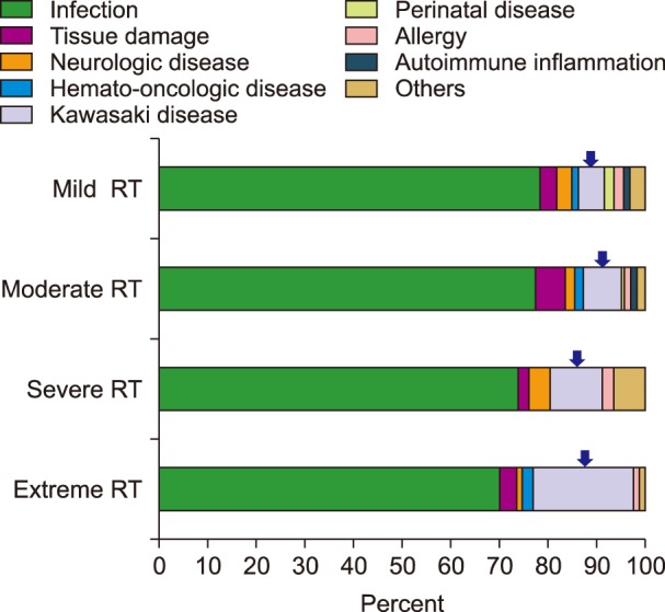
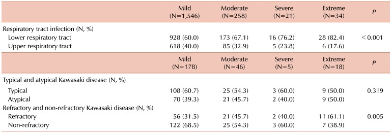
In cases of respiratory tract infection, the proportions of lower and upper respiratory tract infections were significantly different according to the severity of RT (Table 4). The highest proportion of lower respiratory tract infection was observed in the extreme RT group (P<0.001). In cases of KD, the proportions of RT cases that involved KD were 5.2% (178/3,400) for mild RT, 7.9% (46/580) for moderate RT, 10.9% (5/46) for severe RT, and 20.7% (18/87) for extreme RT (Fig. 2). The differences according to RT severity were significant, and the highest proportion was observed in the extreme RT group (P<0.001). The proportions of refractory KD were significantly different according to RT severity (P=0.005) (Table 4). The proportions of atypical KD were not significantly different according to the RT severity (P=0.319). In cases of children with RT who had KD, correlation analysis revealed a positive correlation between platelet count and fever duration (r=0.175, P=0.006). Non-KD autoimmune inflammation (Henoch-Schönlein purpura, juvenile idiopathic arthritis, Crohn disease, or ulcerative colitis) was not involved in cases of severe or extreme RT, and was limited to mild RT cases (1.4%, 46/3,400) and moderate RT cases (1.7%, 10/580) (Fig. 2).
Age-related distributions of underlying diseases
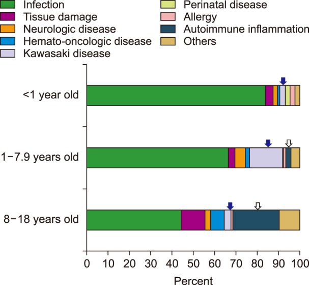
Children were classified by age into the following groups: children <1 year old, those 1–7.9 years old, and those 8–18 years old. The proportions of KD were 2.6% (77/2,922) in the <1-year-old group, 15.6% (166/1,066) in the 1–7.9-year-old group, and 3.2% (4/124) in the 8–18-year-old group. The proportions of non-KD autoimmune inflammation (Henoch-Schönlein purpura, juvenile idiopathic arthritis, Crohn disease, or ulcerative colitis) were 0% (0/2,922) in the <1-year-old group, 2.4% (26/1,066) in the 1–7.9-year-old group, and 21.8% (27/124) in the 8–18-year-old group. The proportion of KD was significantly higher in the 1–7.9-year-old group and the proportion of autoimmune inflammation was significantly higher in the 8–18-year-old group (both P<0.001) (Fig. 3).
Treatment and clinical outcome of children with RT
Treatment of underlying disorders was performed in participants, and platelet counts gradually decreased after the underlying cause was resolved. Among the 4,113 children, 2.1% of cases had extreme RT. Approximately 40% of children with extreme RT received low-dose acetylsalicylic acid; of these children, over 50% had KD and the remainder did not (Table 2). None of the patients experienced clinical complications (such as thromboembolic events or bleeding) that were associated with extreme RT.
DISCUSSION
Among the children who were admitted to our center, the prevalence of RT was 13.5% (4,113/30,355), which is similar to the prevalence in other countries [123]. The incidence of RT is age-dependent, with much higher rates among young children, especially those who are <1–2 years old [236791011] and much lower rates among children aged >11–15 years [23]. In the present study, infants who were <1 year old accounted for >70% of all RT cases. This phenomenon is related to the correlation between serum thrombopoietin (TPO) levels and age-dependent platelet changes [15], with serum TPO levels being highest during the neonatal period and gradually decreasing with age [15]. Despite the prevalence of RT among children, extreme RT is very rare and accounts for approximately 2% of RT cases [36], which is similar to the rate in the present study (2.1%, 87/4,331). As previously reported [3], extreme RT was associated with low Hb levels, high WBC counts, and a prolonged admission period. Platelet counts were not correlated with C-reactive protein levels, as has been previously reported [3], although extreme RT was associated with high ESR, and platelet count was positively correlated with ESR in the present study.
Respiratory tract infections are the most common causes of RT and extreme RT [81016], with upper respiratory tract infections being common in cases of mild thrombocytosis and lower respiratory tract infections being common in cases of severe thrombocytosis [8]. In the present study, the highest proportion of lower respiratory tract infection was observed in the extreme RT group. In other words, more severe RT was associated with more severe respiratory tract infection. In the present study, respiratory syncytial virus (RSV) and rhinovirus were the most common pathogens in cases of respiratory tract infection with extreme RT (Table 2), and similar results have been previously reported [17]. Levels of interleukin (IL)-6 or IL-8 can increase during RSV infection [18], which is related to the activation of bronchial epithelial cell secretion and stimulation of platelet production [18].
In this study, KD was the second most common cause of all cases of RT (6.0%, 247/4,113) and extreme RT (20.7%, 18/87). Similarly, KD causes RT in approximately 9.4% of Japanese pediatric cases [2]; these results are markedly higher than those in other countries [67891011], which is likely related to the high prevalence of KD in Asian countries, including Korea and Japan [1920]. Interestingly, the proportions of both KD and refractory KD were highest in the extreme RT subgroup in this study. Further, correlation analysis revealed a positive correlation between platelet count and fever duration in children with KD and RT. In other words, more severe RT was associated with more severe KD.
Thrombocytosis in cases of KD is an important condition, as it increases the risk of coronary aneurysm thrombosis, myocardial infarction, and death [21]. Among patients with KD, RT can be affected by the levels of various cytokines, such as IL-1, IL-6, TPO, and tumor necrosis factor α[21], with TPO being the main cytokine that affects platelet production and megakaryocyte development [21]. The liver and kidneys produce TPO, and serum concentrations are regulated through binding to c-Mpl on platelets and megakaryocytes [22]. Interestingly, TPO levels are usually elevated during the first week of the acute infection phase, with subsequent decreases that correspond to improvement in the symptoms of infection [2324]. However, platelet counts increase during the second and third weeks of infection [2324], which may reflect a gradual effect of TPO levels on platelet production through the c-Mpl ligand mechanism [2324].
In a Japanese study, autoimmune diseases (juvenile idiopathic arthritis, inflammatory bowel diseases, and KD) accounted for 4–11% of pediatric RT cases [2], with KD being the principal cause of RT among children <7 years old and other diseases being the main causes among those aged >11 years [2]. In the present study, the cause of RT was classified as KD or non-KD autoimmune inflammation, which included Henoch-Schönlein purpura, juvenile idiopathic arthritis, and inflammatory bowel diseases. Similar to the findings of the Japanese study [2], the present study revealed that the proportion of KD was highest in the 1–7.9-year-old group and the proportion of non-KD autoimmune inflammation was highest in the 8–18-year-old group (P<0.001).
In most cases, RT is benign and the platelet count is normalized after treatment of the primary disease, in the absence of complications [1025]. Furthermore, most patients with RT do not require prophylactic treatment using anticoagulants or platelet aggregation inhibitors [12]. Even in extreme cases of RT, there is no established prophylactic treatment or evidence to support the use of prophylactic drugs for asymptomatic children [12]. Thus, if thrombocytosis persists after treatment of RT that is based on the underlying etiology of RT, treatment can be considered for thrombocytosis [25].
There are some limitations in our study. Most patients with RT did not undergo blood testing until platelet counts had dropped to completely normal levels. Therefore, the exact duration of the platelet increase could not be calculated and was insufficient to be included as statistical data.
In conclusion, infection, especially respiratory tract infection, was the most common cause of RT among Korean children in this study, and KD was the second most common cause. Extreme RT was associated with leukocytosis, anemia, increased ESR, prolonged admission duration, more severe respiratory tract infection, and more severe KD. Therefore, RT could be a factor in evaluating the severity of disease and predicting hospitalization duration. In addition, the present study revealed that KD was an important cause of extreme RT and all RT among 1–7.9-year-old patients, whereas non-KD autoimmune inflammation was more likely to cause mild-to-moderate RT, especially among children ≥8 years old. However, the present study is limited by its single-center design, and further evaluation is needed to better understand the status of RT in Korea.
 XML Download