

 PDF
PDF ePub
ePub Citation
 Print
Print


Dear Editor:
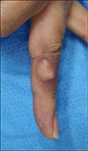
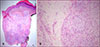
A 46-year-old man presented with a 5-year history of a nodule on his finger. Physical examination revealed a tender, solitary skin-colored irregular-shaped nodule measuring 1.4×1.4 cm, located on the left fourth finger (Fig. 1). Histological examination revealed a spindle-cell tumor in an admixture of fibromyxoid matrix in the dermis (Fig. 2A). The tumor consisted of alternating hypercellular myxoid and hypocellular fibrous stroma with an abrupt transition from one to the other. The tumor cells were bland spindle-shaped cells and stellate cells, arranged in a swirling or fascicular growth pattern (Fig. 2B). A few curvilinear vessels were observed. Mitotic figures were detected in 5/50 high power fields. Immunohistochemical studies showed reactivity for vimentin, CD99, CD68, and epithelial membrane antigen (EMA), whereas the tumor cells were negative for CD34, S100, smooth muscle actin, and desmin. These findings were consistent with low-grade fibromyxoid sarcoma (LGFMS).
LGFMS is a rare soft tissue tumor, which typically occurs in the deep soft tissues of the proximal extremities or trunk in young to middle-aged adults12.
Histologically, LGFMS consists of bland fibroblasts with a whorled or linear arrangement, alternating between paucicellular fibrous zones and myxoid zones123. There is a characteristic abrupt transition between fibrous and myxoid zones123. The myxoid zones of LGFMS are composed of bland spindle to stellate tumor cells in a fibromyxoid stroma, often with branching to curvilinear vessels123. The paucicellular fibrous zones of LGFMS typically consist of bland spindle tumor cells, frequently arranged in fibromatosis-like fascicular growth patterns3. Tumor cells tend to be small, with poorly defined, palely eosinophilic cytoplasm and round to ovoid nuclei3. Mitotic figures are primarily absent or sparse2.
The tumor cells consistently show positivity for vimentin and CD99 in many reports12. In the majority of cases, tumor cells are negative for S100, neuron-specific enolase, EMA, actin, desmin, cytokeratin, and CD3412.
The main differential diagnosis of LGFMS includes low-grade myxofibrosarcoma, desmoid fibromatosis, myxoid neurofibroma, perineurioma, and superficial acral fibromyxoma (SAFM)1. Low-grade myxofibrosarcoma arises in older patient populations, and has more atypical cells with bizarre nuclei and abnormal mitotic figures than LGFMS14. Desmoid fibromatosis has more fascicular architecture and is positive for SMA3. Myxoid neurofibroma, perineurioma, and SAFM are positive for CD34. SAFM typically occurs on the hands and feet in middle-aged women, and consists of spindle-shaped and stellate fibroblast-like cells with random, loose storiform, and fascicular growth patterns5. It is embedded in myxoid, myxocollagenous or collagenous matrix, often with mildly to moderately accentuated vasculature, which is very similar to LGFMS5. However, an abrupt transition from one type of area to the other is not a feature of SAFM, and mast cells are easily identified in SAFM5.
LGFMS exhibits an indolent morphology; however, it has a malignant clinical course3. Wide local excision of primary and metastatic lesion is the preferred treatment.
In conclusion, we report a rare case of LGFMS arising in the superficial localization of the finger. Considering that this disease has a malignant clinical course despite its bland histological features, it is important to be aware of the disease and perform early diagnosis using skin biopsy.
 XML Download