

 PDF
PDF ePub
ePub Citation
 Print
Print


Dear Editor:
Nevus lipomatosus cutaneous superficialis (NLCS) is a rare form of connective tissue disease, in which mature adipocytes are present ectopically in the dermis1. In general, lipomas and other tumors originating from the mesenchyme are known to be accompanied by mucin deposition. However, it is rare to observe mucin deposition in NLCS, and mucinous nevus in NLCS has not been reported yet2.
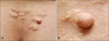
A 41-year-old man presented to our department with skin lesions on his buttock. The history and physical examination revealed localized yellowish plaques in clusters and 0.3×0.3 cm pedunculated skin-colored papule on his buttocks (Fig. 1). Without symptoms and trauma history, the plaques were found incidentally about 10 years prior, and the papule that had gradually increased in size over time was found several years ago.
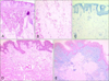
A skin biopsy specimen taken from one of the plaques showed hyperkeratosis and acanthosis of the epidermis, and groups of mature adipose tissues among collagen fibers was observed in the middle and lower part of the dermis, connecting to the subcutaneous fat (Fig. 2A∼C). Biopsy results of the pedunculated papule revealed hyperkeratosis, mild acanthosis, and mucin deposition limited to the papillary dermis (Fig. 2D). And mucin was positive for Alcian blue stain at pH 2.5 (Fig. 2E).
Mucinous nevus is a rare form of connective tissue nevus and was first reported by Redondo Bellón et al.3 in 1993. The term mucinous nevus was used because histological and clinical findings showed a nevoid appearance, with deposition of mucin in the papillary dermis. On histological examination, it shows a ribbon of mucin deposition in the upper dermis. It can be accompanied by acanthosis, extended rete ridges, and various degrees of hyperkeratosis in the epidermis4. The origins of the mucin and the mechanism of its development are not known, but previous studies suggested that mucin formation might increase as a result of fibroblast upregulation5. Because the present case showed a decrease in fat composition and an increase in fibrosis in the NLCS lesion over time, it is considered to be an interesting case supporting this correlation. Apart from fibroblast upregulation, external stimulation by constant friction, and coincidental co-occurrence of mucinous nevus and NLCS should be regarded as possible hypotheses, and more cases and study will be needed to better understand the relation between two diseases and mechanism of mucin deposition.
The clinical features of the present case were similar to those of soft fibromas or neurofibromas, so histopathological examination and special staining were required for differential diagnosis. In conclusion, we report a rare case of acquired mucinous nevus in NLCS. Our case provides additional support for the hypothesis that mucin deposition may develop in association with fibroblast.
 XML Download