

 PDF
PDF ePub
ePub Citation
 Print
Print


Dear Editor:
Porokeratosis is a chronic skin disorder of aberrant epidermal keratinization, clinically manifesting as patches with an elevated peripheral keratotic ridge that corresponds histologically to the cornoid lamella. The lesions have a tendency toward peripheral expansion1. The cornoid lamella is a column of tightly packed parakeratotic cells with pyknotic nuclei. In the cornoid lamella, the granular layer is usually lost and the keratinocytes beneath the parakeratotic column show vacuolated or eosinophilic degenerative cytoplasm in association with mild superficial dermal mononuclear cell infiltration1. According to the number, size, and distribution of the lesions, at least six clinical variants have been described, including disseminated superficial porokeratosis (DSP) and porokeratosis of Mibelli (PM)1. The malignant transformation of porokeratosis into Bowen's disease and squamous cell carcinoma has been described, with a reported incidence of 7.5% to 11%1. In line with this, the porokeratotic cells revealed abnormal DNA ploidy with increased DNA indices1. Although it is believed that the clonal expansion of abnormal keratinocytes leads to the development of porokeratosis, the pathogenesis remains unknown1.
Cyclin-dependent kinases 4 and 6 are critical regulatory enzymes that drive cell cycle progression from the G0 or G1 phase into the S phase, so their activity is under stringent control to ensure successful cell division23. The protein 16INK4a (p16INK4a) downregulates the kinase activity of cyclin-dependent kinases 4 and 6 and acts as an inhibitor of cell cycle progression2. Moreover, p16INK4a is strongly associated with the cellular senescence process and a diverse array of aging stimuli upregulate its expression23. To the best of our knowledge, only one report has described the transcriptional overexpression of p16INK4a in a single case of congenital unilateral linear porokeratosis4. However, the immunohistological localization of p16INK4a has never been reported.
We examined formalin-fixed and paraffin-embedded tissues of five cases of normal skin and seventeen cases of porokeratosis (DSP, n=9; PM, n=8). Sections were deparaffinized with xylene for 10 min and rehydrated through a graded ethanol series. Antigen retrieval was performed using Heat Processor Solution pH6 (Nichirei Biosciences Inc., Tokyo, Japan) at 100℃ for 40 min, and endogenous peroxidase was blocked by incubating the sections with 3% H2O2 (Nichirei Biosciences Inc.). The sections were then incubated with monoclonal antibody against p16INK4a (E6H4, CINtec; Roche MTM Laboratories, Westborough, MA, USA) at 4℃ overnight, followed by incubation with secondary antibody, N-Histofine® Simple Stain MAX-PO MULTI (Nichirei Biosciences Inc.). Immunodetection was conducted with 3,3-diaminobenzidine as a chromogen, followed by light counterstaining with hematoxylin. Sections stained without primary antibody served as a negative control. All of the sections were negatively stained with anti-human papilloma virus antigen antibody (K1H8; Abcam, Cambridge, MA, USA). This study was approved by the ethical committee of Kyushu University Hospital (IRB no. 27-157).
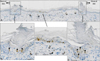
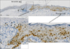
As has been reported previously5, the expression of p16INK4a was not detected in the perilesional normal epidermis (Fig. 1A, 2A). In all cases of both DSP (Fig. 1) and PM (Fig. 2), parakeratotic cornoid lamella was negative for p16INK4a. However, a population of keratinocytes beneath and around the cornoid lamella were positively stained with p16INK4a (Fig. 1B, D, 2B, Supplementary Fig. 1). Some of them were vacuolated cells (Fig. 2B). In the central area of porokeratosis, variable numbers of keratinocytes were also immunolabeled with p16INK4a (Fig. 1C, 2C). The central p16INK4a immunoreactivity was separate from (Fig. 1) or continuous with (Fig. 2) the cornoid lamella area. The staining pattern of p16INK4a was both nuclear and cytoplasmic. Central immunostaining for p16INK4a was detected in 6 of 9 DSP cases and 7 of 8 PM cases.
The present study is the first to reveal the immunolabeling of p16INK4a in porokeratosis. p16INK4a immunopositivity was observed in the keratinocytes underlying the cornoid lamella as well as those in the central portion of the lesion. These results coincide with the upregulated expression of p16INK4a mRNA in a single case of congenital unilateral linear porokeratosis4. Besides p16INK4a, another molecular effector to regulate senescence is p532. Previous studies have revealed that the p53 expression is also upregulated in the keratinocytes underlying and surrounding cornoid lamella67. These results prompted us to speculate that the abnormal porokeratotic clones are senescence-prone keratinocytes which are generated beneath the cornoid lamella extending into the central lesion during lesional expansion to the periphery, which is in sharp contrast to the complete negativity of p16INK4a in the perilesional normal epidermis.
Although p16INK4a immunolabeling has been validated as an accurate surrogate marker for determining the concomitant infection of human papilloma virus in vulvar squamous cell carcinoma8, none of the present cases exhibited immunopositivity to human papilloma virus antigen. In accordance with a pivotal role of p16INK4a in cell cycle arrest and senescence, melanomas expressing p16INK4a exhibit slower growth than those without p16INK4a expression9. In addition, the loss of p16INK4a in affected T cells correlates with disease progression in mycosis fungoides10. Senescence and carcinogenesis are paradoxical phenomena. The loss of proliferative potential with age should suppress cancer, but cancer incidence, like other degenerative diseases of aging, increases nearly exponentially with age. There is now increasing evidence that the increase in cancer incidence is due to senescent cells secreting factors that create a tissue microenvironment that promotes tumor formation3. The aberrant expression of p16INK4a, as well as p53, in affected keratinocytes may be related to the pro-oncogenic nature of porokeratosis. However, further studies are required to confirm this hypothesis.
 XML Download