

 PDF
PDF ePub
ePub Citation
 Print
Print


Dear Editor:
Acne is a very common dermatologic disorder in humans. It is a multifactorial disorder associated with follicular hyperkeratosis, sebaceous lipids, Propionibacterium acnes, and perifollicular inflammation. Excessive production and abnormal composition of sebaceous lipids contribute to the formation of inflammatory acne lesions1. Additionally, the upregulation of inflammatory biomarkers in sebocytes by P. acnes and ultraviolet B (UVB) irradiation can lead to inflammatory acne2. Sebocytes have been identified as bioactive vitamin D-responsive cells, suggesting that vitamin D analogues may be an effective therapeutic agent for acne3. This study was conducted to determine the effects of vitamin D on an increase in the expression of inflammatory cytokines after treatment with human sebocytes with P. acnes or UVB irradiation.
Primary sebocyte culture from occipital hair follicle was performed using Dulbecco's modified Eagle's medium (DMEM; Hyclone Laboratories, Logan, UT, USA) and Epilife (MEPI500CA; Gibco BRL, Grand Island, NY, USA). The second passage sebocytes were obtained for the study after identification with hematoxylin and eosin (Muto Pure Chemicals Co., Tokyo, Japan) and Oil Red O (Sigma; St. Louis, MO, USA) staining, and immunocytofluorescence with cytokeratin 1 and 7 (Chemicon, Billerica, MA, USA). The sebocytes were treated with 10−8 to 10−6 M 1, 25-dihydroxyvitamin D3 (vitamin D) for 24 h as a control group. The concentrations of vitamin D were decided based on Cell Counting Kit-8 (CCK-8; Dojindo Laboratories, Kumamoto, Japan) assays. In addition, the sebocytes were treated for 5 days with 1010 CFU/µl P. acnes (ATCC1182) or a combination of vitamin D (10−6 M) and P. acnes (1010 CFU/µµl). The sebocytes were also treated with vitamin D (10−8 or 10−6 M) and 40 mJ/cm2 or 70 mJ/cm2 UVB irradiation with Dermapal (Daavin, Bryan, OH, USA). The sebocytes were prepared for the evaluation of protein 5 days after treatment with vitamin D±P. acnes and 1, 3, and 5 days after treatment with vitamin D+UVB. Analysis of interleukin (IL)-1β, IL-6, IL-8, and tumor necrosis factor (TNF)-α protein expression was performed with ELISAs (R&D Systems, Shanghai, China), following the manufacturer's advices. Briefly, samples were added to each well in triplicate. After then, 200 µl of prepared cytokine conjugate and 200 µl of premixed TMB substrate solution were mixed to each well in that order. The plates were developed in the dark at room temperature for a half hour, and the reaction was stopped by mixture of 50 µl stop solution to each well. Lastly, absorbance was measured with a VersaMax Microplate Reader (Molecular Devices, Sunnyvale, CA, USA). Cultured sebocytes were also seeded on 60 mm dishes in quadruplicate for sebum lipid analysis. After 5 days, PBS was mixed and cells were collected with centrifugation (1,300g, 5 min). Lipid extraction solution (0.9% NaCl and 1% Triton X-100) was mixed to the precipitate and the mixture was homogenized with vortexing. The homogenized specimen was centrifuged (13,000g, 15 min). Lipid levels were measured twice with an enzymatic method (ASAN Co., Seoul, Korea) and corrected for protein levels measured with the Bradford method. Data were evaluated by ANOVA. A p-value of <0.05 was considered as statistical significance.
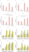
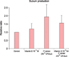
Upregulation of IL-1β, IL-6 and IL-8 (p<0.05) in the sebocytes by P. acnes (1010 CFU/µl) was inhibited by vitamin D at 10−6 M (Fig. 1A~C). Upregulation of TNF-α in the sebocytes by P. acnes (1010 CFU/µl) was not inhibited by vitamin D at 10−6 M (Fig. 1D). Upregulation of IL-1β, IL-6, IL-8 and TNF-α in the sebocytes by 40 mJ/cm2 UVB showed more decreasing tendency after treatment with 10−6 M vitamin D compared with 10−8 M vitamin D (Fig. 1E~H). Upregulation of IL-1β, IL-6 and TNF-α in the sebocytes by 40 mJ/cm2 UVB was inhibited 1 day after treatment with 10−6 M vitamin D compared with control (Fig. 1E, F, H). Upregulation of IL-1β, IL-6 (p<0.05), IL-8 and TNF-α in the sebocytes by 70 mJ/cm2 UVB showed much more decreasing tendency after treatment with 10−6 M vitamin D compared with 10−8 M vitamin D (Fig. 1E~H). Sebum production of cultured sebocytes after treatment with 10−6 M vitamin D or P. acnes (1010 CFU/µl) was increased. However, sebum production of cultured sebocytes after treatment with P. acnes (1010 CFU/µl) was decreased by the addition of 10−6 M vitamin D (Fig. 2).
P. acnes plays a key role in the initiation of inflammatory acne3. Previous studies have demonstrated proliferation of the sebaceous glands and cultured sebocytes after UV irradiation45. Sebocytes have shown the upregulation of inflammatory cytokines following treatment with UVB irradiation67. Vitamin D has been reported to stimulate the proliferation of sebocytes and to inhibit their differentiation and lipid synthesis4. Furthermore, vitamin D decreases the production of inflammatory biomarkers, especially IL-6, IL-8, and MMP-9, from cultured sebocytes. Krämer et al.8 also reported that vitamin D reduced the secretion of IL-6 and IL-8 in SZ95 sebocytes. On the basis of these, we investigated the effect of vitamin D on the inflammatory reaction of sebocytes treated with P. acnes or UVB irradiation. In this study, as reported previously, vitamin D decreased the expression of the inflammatory cytokines, such as IL-1β, IL-6, IL-8, and TNF-α. In addition, vitamin D inhibited the upregulation of IL-1β, IL-6 and IL-8 in sebocytes after treatment with P. acnes. Furthermore, higher concentration (10−6 M) of vitamin D inhibited the upregulation of IL-1β, IL-6, IL-8, and TNF-α in sebocytes after treatment with 40 mJ/cm2 or 70 mJ/cm2 UVB irradiation compared with lower concentration (10−8 M) of vitamin D. Vitamin D decreased sebum production after treatment of sebocytes with P. acnes in our study. It was reported that treatment of slowly proliferating SZ95 sebocytes with vitamin D results in a statistically significant time- and dose-dependent reduction of sebum lipids8. Unlike our expectation and a previous report, the treatment of sebocytes with vitamin D only showed a mild increase in sebum production in this study. However, there was not statistically significant. Like this study, P. acnes extracts usually increase sebum production in hamster sebaceous glands both in vivo and in vitro9.
In conclusion, the treatment of sebocytes with vitamin D shows a tendency to inhibit the upregulation of inflammatory biomarkers by P. acnes and UVB irradiation. On the basis of these findings, the use of vitamin D for inflammatory acne may be promising. This is supported by the report that in severe acne patients vitamin D deficiency significantly potentiates the inflammatory process10. In addition, the treatment of acne with vitamin D has been tried since long before. However, because of liphophilic property and high molecular weight of vitamin D, it should be considered that vitamin D in topical agents do not easily penetrate into the deep dermis, especially sebaceous gland.
 XML Download