

 PDF
PDF ePub
ePub Citation
 Print
Print


Dear Editor:
Diffuse cutaneous melanosis (DCM) is a rare condition affecting a minority of advanced melanoma patients. It is often described in the setting of metastatic disease of the liver and was first defined by the German pathologist Wagner1, who reported on a patient with melanoma arising from a congenital nevus with subsequent gray skin coloration.
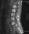
We report on an otherwise healthy 45-year-old Caucasian woman with a 3-mm-thick nodular melanoma (pT3a) on the right flank. At the time of the first diagnosis (2011), there were no clinical/radiological signs of metastatic disease. After the positive sentinel-lymph-node biopsy (sn1/3), she underwent a complete lymphadenectomy. Two years later and upon negative clinical and radiological follow-up, she complained of intense lower-back pain radiating down to the left leg. Magnetic resonance imaging of the spine showed diffuse bone metastases with pathological fractures and multiple metastases in L5 with penetration of the rear edge and epidural fat tissue with compression of the S1 nerve. A computed tomography-controlled percutaneous aspiration biopsy was performed for routine molecular diagnostics. The melanoma tissue showed the presence of wild-type BRAF. The patient was treated with combination chemotherapy (vindesine/cisplatin) and symptomatic palliative radiotherapy of the spinal cord with five 4-Gy sessions, amounting to a total dose of 20 Gy. After two cycles of chemotherapy, she was hospitalized because of pain exacerbation. Subsequent imaging studies showed disease progression with new liver metastasis and progressive spinal metastasis (M1c) (Fig. 1). High levels of lactate dehydrogenase (1,264 U/L) and S-100 (18.3 µg/L) were detected. Furthermore, she developed diffuse gray hyperpigmentation affecting the entire skin and sclera corresponding to DCM (Fig. 2). No pigment changes of the hair were noted. The patient was treated symptomatically and was transferred to a palliative care unit. Four months after the onset of DCM, she died.
The clinical presentation of DCM consists of diffuse blue-grayish pigmentation of the skin and sclera, with a descending cephalo-caudal progression2. Additional clinical features include melanuria, hair pigment changes, and darkening of the serum or peritoneal fluids. Histologically, in most of the published cases, the presence of dermal pigment, deposition of melanin in histiocytes, and free melanin in dermal connective tissue are reported3. DCM has a poor outcome with a mean time to death of approximately 4~5 months after disease onset4.
The pathogenesis of DCM is yet unclear. One current proposed theory is that elevated melanocytic growth factors such as α-melanocyte-stimulating hormone, endothelin-1, and hepatocyte growth factor activate the melanogenesis, proliferation, and motility of normal and malignant melanocytes5. Nevertheless, this hypothesis is contradictory to the fact that no alteration in melanocyte numbers or keratinocyte pigmentation was observed in most cases of DCM.
Oncologists and dermato-oncologists should be familiar with DCM and its underlying poor prognosis. Taking into account the anticipated survival of these patients, an interdisciplinary palliative care treatment aimed at providing good individualized symptom control and relief in all areas of the patient's life should be offered in experienced centers.

 XML Download