

 PDF
PDF ePub
ePub Citation
 Print
Print


INTRODUCTION
Tinea incognito is a cutaneous fungal infection induced by systemic or topical steroids, or other immunosuppressive agents. The clinical manifestation of tinea incognito varies and can mimic those of various skin diseases such as lupus erythematosus and eczema1. This characteristic makes its diagnosis difficult and can easily lead to misdiagnosis, preventing prompt and appropriate treatment2. Here, we report an interesting case of tinea incognito after topical steroids administration in an immunosuppressed patient with dermatitis artefacta.
CASE REPORT
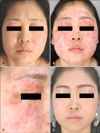
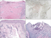
A 40-year-old female patient presented with a 2-month history of moderately itchy multiple erythematous erosive lesions on the face and upper chest (Fig. 1A). She had a multi-year history of chronic inflammatory demyelinating polyneuropathy (CIDP) and had been taking systemic glucocorticoids for 4 years. Punch biopsy and immunofluorescence stain were performed on the upper chest lesion, which was suspicious of pemphigus foliaceus. Histologic examination yielded no specific results (Fig. 2A), and immunofluorescence staining showed no deposition of immunoglobulin G (IgG), IgA, IgM, C3, C4, or C1q. While she denied any role in causing the lesions, she presented with multiple severely inflamed mosquito bite-like lesions on her arms; the patient stated that she had a habit of scratching her skin. A diagnosis of dermatitis artefacta was made, and the skin lesions showed improvement with topical steroid administration.
Two months after starting treatment, the facial skin lesions became aggravated. At that time, CIDP also became aggravated, and immunosuppressant azathioprine was prescribed at the Department of Neurology. The clinical feature of the facial lesions became polycyclic and erythematous over time and differed from the preexisting ones (Fig. 1B, C).
Because of the possibility of lupus erythematous, skin biopsy was performed on the forehead lesion. Histopathology revealed no evidence of lupus erythematosus but showed fungal hyphae in the cornified layer (Fig. 2B, C). Blood tests including antinuclear antibody were performed, but the results were normal. Fine scale was observed concurrently, and the KOH test result on the fine-scale lesion was positive (Fig. 2D). Therefore, the patient was diagnosed with tinea faciei (i.e., tinea incognito). After diagnosis, topical steroid was discontinued and oral terbinafine was administered. Complete remission was achieved after 1 week of oral terbinafine and topical amorolfine administration (Fig. 1D).
Written informed consent was received from patient to participate in this report.
DISCUSSION
Tinea incognito is a dermatophytic infection in which systemic or topical steroids may actually spread the infection and modify the clinical manifestations such that they are atypical. Tinea incognito usually lacks the classic features of a typical fungal infection, being less scaly and with less raised margins, and appears more pustular and extensive with substantial irritation. In a large retrospective study of 200 cases of tinea incognito, the disease clinically resembled lupus erythematosus, eczema, or rosacea on the face and impetigo- and eczema-like on the trunk and limbs1. There are reports of tinea incognito mimicking purpura, seborrheic dermatitis, lichen planus, contact dermatitis, psoriasis, and erythema migrans3,4. Treatment is somewhat simple: withdrawing anti-inflammatory agents and administering systemic and/or topical anti-fungal agents such as terbinafine, itraconazole, and fluconazole5.
Although the treatment of tinea incognito is simple and relatively easy, proper diagnosis and treatment tend to be delayed, because the disease usually masquerades as other skin disorders. Misinterpretation as an aggravation of a pre-existing skin disorder is also frequent, which may also lead to prolonged treatment with topical steroids.
Although dermatophyte infection has some characteristic histological findings such as neutrophils in the stratum corneum, compact orthokeratosis, papillary dermal edema, and the presence of fungal hyphae between 2 zones of cornified cells6,7, the sensitivity of H&E staining for detecting hyphae may be insufficient to conclude or exclude a diagnosis of fungal infection. Therefore, periodic acid-Schiff staining should be performed to confirm hyphae, as routine H&E staining may easily miss hyphae and delay correct diagnosis8.
Frequent skin examinations are necessary in vulnerable patients such as those taking immunosuppressants. If any skin lesions are resistant to conventional treatment, tinea incognito should be considered and ruled out even the lesions lack the clinical manifestations of a fungal infection, such as scales.
 XML Download