

 PDF
PDF ePub
ePub Citation
 Print
Print


INTRODUCTION
The macrophage-derived chemokine (MDC)/CC chemokine ligand 22 (CCL22) is one of the functional ligands for CC chemokine receptor 4 (CCR4) and is a chemoattractant for the CCR4-expressing cells such as Th2 cells. We and other groups reported that keratinocytes produced CCL22 after stimulation with tumor necrosis factor (TNF)-α and interferon (IFN)-γ in vitro, suggesting that keratinocytes are a main source of CCL221,2. In addition, inhibitors for nuclear factor-kappa B (NF-κB) and p38 mitogen-activated protein kinase (MAPK), but not for extracellular signal-related kinase (ERK), inhibited TNF-α and IFN-γ induced production of thymus- and activation-regulated chemokine CCL17, another Th2-type chemokine and CCR4 ligand, by the human keratinocyte cell line, HaCaT. Further, an inhibitor of epidermal growth factor receptor (EGFR) tyrosine kinase enhanced the CCL17 production by these keratinocytes3. However, the mechanism of CCL22 production by HaCaT cells has not been fully identified. Herein, we investigated the signal transduction pathways by which TNF-α and IFN-γ stimulate HaCaT cells to produce CCL22 by adding various inhibitors and comparing expression of CCL22 with that of CCL17. We also investigated the signal transduction pathways of CCL22 production using normal human epidermal keratinocytes (NHEKs) and compared these pathways with those identified using HaCaT cells.
MATERIALS AND METHODS
Cytokines and antibodies
Recombinant human TNF-α and IFN-γ were purchased from PeproTech (Rocky Hill, NJ, USA). PD98059 (ERK inhibitor) was from Alexis Biochemicals (San Diego, CA, USA). PD153035 (EGFR inhibitor), Parthenolide (NF-κB inhibitor), Bay11-7085 (NF-κB inhibitor), SB202190 (p38 MAPK inhibitor), c-Jun N-terminal kinase (JNK) inhibitor II, and Janus kinase (JAK) inhibitor 1 were from Calbiochem (San Diego, CA, USA).
Cell cultures
HaCaT cells were a generous gift from Dr. T. Kuroki (Showa University, Tokyo, Japan) with the permission of Dr. N. E. Fusenig (Institute for Cell and Tumor Biology, German Cancer Research Center, Heidelberg, Germany). They were grown routinely in Eagle's minimum essential medium (MEM; SIGMA, St. Louis, MO, USA) with 10% fetal calf serum (FCS). Cells of the 30th to 50th passage were used for experiments. Cells were incubated in MEM without FCS for 24 hours before stimulation3.
NHEKs were purchased from Kurabo (Osaka, Japan), and cultured in keratinocyte serum-free media (K-SFM; GIBCO, Invitrogen, Carlsbad, CA, USA) supplemented with 40 µg/ml of bovine pituitary extract (BPE; Kyokuto Seiyaku, Tokyo, Japan) and 5 ng/ml epidermal growth factor (EGF; R&D Systems, Minneapolis, MN, USA). Cells passaged 3~6 times were used for this study. Cells were incubated in K-SFM without BPE and EGF for 24 hours before stimulation4.
Several signal transduction inhibitors, PD98059 (50 µM), PD153035 (5 µM), Parthenolide (5 µM), Bay 11-7085 (2 µM), SB202190 (5 µM), JNK inhibitor II (50 nM), and JAK inhibitor 1 (5 µg/ml), were added 1 hour before stimulation with 50 ng/ml of recombinant human TNF-α and IFN-γ Supernatants were harvested after 24 or 48 hours after stimulation.
RESULTS
CCL22 production levels by HaCaT cells in 24-hour culture
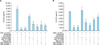
As shown in Fig. 1A, TNF-α- and IFN-γ-induced CCL22 production in HaCaT cells was inhibited by inhibitors PD98059 (ERK inhibitor), PD153035 (EGFR inhibitor), Parthenolide (NF-κB inhibitor), Bay 11-7085 (NF-κB inhibitor), SB202190 (p38 MAPK inhibitor), JNK inhibitor II, and JAK inhibitor 1 by 90%, 90%, 35%, 70%, 80%, 70%, and 75%, respectively, in 24-hour culture.
CCL22 production levels by HaCaT cells in 48-hour culture
When cultured for 48 hours, the CCL22 production was inhibited by inhibitors, PD98059, PD153035, Bay 11-7085, SB202190, JNK inhibitor II, and JAK inhibitor 1 by 85%, 80%, 55%, 70%, 40%, and 30%, respectively (Fig. 1B).
CCL22 production levels by NHEKs
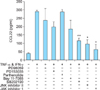
As shown in Fig. 2, TNF-α- and IFN-γ-induced CCL22 production in NHEKs was blocked by inhibitors SB202190, JNK inhibitor II, and JAK inhibitor 1 by 60%, 65%, and 80%, respectively, in 24-hour culture.
DISCUSSION
Our results indicate that CCL22 production in HaCaT cells is dependent on ERK, EGFR, NF-κB, p38 MAPK, JNK, and JAK, and production of CCL22 is mediated by different signal pathways than those regulating production of CCL173. Collectively, our previous and present results suggest that EGFR activation represses CCL17 but enhances CCL22 production by these cells. Furthermore, we performed an ELISA both 24 and 48 hours after stimulation to evaluate the possibility that signal pathway inhibitors may work on different time scales, which may cause different CCL22 production in HaCaT cells 48 hours after culture treatment. The data obtained at both time points showed almost the same results except that of Parthenolide, an NF-κB inhibitor, inhibited CCL22 production in a 24-hour but not in a 48-hour culture. In contrast, another NF-κB inhibitor, Bay 11-7085, inhibited the CCL22 production in both 24- and 48-hour cultures (Fig. 1, 2). The different results between these two NF-κB inhibitors may be due to their difference in specificity or potency.
Because HaCaT cells and NHEKs differentially produce CCL17 and CCL222,5, we compared the CCL22 production of both cell types in 24-hour cultures (Fig. 1A, 2). ELISA data from both cell types showed the same tendency; inhibitors for p38 MAPK, JNK, and JAK inhibited the CCL22 production by both cell types. Inhibitors for ERK, EGFR, and NF-κB inhibited the CCL22 production by HaCaT cells and tended to inhibit production by NHEKs, but not to significant levels. The differing results may be partly due to the difference in CCL22 production levels between these two cell lines; HaCaT cells produce much more CCL22 than NHEKs, as also reported previously2. Because CCL17 is not produced by NHEKs6, we evaluated the CCL22 production levels using HaCaT cells with various inhibitors and compared them with those of CCL17.
TNF-α causes the activation of signaling molecules such as ERK, NF-κB, p38 MAPK, JNK in HaCaT cells and that IFN-γ activates ERK, p38 MAPK, and JAK in several cell types7,8,9,10, indicating consistency with our results. It seems more relevant to analyze the underlying signaling pathway for each cytokine treatment by TNF-α or IFN-γ, but each cytokine separately stimulates CCL22 production weakly while their combination synergistically stimulates it; this was shown by Kwon et al.11, and also confirmed by our preliminary experiment (data not shown). Furthermore, because we wanted to compare the mechanism of CCL22 production with that of CCL17 production, we investigated the signal transduction pathways by which TNF-α and IFN-γ stimulate HaCaT cells to produce CCL22 by adding various inhibitors, as was done in other reports. Qi et al.12 demonstrated that secretion of both CCL17 and CCL22 by HaCaT cells was inhibited by p38 MAPK inhibitor, but not by PD98059 (10 µM) or SP600125 (10 µM), inhibitors for ERK and JNK, respectively. In addition, Kwon et al.11 showed that p38 MAPK inhibitor significantly inhibited CCL17 and CCL22 production at protein and mRNA levels, whereas inhibitors for ERK (PD98059, 20 µM) and JNK (SP600125, 20 µM) had the most minimal effect. There are discrepancies between their results and ours with regard to the effect of ERK and JNK inhibitors. These discrepancies may partly be due to the differences in cell origins or culture conditions, or kinds and concentrations of inhibitors used. We must be careful in interpreting these results.
Our previous and present results showed that the EGFR inhibitor enhanced CCL17 production but reduced CCL22 production by HaCaT cells. A similar phenomenon has been reported by Mascia et al.13: the EGFR signaling blockade increased CCL2, CCL5, and CXCL10, but decreased CXCL8 expression in NHEKs. EGFR is known to activate various signaling cascades such as ERK and phosphatidylinositol 3-kinase3. The finding that both EGFR and ERK inhibitors reduced CCL22 production suggests that the activated EGFR exerts its signal at least in part through ERK to enhance CCL22 production. Although the mechanism of CCL17 production enhancement and CCL22 reduction by the EGFR inhibitor needs further investigation, it is plausible that EGFR activation suppresses CCL17 production, while it enhances CCL22 production. Cultured growing epidermal keratinocytes show activated EGFR even in the basal condition14, which may affect the production of CCL17 in cultured NHEKs, while in vivo expression of CCL17 has been shown in several reports15. The differential EGFR involvement in the production of these two Th2 chemokines may be reflected in their expression patterns in the epidermis and may be important in differential regulation of CCR4-positive cell infiltration into the epidermis. Recently, the frequent use of chemotherapeutic regimens of EGFR inhibitors has been associated with some cutaneous toxicities, including acne-like rashes and paronychia. Very recently, Paul et al.16 investigated the underlying mechanism of EGFR inhibition-associated chemokine production in keratinocytes as well as in patients after treatment with epidermal EGFR inhibitors. Increased CCL2, CCL5, and decreased CXC chemokine ligand 8 (CXCL8) expression was observed in keratinocytes, consistent with the previous report13. Furthermore, they showed that in EGFR-treated patients, low levels of serum CXCL8 corresponding to stronger EGFR inhibition were associated with a higher grade of skin toxicity and a prolonged overall survival16. Further studies will be necessary to determine the underlying mechanism of EGFR inhibition-associated chemokine production including CCL17 and CCL22 in patients after EGFR inhibitor treatment. In summary, our results indicate that CCL22 production in HaCaT cells is mediated by signal pathways that are differ from those regulating production of CCL17. This study may shed new insight on the mechanism underlying Th2-dominant skin diseases such as atopic dermatitis.
 XML Download