

 PDF
PDF ePub
ePub Citation
 Print
Print


Dear Editor:
Leprosy, a chronic granulomatous infection caused by Mycobacterium leprae, commonly affects the skin and nerves. Because of its many different manifestations, leprosy can be easily misdiagnosed as syphilis, sarcoidosis, lymphoma, leishmaniasis, and cutaneous tuberculosis1. Since multidrug therapy (MDT), the prevalence of leprosy has decreased to <1 in 10,000. However, there are still newly diagnosed and relapsed cases. We report an unsuspected case of relapsed multibacillary leprosy.
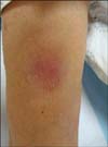
A 63-year-old Korean man visited a neurologist because of tingling sensations in his hands and arms for 8 months. His medical history revealed congenital syphilis, claw hand deformity, uveitis, hypertension, and diabetes mellitus. Additionally, he was found to have prurigo 1 year before his visit, presenting as generalized pruritic nodules; however, these were not present at his visit. Neurologic examination revealed thenar/hypothenar muscle atrophy, ankle/knee jerk areflexia, hypoesthesia, and motor weakness in the fingers. Provisional diagnosis included neurosarcoidosis, polyneuropathy from diabetes mellitus, and tabes dorsalis. The laboratory findings were within the reference ranges, except for increased angiotensin-converting enzyme (ACE, 85.8 U/L; reference range, 18~55 U/L). His chest radiograph and cerebrospinal fluid were normal. Nerve conduction results showed distal, symmetric, sensorimotor polyneuropathy. He was referred to us for a newly developed single erythematous nodule on the right upper arm (Fig. 1). A punch biopsy was done to rule out sarcoidosis. Histopathologic examination revealed foamy histiocytes throughout the dermis, with perivascular lymphocytic infiltration (Fig. 2A, B). Acid-fast staining revealed many bacilli (Fig. 2C). Only then did the patient mention that he had developed lepromatous leprosy in 1973. The Korean Hansen Welfare Association was contacted, which stated that he had been treated for a year, remained disease-free, and was lost to follow-up in 1995. Finally, his condition was diagnosed as relapsed multibacillary leprosy. Previous cutaneous lesions, diagnosed as prurigo, could not be differentiated from manifestations of relapsed leprosy because biopsies and clinical photographs were missing. He is being treated with MDT (rifampicin, dapsone, and clofazimine) for another year.
Typical skin lesions of relapsed multibacillary leprosy are multiple, soft, shiny, pink papules or nodules on the forehead, lower back, dorsa of hands and feet, posterior arms, thighs, and buttocks2. Like sarcoidosis, ACE frequently increases in leprosy, indicating extensive macrophage activity3. Modern clinicians with little experience of leprosy may not recall the disease even when circumstantial evidences including claw hand deformity, thenar and hypothenar muscle atrophy, and tingling sensation indicate leprosy, as in this case. As patients may hide their medical history, suspicion for leprosy is needed. An early and accurate diagnosis is important to start MDT, reduce the risk of transmission, and minimize permanent damage. Proper treatment is important in relapsed cases as relapse usually occurs because of reinfection or growth of persistent organisms4. We report this case from an educational point of view because young dermatologists may impede the diagnosis of leprosy at first. Additionally, because of the increase in global travel and immigration, doctors even in nonendemic areas need to know how to identify leprosy so that no case will be missed.

 XML Download