

 PDF
PDF ePub
ePub Citation
 Print
Print


Dear Editor:
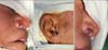
An 82-year-old man presented with a grayish-black papule on the right ala of the nose that had been there for 3 to 4 years, and which bled occasionally. Physical examination revealed that the size of the papule was 0.5×0.5 cm. The patient had no family history of skin cancer; he had a history of heavy cigarette smoking. We suspected basal cell carcinoma and performed a 2-mm punch biopsy. The results of the pathologic examination of the specimen confirmed the diagnosis of basal cell carcinoma.
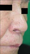
Before the surgery, we ensured that he stopped cigarette smoking to allow better survival of the graft. The tumor was widely excised with a 2-mm surgical margin, resulting in a 0.9×0.8 cm full-thickness defect (Fig. 1A). The defect was reconstructed by using a composite chondrocutaneous graft harvested from the antitragus of the right ear (Fig. 1B). The graft was prepared in such a way that it was approximately 20% larger than the defect in order to compensate for the natural contraction of the graft. The donor site was closed primarily, and after hemostasis of the recipient bed, the defect was repaired with the composite skin graft (Fig. 1C). To lower the metabolic demand, an ice pack was applied to the recipient site postoperatively for surface cooling. At a 5-month postoperative follow-up, desirable aesthetic and functional results were observed (Fig. 2). The patient agreed publication of his case.
Auricular composite grafting is relatively simple, and can be performed in a single surgical session for the repair of all 3 layers of the nasal ala. The donor tissue provides mechanical stability to the repaired ala for preventing collapse on inhalation1, as well as acceptable color and contour match. In addition, the donor site can be easily repaired without noticeable scarring.
One drawback of the composite graft is that it cannot be used for defects >2 cm, because of its high metabolic demand. Moreover, the procedure is less reliable than either the local or regional flap techniques2,3, and can only be used on healthy recipient sites3. Smoking increases the risk of postoperative ischemia, which can result in poor wound healing or tissue necrosis1. In smokers, like our patient, other reconstructive methods or complete cessation of smoking should be considered.
Several procedures have been introduced to increase the survival of composite chondrocutaneous grafts and overcome graft failure. With regard to postoperative procedures, cooling can be used to lower the metabolic demands of the graft4. Hyperbaric oxygen can also be supplied to enhance fibroblast replication, which increases collagen formation and neovascularization5. Finally, scarification with a thin needle (20~26 gauge) coupled with heparin injections can be used for decongestion of the graft1.
In conclusion, we successfully reconstructed a full-thickness defect on the free margin of the nasal alar rim with a composite chondrocutaneous graft from the patient. We believe that reconstructive surgeons can give preference to this technique when reconstructing nasal alar defects, especially small defects. In addition, surgeons should consider the use of appropriate postoperative methods to increase graft survival.
 XML Download