

 PDF
PDF ePub
ePub Citation
 Print
Print


Dear Editor:
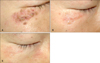
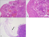
A 30-year-old-man presented with a 2-month history of a facial lesion. He was examined by general practitioners, and the eruption was diagnosed as eczema. Topical corticosteroid was applied for 1 month, and the eruption initially seemed to improve with this treatment; but later on, it persisted, and gradually extended in size. Dermatological examination revealed the presence of grouped erythematous papules, vesicles and crusts on erythematous bases, on the right lower eyelid (Fig. 1A). He had no medical history, and no family member who had had similar skin eruptions or symptoms. The initial clinical differential diagnosis included herpes simplex, herpes zoster, and allergic contact dermatitis, caused by an antibiotic eye drop. We prescribed oral and topical acyclovir, and performed a skin biopsy, to reveal the exact diagnosis. Skin lesions did not respond to 5 days of acyclovir therapy, and histologic examination showed infiltration of various inflammatory cells from the upper to lower dermis, parakeratosis, irregular acanthosis, intraepidermal exocytosis of neutrophils, and extravasation of erythrocytes (Fig. 2A, B). Fungal hyphae and spores in the stratum corneum were identified on the periodic acid Schiff stained section (Fig. 2C). These findings led to the diagnosis of superficial fungal infection that had lost its typical clinical appearance, because of the use of steroids. The cause of infection might be dermatophytes, but non-dermatophytic fungi could be possible. Afterwards, the history that he had had contact with his cat was verified. He was administered oral terbinafine 250 mg daily, and topical terbinafine cream. After 8 weeks, the number of papules decreased, and the inflammatory reaction improved (Fig. 1B). Treatment was continued a month longer, and the facial eruption finally cleared (Fig. 1C).
The clinical features of tinea faciei are characterized by various morphology, and because of that, the entity can mimic many other cutaneous disorders1. Moreover, because tinea faciei is relatively uncommon when compared with other forms of superficial fungal infection, it is often misdiagnosed; and treated with glucocorticosteroids, not antifungal agents1. Imprecise use of topical or oral corticosteroids in tinea faciei can modify their clinical features, and make the correct diagnosis more difficult2; therefore, tinea faciei is one of the considerable examples of tinea incognito1. One retrospective study showed 35.7% cases of tinea incognito among tinea faciei, because of improper diagnosis, and inappropriate therapy3. Other authors revealed that 50% to 70% of patients with tinea faciei are initially misdiagnosed as having other dermatoses2. The pathomechanism of tinea incognito is thought to be closely associated with a steroid-modified response of the host to cutaneous fungal infection4. Topical corticosteroids allow fungi to grow readily, and alter the clinical feature of the lesions, because they have immunosuppressive activity5. Regretfully, glucocorticosteroids are extensively used by patients and non-dermatologists, and lead to long-lasting fungal infections1, similar to that which occurred in our case.
Atypical presentations of tinea faciei can lead to misdiagnosis. The present case underlines that physicians should keep in mind that clinical features of superficial fungal infection can be substantially modified by incorrect treatment, to mimic even herpes simplex virus infection. Fungal infection should be on the list of differential diagnoses of facial eruptions, especially in cases not responding to preceding management.
 XML Download