

 PDF
PDF ePub
ePub Citation
 Print
Print


INTRODUCTION
Insulin-like growth factor-I (IGF-I) is a growth factor with sequence homology to pro-insulin. It has been identified as an important growth factor in many biological systems1. IGF-I is critically involved in promoting hair growth by regulating cellular proliferation and migration during the development of hair follicles (HFs)2. The proliferating function of IGF-I in skin may be important to the development of HFs. Transgenic mice overexpressing IGF-I in the skin have earlier HF development3. IGF-I receptor knockout mice have fewer, smaller and earlier HF development4. Itami and Inui5 demonstrated that IGF-I is produced by dermal papilla cells. Since IGF-I receptors were detected in keratinocytes, it is possible that IGF-I produced by dermal papilla cells might act on keratinocytes, thereby promoting hair growth through stimulation of the proliferation of keratinocytes in the HFs6.
To exert its biological effects, IGF-I must activate cells by binding to specific cell-surface receptors. The type I IGF receptor (IGF-IR) is the only IGF receptor to have IGF-mediated signaling functions1. In tissues, IGF-I is produced by mesenchymal type cells and acts in a paracrine fashion and/or an autocrine fashion by binding to the IGF-IR. This binding activates the receptor tyrosine kinase that triggers the downstream responses and finally stimulates cell division7. IGF-I may therefore be able to stimulate the proliferation of HF cells through cellular signaling pathways of its receptors.
The growth of hair is a cyclic process in which every follicle proceeds from an active phase (anagen) through a regression phase (catagen) to a resting phase (telogen). During catagen, HFs undergo apoptosis and there is a decline in an apoptotic protein, Bcl-2, and an increase in a pro-apoptotic protein, Bax8. In several reports, IGF-I and IGF-II have prevented the follicle from developing catagen-like status9.
In many experimental reports, pro-inflammatory cytokines, such as IL-1α,β, TNF-α, and INF-γ, have been reported to induce apoptosis of hair bulb keratinocytes10-12.
Platelet-derived growth factor (PDGF) is a potent mitogen produced in a variety of cell types including keratinocytes and endothelial cells, and is important for cell growth. Expression of PDGF isoforms in HFs in vitro can be influenced by treatment with cytokines that are known to be positive and negative regulators of HF growth activity13. Recently, PDGF also induced and maintained anagen phase in mouse hair cycling14. The effects of PDGF on HFs, that is induction and maintenance of anagen, is very similar to the effect of IGF-I on hair growth.
In the current study, expression of hair growth factors & cytokines (PDGF-A, PDGF-B, transforming growth factor [TGF]-β, interleukin [IL]-1α, IL-1β, tumor necrosis factor [TNF]-α, interferon [INF]-γ) and apoptosis-related molecules (Bax, Bcl-2, c-myc) were determined by reverse transcription polymerase chain reaction (RT-PCR), after treatment of cells with IGF-I using an organ culture model of human HFs. This experiment was done in order to examine the molecular mechanism underlying the effect of IGF-I on HFs.
MATERIALS AND METHODS
Specimens
Human occipital scalp skin specimens were obtained from hair transplantation surgery, after informed consent. The medical ethical committee of the Yonsei University Wonju College of Medicine, Wonju, Korea, approved all described studies. The study was conducted according to the Declaration of Helsinki principles. Skin samples, used for HF microdissection and organ culture, were collected and stored in a refrigerator at 4℃.
Isolation and culturing of human HFs
Human HFs in the Anagen VI stage were isolated as previously described15. Briefly, after separation of epidermis and dermis from the dermo-subcutaneous interface, anagen HFs were isolated from subcutaneous fat under a binocular microscope with watchmaker's forceps. The total number of HFs used in this study was 140, derived from 3 different individuals. Each one of the anagen HFs was carefully transferred to a per well of a 24-well plate (Corning Incorporated, Corning, NY, USA). In the control group, isolated anagen HFs were maintained in 500 ul of William's E medium (Gibco BRL, Gaithersburg, MD, USA) supplemented with 10 ng/ml hydrocortisone (Sigma, St. Louis, MO, USA), 2 mM L-glutamine (Gibco BRL), 100 IU/ml penicillin and 100 ug/ml streptomycin (Gibco BRL) and without insulin. In the IGF-I group anagen HFs were incubated under the same conditions. The only difference was that 10-7 M IGF-I was added to the experimental group.
HFs were maintained free-floating at 37℃ in an atmosphere of 5% CO2 and 95% air in a humidified incubator. At least 20 HFs were cultured in each type of medium. Briefly, control HFs were cultured with vehicle for 12 days. The culture medium was changed every 2 days and the follicles were cultivated in the culture medium as they were suspended freely. The same experiment was repeated three times.
Measurement of shape and growth in follicle length
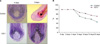
For measurement of follicle length, the entire length from the base of the hair bulb to the tip of the hair shaft was measured using the measuring scales attached to the objective lens of the microscope. This was done until the 12th day of cultivation in 2 day intervals, and the results were statistically analyzed. At the same time, follicle shapes (anagen phase, catagen phase, telogen phase) were observed and the duration of anagen phase for each follicle was examined.
Semi-quantitative RT-PCR analysis
Total RNA was extracted from isolated human anagen VI stage HFs using a monophasic solution of phenol and guanidine isothiocyanate (TRIzol Reagent; Gibco BRL). The concentration of RNA was determined using a UV spectrometer at 260 nm. Aliquots (1.0 ug) of RNA were reverse transcribed using Moloney murine leukemia virus reverse transcriptase (MML-V RTase, Promega, Madison, WI, USA). Briefly, RNA samples were incubated at 70℃ for 10 minutes with molecular biology grade water. After chilling on ice, primer extension and reverse transcription were done by the addition of 1X RT-buffer, 5 mM MgCl2 Solution, 1 mM deoxynucleotide triphosphates, 2.5 uM Oligo d(T)16 (Roche Molecular Biochemicals,. Indianapolis, IN, USA) and MML-V RTase (2.5 units/ul) in 20 ul reaction volumes. Samples were then incubated at 42℃ for 45 minutes before storage at -20℃. One ul cDNA was then subjected to PCR cycles as follows: a 95℃ denaturation phase for 5 minutes, followed by 35 cycles of 95℃ for 1 min, 60℃ for 1 minute, 72℃ for 1 min; and an additional extension for 10 min at 72℃. The primers used for amplifying the respective fragments are listed in Table 1. The PCR product was visualized on a 2% agarose gel.
RESULTS
Effect of IGF-1 on cumulative human HF elongation over 12 days
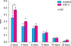
Grossly, the IGF-1 treated group showed more remarkable hair growth (approximately 0.10 mm/day) compared with the control group (approximately 0.08 mm/day).
On each experimental day, the IGF-1 group showed more significant hair growth than control (Fig. 1). This tendency was most prominent during the second day of the experiment, with the results showing notable numerical differences between the two groups (Fig. 1). There was a significant difference (p<0.05) in cumulative HF elongation between the control group (0.97±0.09 mm) and the 10-7 M IGF-1 treated HFs (1.24±0.09 mm) (Fig. 2).
Proportion of anagen HFs in control and experimental groups
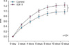
Both experimental and control groups consisted of 24 anagen hairs. Until the 4th day of the experiment, in both groups, all HFs remained in the anagen hair stage (24/24). On the 6th day, a difference in the anagen proportion was found. The IGF-I treated group showed more anagen HFs (21/24) than controls (17/24). The IGF-I group showed a larger anagen proportion from the 6th to the 12th day. On the 8th and 10th days, the control group showed eleven anagen HFs (11/24) and the IGF-I group showed seventeen anagen HFs (17/24).
On the 12th day, the control group showed 45.8% anagen HFs (11/24) and the IGF-I group showed 70.8% anagen HFs (17/24) (Fig. 3).
Change in the expression by HFs of hair growth related factors
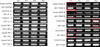
Increasing evidence suggests that several factors, such as cytokine and apoptosis related molecules, are involved in the hair growth cycle15-18. In this study, semi-quantitative RT-PCR analysis was used to search for factors that were affected by IGF-I. We measured expression levels of PDGF-A (platelet derived growth factor-A), PDGF-B, TGF-β, IL-1α, IL-1β, TNF-α, INF-γ, and apoptosis-related molecules (Bax, Bcl-2, c-myc).
Pro-inflammatory cytokines such as IL-1α, IL-1β, INF-γ, and TNF-α are known to promote the apoptosis of follicular keratinocytes. In both the experimental and the control groups, baseline differences in these pro-inflammatory cytokines were insignificant, as was TGF-β. However, on the 2nd day, there was a significant difference in PDGF-A & B expression between the two groups. On the 2nd day, the IGF-I treated group showed more prominent expression of PDGF-A, and PDGF-B also showed a significant increase in expression. Among the apoptosis related molecules, Bax and Bcl-2 showed differences in expression. On the 8th day, Bax was weakly expressed in the IGF-I treated group. On the 2nd day, Bcl-2 was more strongly expressed in the IGF-I treated group compared with the control group. Another apoptosis related molecule, c-myc, did not show a significant difference in expression (Fig. 4).
DISCUSSION
The HF is a cutaneous organ that shows cyclic activity in postnatal life and transits through periods of active hair growth (anagen), apoptosis-driven involution (catagen), hair shedding (exogen), and relative rest (telogen)14. During HF growth and hair production, the activity of factors promoting the proliferation, differentiation, and survival predominates, whereas HF regression is characterized by the activation of a variety of signaling pathways that induce apoptosis in HF cells8,15.
IGF-I is known as an important growth factor in many biological systems and it has also been shown to play a critical role in promotion of hair growth19,20. Since IGF-I receptor mRNA was detected in keratinocytes14, it is possible that IGF-I produced by dermal papilla cells acts on keratinocytes, thereby promoting hair growth through stimulation of the proliferation of keratinocytes in HFs20. In several reports, IGF-I and IGF-II prevented the follicle from developing catagen like status9.
In this study, we showed that IGF-I has a significant effect on the rate of linear hair growth and extended the overall anagen phase, making it longer than that of the control group. On the 2nd day, both the IGF-I treated group and the control group showed the most significant growth. We also demonstrated that these growth patterns are correlated with the molecular findings - the expression of PDGF-A and PDGF-B were remarkably increased compared to control and the expression of Bcl-2/bax ratio was higher than control on the 2nd day. According to Tomita et al.14, PDGF-A and PDGF-B induced and maintained the anagen phase of murine HFs. These effects were very similar to effects of IGF-I on hair growth.
Anti-apoptotic molecules have a critical role in hair cycles. For example, bcl-2 was found in the dermal papilla throughout the murine hair cycle, but its expression in the follicular epithelium increases in anagen, decreases in catagen and disappears in telogen21. Lindner et al.8 also demonstrated that during catagen, follicle cells in the bulb region undergo apoptosis and this is correlated with a down-regulation of bcl-2, while these apoptotic processes do not occur in the dermal papilla. It was believed that changes in the expression level of bcl-2 genes before catagen may be the signals for inducing apoptosis, while their reduced expression during catagen may be due to the apoptotic process22.
Regulation of bcl-2 through IGF signaling pathways have been studied in other cell types. For example, apoptosis prevented by IGF-I in promyeloid cells during aging was reported to be associated with activation of phosphatidylinositol 3'-kinase (PI3K)23. The latter function is correlated with high levels of Bcl-222. In this study, we also found that on the 2nd day of the experiment the expression of Bcl-2 was stronger in the IGF-I treated group than in the control group. On the 8th day, the expression of Bax was weaker in the IGF-I treated group. These changes in expression patterns were similar to those for PDGF-A and PDGF-B: both PDGF-A and B increased in the IGF-I treated group on the 2nd day.
IGF-I activates two signaling pathways: 1) the extracellular signal related kinase/mitogen activated kinase pathway and 2) the phosphatidylinositol 3 kinase (PI3K)/Akt pathway. Both are implicated in survival.
In conclusion, IGF-I has a significant effect on the rate of linear hair growth and extended the overall anagen phase. As stated above, the IGF-I treated group showed a significant difference in hair growth on the 2nd day of the experiment. This result was similar to the increase in expression of PDGF-A, B and the Bcl-2/Bax ratio in experimental groups that took place on the 2nd day of the experiment. According to these finding, we suggest that the effect of IGF-I on hair growth is associated with upregulation of PDGF-A and PDGF-B and the anti-apoptotic effects of IGF-I.
 XML Download