

 PDF
PDF Citation
 Print
Print


INTRODUCTION
Birt-Hogg-Dubé syndrome1 (BHDS) is a rare autosomal dominant disease characterized by a triad of benign skin tumors consisting of multiple fibrofolliculoma, trichodiscoma, and acrochordon on the face, neck, and upper trunk. The lesions clinically appear as multiple, 2~4 mm, white to skin-colored, and dome shaped papules, affecting the face and upper trunk. In further studies, an association between BHDS and renal neoplasm and lung cyst/spontaneous pneumothorax was also suggested2,3. BHDS is caused by a mutation in the gene folliculin (FLCN) which is located on chromosome 17p11.24. We report a patient with BHDS for whom molecular analysis revealed a mutation in folliculin gene.
CASE REPORT
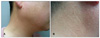
A 43-year-old man presented with a 15-year history of intermittent pruritic, multiple, skin-colored papules on the face and neck. The lesions developed on the face at first, and then slowly progressed to the neck, covering a 15-year period. Physical examination revealed multiple firm, skin-colored, 2~4 mm sized, papular lesions distributed on the face and neck area (Fig. 1). Except for the cutaneous findings, the patient was otherwise well and had no past history of kidney disease, pneumothorax, or other tumors. The result of the routine laboratory studies and chest x-ray were either normal or negative. His father died after suffering from colon cancer and his younger brother was ill with a pneumothorax. He also stated that his daughters had similar symptoms to his skin lesions.
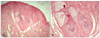
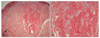
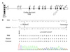
A biopsy of one of the facial papules was performed, and the lesion was reminiscent of a fibrofolliculoma by concentric fibrous tissue around follicular epithelium with thin and anastomosing bands separating from adjacent fibrovascular proliferation (Fig. 2). Meanwhile, another section of the same sample showed a relatively well-defined fibrovascular lesion with vascular proliferation and hair follicle at the margin of the lesion, suggesting trichodiscoma (Fig. 3). We studied the molecular genetic status of the FLCN gene after informed consent from the patient. We performed direct amplification of all exons (exon 1-14) of FLCN using previously reported primer sets and PCR conditions5. Direct amplification of FLCN from EDTA-treated whole blood was performed using AnyDirectMaxTM Taq REDMix (BioQuest, Seoul, Korea)6. After gel purification of direct amplicon using a Gel Purification Kit (Solgent, Daejeon, Korea), direct sequencing was performed by ABI 3730XL Sequencer (Applied Biosystems, Foster, CA, USA). Sequencing analysis revealed a heterozygotic c.1285dupC mutation (accession No. NM_144997) in the FLCN gene exon 11 (Fig. 4). This mutation was the same as the previously described 1733insC mutation. This mutation results in producing the truncated protein folliculin, which is caused by a frameshift of the amino acid sequence in the FLCN gene product (Fig. 4). Based on this, we were able to diagnose this patient as BHDS, with support from his clinical and histological findings, family history, and gene mutation revealed by sequencing analysis at genomic DNA level.
DISCUSSION
BHDS is a rare autosomal dominant disorder that was first described in 1977 by Birt, Hogg, and Dubé1. Cutaneous lesions characterized by small, firm, dome shaped, white or skin-colored papular skin lesions distributed over the scalp, forehead, face, and neck were diagnosed as fibrofolliculoma and trichodiscoma, which are difficult to distinguish clinically. With additional noted acrochordon7, this triad became known as BHDS. Also, recent reports revealed that affected patients are associated with lung cysts, recurrent spontaneous pneumothorax3,8, and renal neoplasms9, but we failed to detect any visceral lesions in our patient. In 2002, Zbar et al.2 reported that the risk of BHDS developing was about 9 and 32 times more likely to develop with renal tumor and spontaneous pneumothorax compared to unaffected persons, respectively. However, they found no increase in the risk for colorectal polyps and cancer, although some patients also may suffer from these. The cutaneous lesions usually appear in the third or fourth decades10,11. Clinical diagnosis of BHDS is usually defined when over 10 skin lesions are clinically compatible with fibrofolliculoma and at least 1 lesion histologically compatible with fibrofolliculoma5,12. Although fibrofolliculoma and trichodiscoma in BHDS are indistinguishable clinically13, some would distinguish these diseases histologically7. Fibrofolliculoma exhibits a central distorted hair follicle that is surrounded by a mantle of basophilic fibrous stroma1,14. Dilation or distortion of the hair follicle and anastomosing septa of follicular epithelium classically distinguish fibrofolliculoma from trichodiscoma15. On the other hand, trichodiscoma can be seen in the dermis as an area of fine fibrillary connective tissue containing ectatic blood vessels. Epithelial hyperplasia resembling follicular structures as seen in fibrofolliculoma and hair follicles at the margin of the lesion are often seen. Thus, some authors maintained that fibrofolliculoma and trichodiscoma are separate entities based on the histological criteria originally outlined by Pinkus et al. in 197416. However, many recent authors considered that fibrofolliculoma and trichodiscoma in patients with suspected BHDS were actually variations of the same disorder17. Steffen and Ackerman18 argued that fibrofolliculoma and trichodiscoma are a single pathologic condition. They explained that both diseases share the same epithelial and mesenchymal components, differing only in the predominance of the epithelial component in fibrofolliculoma and of the connective tissue in trichodiscoma. In addition, they offered that characteristics of both can be found on the same biopsy, which confounds the possibility for definitive diagnosis if care is not taken to examine the biopsy specimen thoroughly. Collins et al.19 also found that fibrofolliculoma and trichodiscoma that occur with BHDS show microscopically contiguous histomorphologic features as compared with those that occur sporadically. They add that, despite subtle histomorphologic differences, trichodiscoma and fibrofolliculoma are immunophenotypically similar, and are thus likely derived from a similar histogenic precursor. Likewise, we believe that both diseases are part of a spectrum of conditions, because the serial sectioning of our patient's specimen showed each histopathologic finding of fibrofolliculoma and trichodiscoma. Histopathologic findings showed concentric fibrous tissue around follicular epithelium with thin and anastomosing bands separated from adjacent fibrovascular proliferation, and fibrovascular lesion with vascular proliferation and hair follicle at the margin of the lesion, respectively.
The responsible gene has been mapped to chromosome 17p11.2, recently identified as the FLCN gene encoding genetic information of folliculin protein10,21. FLCN is composed of a total 14 exons and 11 coding exons (Fig. 4). The function of folliculin is still unknown, although it is speculated to be a tumor suppressor gene.
A germline mutation in the tumor suppressor BHDS gene is underlying pathology. Several BHDS germline mutations have been identified in the translated exons 4-14 except exon 8 and 10. The most common mutations are c.1285dupC and c.1285delC located in exon 11, representing about 50% of reported mutations22. In particular, the c.1285dupC mutation is a hot spot in Asian BHDS patients21. However, novel mutations have been detected continuously4,23,24.
The treatment of BHDS includes screening and management of kidney, lung, and colon findings. For cosmetic needs of the skin lesions, laser therapy or shave excision can be made available.
This case emphasizes that a dermatologist need to be aware of BHDS when evaluating patients with multiple flesh-colored papules on the face, neck, and upper trunk. If the skin findings of BHDS are misdiagnosed as simply acrochordon, we may fail to have any chance at detecting early stages of associated cancers. Therefore, if BHDS is suspected, we believe that the patient and their family members should undergo a medical investigation of the kidney, lung, and colon and a genetic testing for closed follow up to genetic carrier.
We report a patient with BHDS who presented multiple papules on the face and neck and revealed a mutation in the folliculin gene.
 XML Download